. 2025 May 15;39(9):e70597.
doi: 10.1096/fj.202402329RR.

Interleukin-1β Stimulates Matrix Metalloproteinase 10 Secretion: A Possible Mechanism in Trophoblast-Dependent Spiral Artery Remodeling

Affiliations

Interleukin-1β Stimulates Matrix Metalloproteinase 10 Secretion: A Possible Mechanism in Trophoblast-Dependent Spiral Artery Remodeling

Holly Tinsley et al. FASEB J. .

Abstract

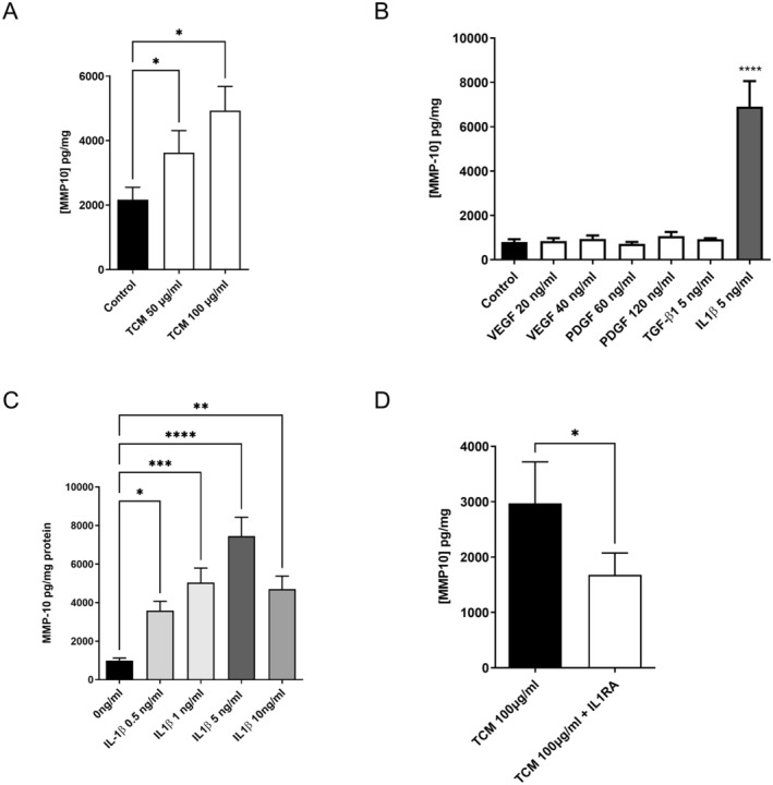
Maternal uterine spiral arteries (SpA) undergo significant structural changes in early pregnancy, resulting in increased blood flow to the developing fetus. Endothelial cells (EC) and vascular smooth muscle cells (VSMC) are lost from the SpA wall and are replaced by trophoblasts. We have previously shown that matrix metalloproteinase 10 (MMP-10) and Heparin binding-EGF like growth factor (HB-EGF) gene expression is increased in a 3D EC/VSMC co-culture system in response to trophoblast secreted factors. This study investigated trophoblast mediated MMP-10 and HB-EGF expression and determined if there was a relationship between the secretion of MMP-10 and the release of soluble HB-EGF (sHB-EGF) from EC. MMP-10 was widely expressed in first trimester decidual tissue including trophoblast, and EC, but not VSMC. MMP-10 expression was significantly lower in decidual tissue from pregnancies at increased risk of developing pre-eclampsia compared to low-risk pregnancies. In vitro, SGHEC-7 cells, a human EC line, but not SGHVMC-9, a human VSMC cell line, secreted MMP-10 in response to trophoblast conditioned medium (TCM). TCM contains several growth factors and cytokines, but only interleukin-1β (IL1β) significantly stimulated MMP-10 secretion by SGHEC-7 cells. Interleukin-1 receptor antagonist (IL-1Ra) significantly inhibited TCM-induced MMP-10 secretion. Interrogation of intracellular pathways established the involvement of MEK and JNK in TCM and IL-1β stimulated MMP-10 secretion. Although IL-1β also significantly increased sHB-EGF, inhibition of MMP-10 activity using a broad spectrum MMP inhibitor had no effect on sHB-EGF. Western blot analysis indicated that MMP-10 secreted by EC in response to IL-1β stimulation was the enzymatically inactive pro form.

Keywords: MMP‐10 (MMP10); endothelial; spiral artery; stromelysin‐2; trophoblast.


Conflict of interest statement

The authors have nothing to report.

The authors declare no conflicts of interest.

Figures

FIGURE 1
FIGURE 1
Immunofluorescent staining of first trimester placental bed decidua. (A–C) expression of MMP10 in vascular cells (A) Immunofluorescent staining of CK7 (trophoblasts; green) (B) MMP‐10 (orange/red) and CD31 (endothelial cells; green) and (C) MMP‐10 (orange/red) and smooth muscle actin (VSMC; green). White arrows indicate endothelial cells. The inserts panels show the appropriate non‐immune IgG negative control stained. (D–F) expression of (D) CK7 (E) MMP10 and (F) co‐localisation of CK7 and MMP‐10. The trophoblast plug is highlighted by and asterisk while the arrow indicates a dual‐stained extravillous trophoblast. (G) Expression of MMP10 in 1st trimester decidua from normal pregnancies and those at increased risk of developing PE. Decidual tissue was homogenized in RIPA buffer and the protein separated by gel electrophoresis. Membranes were then probed for MMP10 and tubulin which was used as a loading control. The data are expressed as a ratio of the MMP10 to tubulin and are the mean ± SEM (n = 8 per risk group). The level of significance was determined using unpaired t‐test where *p < 0.05.
FIGURE 2
FIGURE 2
The effect of TCM and IL1β on the secretion of MMP‐10 by EC. (A) EC were stimulated with TCM for 2 h before the medium was removed and the cells were washed extensively with PBS. The cells were then incubated with phenol red free RPMI containing 5% (v/v) FCS for 72 h. (B) EC were serum starved overnight and then stimulated with recombinant growth factors and cytokines for 2 h, washed and incubated as above for 72 h. (C) EC cells were stimulated with increasing concentrations of IL‐1β for 2 h, washed and incubated for 48 h as detailed above. (D) EC cells were either incubated alone or with IL‐1RA for 20 min prior and stimulated with TCM for 2 h, washed and incubated as above for 48 h. At the end of the experiments the concentration of MMP‐10 was determined by ELISA. The results were corrected for total cellular protein and expressed as mean + SEM. Where appropriate the results were analyzed by either a Student‐t test, a one‐way ANOVA or a mixed‐effect model using Graph‐pad PRISM. (*p < 0.05, **p < 0.01, ***p < 0.001 ****p < 0.0001).
FIGURE 3
FIGURE 3
Effect of different pathway inhibitors on TCM and IL‐1β stimulation of EC. (A) EC were incubated with either PD98059, SB203580, or DMSO vehicle for 20 min prior to the addition of TCM (142 ng/mL) for 2 h before being replace with phenol red free RPMI containing 5% (v/v) FCS. After 48 h the medium was removed and MMP‐10 determined by ELISA. (B) EC cells were incubated in the presence and absence of 10 μM PD98059 or DMSO for 20 min prior to the addition of IL‐1β at 5 and 10 ng/mL for 2 h before being replace with phenol red free RPMI containing 5% (v/v) FCS. After 48 h the medium was removed and MMP‐10 determined by ELISA. The results were corrected for protein and expressed as the mean + SEM. (n = 3, ns is not significant, *p < 0.05, **p < 0.01, ***p < 0.001 ****p < 0.0001).
FIGURE 4
FIGURE 4
Inhibition of JNK and STAT3 inhibits IL‐1β and TCM‐stimulated MMP‐10 secretion by EC. (A) EC cells were incubated in the presence and absence of STAT3 inhibitor III for 20 min prior to the addition of 50 ng/mL TCM for 2 h. (B) EC cells were incubated in the presence and absence of JNK inhibitor, CC401 or DMSO for 20 min prior to the addition of IL‐1β at 5 for 2 h. After 2 h the medium was removed and replaced with phenol red free RPMI containing 5% (v/v) FCS. After 48 h the medium was removed and MMP‐10 determined by ELISA. This medium was removed and replaced with phenol red free RPMI containing 5% (v/v) FCS. After 48 h the medium was removed and MMP‐10 determined by ELISA. The results were corrected for total cellular protein and expressed as the mean + SEM (A n = 5, B n = 3 independent experiments, *p < 0.05 and **p < 0.01).
FIGURE 5
FIGURE 5
The effect of IL‐1β release of HBEGF and secretion of MMP10 by endothelial cells in culture. (A) EC (0.5 × 106 per dish) were incubated in 0% (v/v) FCS media for 72 h with 5 ng/mL IL‐1β with or without 100 mM NNGH. The concentration of HBEGF was determined by ELISA and corrected for cellular protein. The results are expressed as fold change in HBEGF and is the mean ± SEM of n = 4 independent experiments. The level of significance was determined using one‐way ANOVA with Sidak's multiple comparisons test where ns, not significant, ****p < 0.0005. (B) rh‐MMP3 and rh‐MMP10 (2.5‐10 ng/well) was subjected to PAGE, transferred and probed with the R&D systems antibody to determine specificity. (C) EC (0.75 × 106 cells per dish) were cultured for 48 and the medium then removed and concentrated as detailed in the methods. Lane 1 is the molecular size ladder lane 2 control cells, lane 3 cells stimulated with 10 ng/mL IL1β and lane 4 is recombinant MMP10. MMP10 was detected by chemiluminescence and is representative of n = 3 independent experiments. The molecular weight of bands obtained in this experiment were calculated using Image Lab v6.1 by BioRad.

Similar articles

References

    1. Burton G. J., Woods A. W., Jauniaux E., and Kingdom J. C. P., “Rheological and Physiological Consequences of Conversion of the Maternal Spiral Arteries for Uteroplacental Blood Flow During Human Pregnancy,” Placenta 30 (2009): 473–482. - PMC - PubMed
    1. Smith S. D., Choudhury R. H., Matos P., et al., “Changes in Vascular Extracellular Matrix Composition During Decidual Spiral Arteriole Remodeling in Early Human Pregnancy,” Histology and Histopathology 31 (2016): 557–571. - PubMed
    1. Craven C. M., Morgan T., and Ward K., “Decidual Spiral Artery Remodelling Begins Before Cellular Interaction With Cytotrophoblasts,” Placenta 19 (1998): 241–252. - PubMed
    1. Kam E. P., Gardner L., Loke Y. W., and King A., “The Role of Trophoblast in the Physiological Change in Decidual Spiral Arteries,” Human Reproduction 14 (1999): 2131–2138. - PubMed
    1. Pijnenborg R., Vercruysse L., and Hanssens M., “The Uterine Spiral Arteries in Human Pregnancy: Facts and Controversies,” Placenta 27 (2006): 939–958. - PubMed

MeSH terms

Substances

LinkOut - more resources