. 2025 May 6:13:RP103615.
doi: 10.7554/eLife.103615.

Shc1 cooperates with Frs2 and Shp2 to recruit Grb2 in FGF-induced lens development

Affiliations

Shc1 cooperates with Frs2 and Shp2 to recruit Grb2 in FGF-induced lens development

Qian Wang et al. Elife. .

Abstract

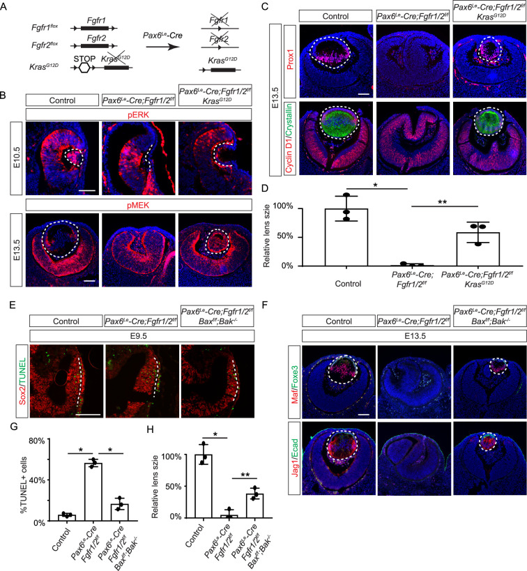
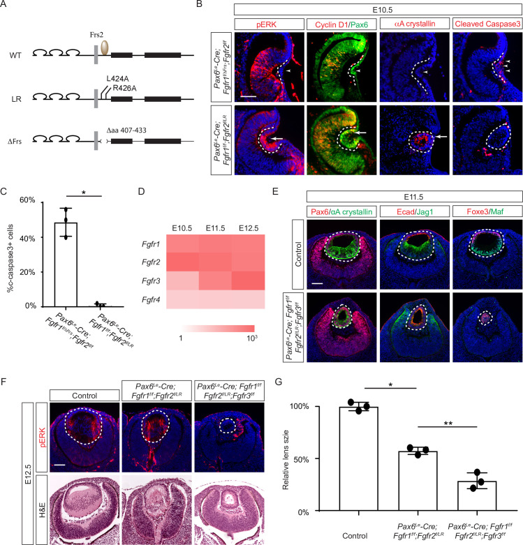
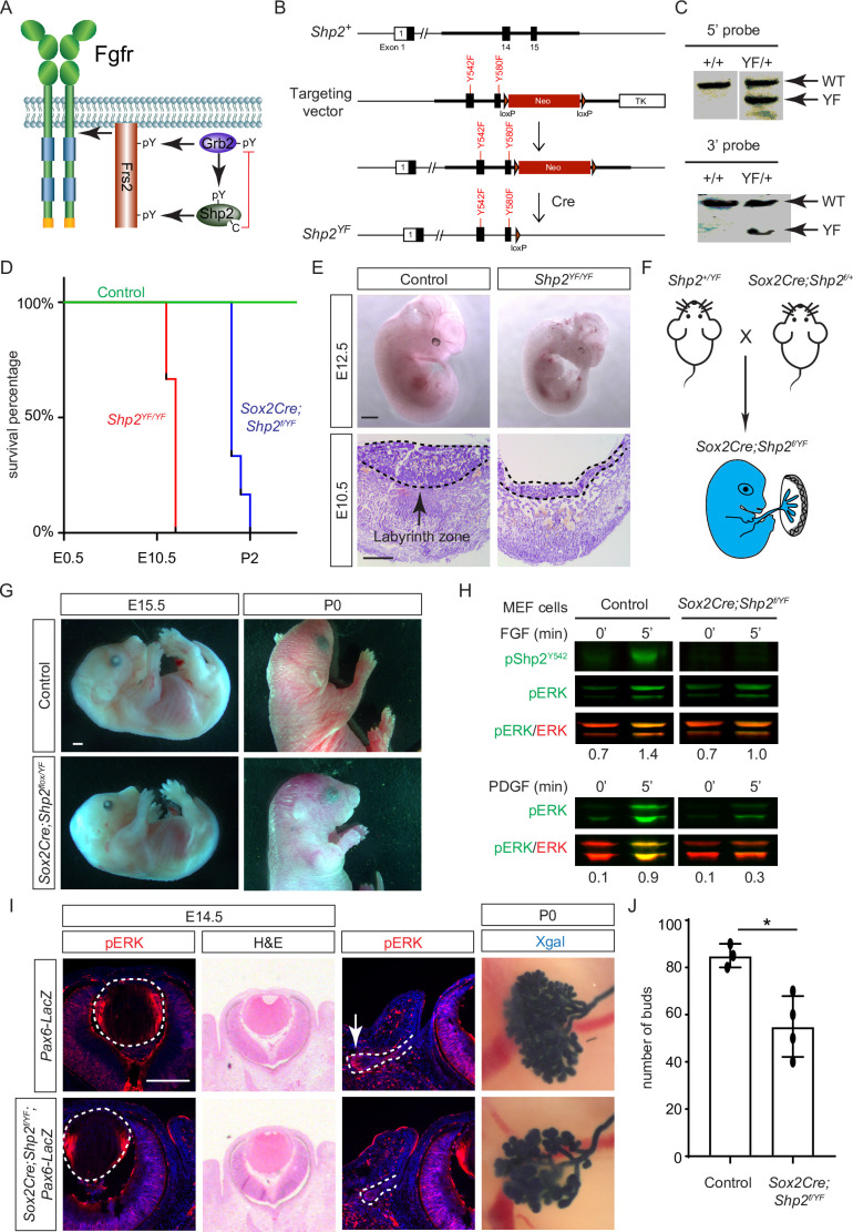
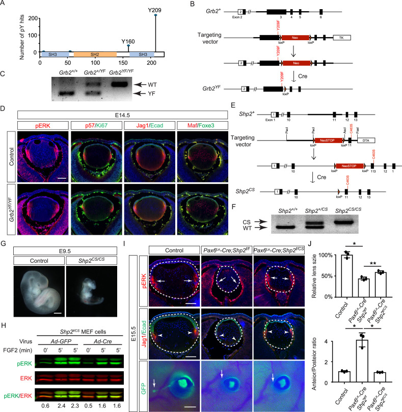
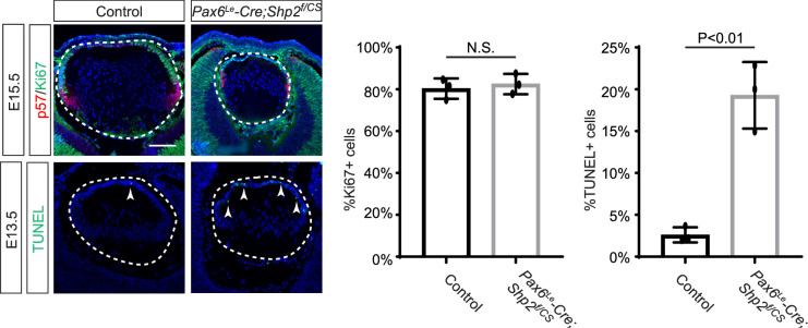
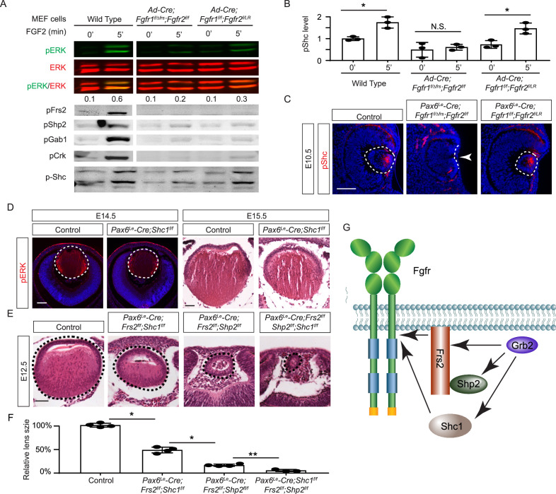
Fibroblast growth factor (FGF) signaling elicits multiple downstream pathways, most notably the Ras/MAPK cascade facilitated by the adaptor protein Grb2. However, the mechanism by which Grb2 is recruited to the FGF signaling complex remains unresolved. Here, we showed that genetic ablation of FGF signaling prevented murine lens induction by disrupting transcriptional regulation and actin cytoskeletal arrangements, which could be reproduced by deleting the juxtamembrane region of the FGF receptor and rescued by Kras activation. Conversely, mutations affecting the Frs2-binding site on the FGF receptor or the deletion of Frs2 and Shp2 primarily impact later stages of lens vesicle development involving lens fiber cell differentiation. Our study further revealed that the loss of Grb2 abolished MAPK signaling, resulting in a profound arrest of lens development. However, removing Grb2's putative Shp2 dephosphorylation site (Y209) neither produced a detectable phenotype nor impaired MAPK signaling during lens development. Furthermore, the catalytically inactive Shp2 mutation (C459S) only modestly impaired FGF signaling, whereas replacing Shp2's C-terminal phosphorylation sites (Y542/Y580) previously implicated in Grb2 binding only caused placental defects, perinatal lethality, and reduced lacrimal gland branching without impacting lens development, suggesting that Shp2 only partially mediates Grb2 recruitment. In contrast, we observed that FGF signaling is required for the phosphorylation of the Grb2-binding sites on Shc1 and the deletion of Shc1 exacerbates the lens vesicle defect caused by Frs2 and Shp2 deletion. These findings establish Shc1 as a critical collaborator with Frs2 and Shp2 in targeting Grb2 during FGF signaling.

Keywords: FGF; Grb2; MAPK; Shc; Shp2; cell biology; developmental biology; lens induction; mouse.

Plain language summary

Cells communicate by releasing proteins that bind to receptors on recipient cells, triggering a cascade of events that alter the cell’s behavior. A family of signaling proteins called fibroblast growth factors (FGFs) is critical for various biological processes, especially during embryonic development. While scientists have a good understanding of how FGFs reach their target cells, less is known about the series of events they activate once they bind to a receptor. Three adaptor proteins – called Frs2, Shp2 and Grb2 – are essential for propagating the FGF signal. First, the activated receptor binds to and adds phosphate groups to Frs2, which then recruits and facilitates the phosphorylation of Shp2 and Grb2. Here, Wang, Li, Mao et al. offer fresh insights into how this complex of molecules transmit the FGF signal through cells during lens development. First, the team genetically modified the structure and activity of FGF receptors in mice to see how this impacted the formation of their lenses. They found that the membrane-embedded portion of the receptor, which includes the binding site for Frs2, is critical for regulating the consecutive steps of lens development. However, the initial stages of lens formation could still occur when only the Frs2 binding site was mutated. Loss of Grb2 produced a similar effect, suggesting that Frs2 and Grb2 are particularly important for the later stages of lens development. Previous studies have suggested that Shp2 acts as a bridge between Frs2 and Grb2. To test this theory, Wang, Li, Mao et al. deleted the two sites in Shp2 that are responsible for binding to Grb2 and stopped phosphorylation interactions between the two adaptors. While these changes affected embryo survival, they had only a modest impact on lens development. Further experiments revealed that another adaptor protein called Shc1 can also mediate Grb2 recruitment and activation, and may be responsible for transmitting the FGF signal later in lens development. This study provides deeper insights into the network of signaling molecules activated by FGFs, uncovering new mechanisms and adaptors involved in this pathway. The findings suggest that the FGF signaling network is highly adaptable, with different components being required at specific stages of development. Future research expanding on this work may lead to the discovery of therapies that target specific organs affected by FGF-related disorders.


Conflict of interest statement

QW, HL, YM, AG, EP, YW, AC, JP, XZ No competing interests declared

Figures

Figure 1.
Figure 1.. Fibroblast growth factor (FGF) signaling regulates lens development in a dose-dependent manner.
(A) Schematic diagram of murine lens development. The head ectoderm is induced by the underlying optic vesicle to become the lens placode, which subsequently folds inwards to become the lens pit. The closure of the lens vesicle sets the stage for the differentiation of the lens epithelium into the lens fibers. (B) Depletion of all four Fgfr1/2/3/4, driven by Pax6Le-Cre and traced with GFP (arrows), led to a thinner lens placode, evident from the absence of pERK signals and the failure to upregulate Sox2 like Pax6 (inserts, arrowheads). (C) Fgfr1/2/3/4 mutants displayed disrupted apical constriction (F-actin accumulation, arrows) and lacked lens-specific expression of Foxe3 and Jag1 (arrowheads). Dotted lines outline the lens pit. (D) Despite Fgfr1/2/3/4 mutations, BMP (pSmad1/5/9 staining, arrowheads) and Wnt signaling (Lef1 expression, arrows) remained unaffected. (E) The absence of Fgfr1/2 alone did not impede the apical buildup of F-actin nor the expression of Foxe3, indicating partial retention of lens development processes. (F) Crucial lens markers, Prox1 and αA-crystallin, were absent in Fgfr1/2 mutants, pointing to a significant developmental defect after the lens induction stage. Biological replicates, n=3. (G) Fgfr1/2 mutants exhibited loss of cell proliferation marker Cyclin D1 (arrows) and widespread apoptosis (TUNEL staining, arrowheads). (H) Quantification of Foxe3+ cells in Fgfr1/2 mutants. Student’s t-test, n=3, p<0.01. (I) Quantification of Cyclin D1+ cells in Fgfr1/2 mutants. Student’s t-test, n=3, p<0.001. (J) Quantification of TUNEL+ cells in Fgfr1/2 mutants. Student’s t-test, n=3, p<0.0001. Scale bars:25 µm.
Figure 1—figure supplement 1.
Figure 1—figure supplement 1.. Lens development in Fgf receptor mutants.
(A) Depletion of all four Fgfr1/2/3/4 did not disrupt the Fibronectin expression at the basal side of the lens placode (arrows). (B) pmTOR and pS6 staining were reduced in Pax6Le-Cre;Fgfr1f/f;Fgfr2f/LR lens. (C) Quantification of pmTOR fluorescent intensity in Fgfr1/2 mutants. Student’s t-test, n=3, p<0.0001. (D) Quantification of pmS6 fluorescent intensity in Fgfr1/2 mutants. Student’s t-test, n=3, p<0.01. Scale bars:25 µm.
Figure 2.
Figure 2.. Restoration of lens development in fibroblast growth factor (FGF) signaling mutants via Kras activation and apoptosis inhibition.
(A) The Pax6Le-Cre driver facilitated the excision of the floxed alleles of Fgf1/2 along with the LSL-STOP cassette at the Kras locus, leading to the expression of the constitutively active KrasG12D allele within the Fgf1/2 mutant background. (B) The activation of Kras signaling in the FGF signaling mutant lenses reinstated pERK activity at E10.5 and pMEK expression at E13.5, indicating restoration of MAPK signaling. (C) The lens-specific expression of Prox1 and αA-crystallin were also recovered, indicating successful lens development rescue. (D) Quantification of the lens size. One way ANOVA, n=3, *p<0.001, **p<0.02. (E) The deletion of pro-apoptotic genes Bak and Bax in Fgf1/2 mutants suppressed apoptosis as shown by TUNEL staining. (F) Inhibiting apoptosis in Fgfr1/2 mutants facilitated lens formation, as indicated by the expression of lens differentiation markers Prox1, Maf, and Jag1. (G) Quantification cell apoptosis at E9.5. One-way ANOVA, n=3, *p<0.001. (H) Quantification of the lens size at E13.5. One-way ANOVA, n=3, *p<0.0001, **p<0.05. Scale bars:50 µm.
Figure 3.
Figure 3.. The Frs2 binding site on fibroblast growth factor receptor 2 (FGFR) is only required for lens vesicle differentiation.
(A) Overview of Fgfr mutant alleles. Fgfr1ΔFrs lacks the Frs2 binding domain (amino acid 407–433), and Fgfr2LR has point mutations disrupting Frs2 binding (L424A and L426A). (B) In Fgf1/2 compound mutants, loss of pERK, CyclinD1, αA-crystallin, and increased cleaved caspase3 were observed with the Fgfr1ΔFrs allele but not the Fgfr2LR allele. (C) Quantification of cleaved caspase3 staining. Student’s t-test, n=3, *p<0.001. (D) Heatmap depicts Fgfr expression levels during lens development. (E) Fgfr2LR mutants in the Fgfr1/2/3 genetic background showed impaired lens vesicle differentiation, with posterior lens epithelial cells failing to elongate and activate lens fiber cell markers Jag1 and Maf. (F) Fgfr1/2/3 triple mutants with Fgfr2LR lost pERK staining and displayed a shallow lens vesicle at E12.5. (G) Quantification of the lens size. One-way ANOVA n=3, *p<0.001, **p<0.05. Scale bars:50 µm.
Figure 4.
Figure 4.. Grb2 is essential for lens vesicle survival, proliferation, and differentiation.
(A) The targeted removal of Grb2 in the lens led to a loss of pERK signaling, reduced CyclinD3 expression, increased apoptosis (TUNEL staining), and disrupted expression of critical lens development genes Maf, Foxe3, Jag1, and γ-crystallin expression at E12.5. (B) Grb2 mutants displayed absent CyclinD1, Prox1, and p57 expression at E11.5 and remained an undifferentiated hollow vesicle at E13.5, failing to undergo normal lens fiber elongation. (C) Quantification of the lens size. Student’s t-test, n=3, *p<0.0001. Scale bars:50 µm.
Figure 5.
Figure 5.. Shp2 C-terminal tyrosine phosphorylation is required for embryonic survival but dispensable for lens development.
(A) Schematic of the core FGF signaling pathway. FGFR activation leads to phosphorylation of the adaptor Frs2 on N- and C-terminal tyrosines, recruiting Grb2 and Shp2, respectively. Shp2 can also bind Grb2 via its own C-terminal phosphotyrosines, and dephosphorylates Grb2 via its catalytic cysteine residue. (B) Generation of the Shp2YF allele by homologous recombination to introduce loxP-flanked Neo and point mutations (Y542F and Y580F) disrupting the Shp2 C-terminal phosphotyrosine sites. The Neo cassette was subsequently excised by Cre-mediated recombination. (C) Validation of the Shp2YF allele targeting was confirmed through Southern blot analysis with both 5’ and 3’ probes. (D) Kaplan-Meier survival curves demonstrate early lethality of Shp2YF embryos (E12.5) and perinatal lethality of Sox2Cre;Shp2f/YF mutants. n=5 for Shp2YF/YF and n=6 for Sox2Cre;Shp2f/YF mutants. (E) Shp2YF/YF embryos displayed reduced body size at E12.5 and thinner labyrinth zones in their placenta at E10.5. (F) Sox2Cre-mediated targeting restricts Shp2 deficiency to the embryonic proper, circumventing placental abnormalities. (G) Sox2Cre;Shp2f/YF mutants appeared grossly normal at E15.5 but failed to survive after birth. (H) While Y542 phosphorylation in Shp2 was lost as expected, Sox2Cre;Shp2f/YF MEFs exhibited a more pronounced reduction in pERK response to PDGF stimulation compared to FGF stimulation. (I) Sox2Cre;Shp2f/YF mutant lens displayed normal pERK staining and morphology, but reduced pERK in lacrimal glands at E14.5 and decreased bud numbers at P0. (J) Quantification of the number of lacrimal gland buds. Student’s t-test, n=3, *p<0.02. Scale bars: 100 µm.
Figure 6.
Figure 6.. Shp2 phosphatase activity is partially required for fibroblast growth factor (FGF) signaling independently of Grb2 dephosphorylation.
(A) PhosphositePlus database indicates that Grb2 predominantly undergoes phosphorylation at Y209 and less frequently at Y160. (B) The Grb2YF allele was constructed by homologous recombination to integrate a loxP-flanked Neo cassette and a Y209F point mutation into the Grb2 locus. (C) PCR genotyping confirmed the presence of the Grb2YF allele. (D) Grb2 mutant lens typical expression patterns of pERK, cell cycle markers p57 and Ki67 and differentiation markers Foxe3, Maf, and Jag1. (E) The Shp2CS allele was generated by inserting the C459S mutation into the Shp2 locus by homologous recombination, followed by Cre-mediated removal of the loxP-flanked Neo cassette. (F) Shp2CS allele validated by PCR genotyping. (G) Shp2CS/CS mutants exhibited growth retardation and died at E9.5. (H) Shp2f/CS MEF cells retained a significant capacity to activate pERK upon FGF stimulation after Cre virus infection. (I) Pax6Le-Cre;Shp2f/f mutants exhibited loss of pERK and Jag1 staining at the lens transition zone (arrowheads), which also shifted posteriorly. Pax6Le-Cre;Shp2f/CS mutant lens, in contrast, maintained staining at the equatorial region. Notably, both mutant types lacked lacrimal gland buds (arrows). (J) Quantification of the lens size. One-way ANOVA, n=3, *p<0.0001, **p<0.001. (K) Quantification of the lens perimeter spanning the anterior epithelium versus that of the posterior lens fiber. One-way ANOVA, n=3, *p<0.001. Scale bars:50 µm.
Figure 6—figure supplement 1.
Figure 6—figure supplement 1.. Cell proliferation and apoptosis inShp2CS mutants.
Pax6Le-Cre;Shp2f/CS lens exhibits normal expression of proliferation marker Ki67, but there is a significant increase in TUNLE+ cells (arrowheads). Student’s t-test, N.S. (not significant) for %Ki67+ cells and p<0.01 for %TUNEL+ cells in the lens epithelium. Scale bar:50 µm.
Figure 7.
Figure 7.. Shc1 complements Frs2 and Shp2 in mediating fibroblast growth factor (FGF) signaling in lens development.
(A) Fgfr1f/f;Fgfr2f/LR infected with Cre virus showed stronger pERK and pShc activation than Fgfr1f/ΔFrs;Fgfr2f/f mouse embryonic fibroblast (MEF) cells, despite both losing Frs2, Shp2, Gab1, and Crk phosphorylation. (B) Quantification of pShc levels. One-way ANOVA, n=3, *p<0.02. N.S. Not significant. (C) pShc staining was lost in Pax6Le-Cre;Fgfr1f/ΔFrs;Fgfr2f/f mutant lens (arrowhead) but preserved in Pax6Le-Cre;Fgfr1f/f;Fgfr2f/LR lens. (D) Shc1-deficient lenses showed a slight decrease in both pERK staining intensity and overall lens size. (E) E12.5 Frs2/Shc1 mutant lenses were smaller than controls, while lenses from Frs2/Shp2 mutants showed a pronounced hollow vesicle structure, a condition that worsened in Frs2/Shp2/Shc1 triple mutants. (F) Quantification of the lens size. One-way ANOVA, n=4, *p<0.001, **p<0.05. (G) Model of FGF signaling network. Frs2 recruits Grb2 directly and indirectly through Shp2, while Shc1 provides an alternate Grb2 recruitment route independent of Frs2. Scale bars:50 µm.

Update of

References

    1. Ackermann JA, Radtke D, Maurberger A, Winkler TH, Nitschke L. Grb2 regulates B-cell maturation, B-cell memory responses and inhibits B-cell Ca2+ signalling. The EMBO Journal. 2011;30:1621–1633. doi: 10.1038/emboj.2011.74. - DOI - PMC - PubMed
    1. Ahmed Z, Lin CC, Suen KM, Melo FA, Levitt JA, Suhling K, Ladbury JE. Grb2 controls phosphorylation of FGFR2 by inhibiting receptor kinase and Shp2 phosphatase activity. The Journal of Cell Biology. 2013;200:493–504. doi: 10.1083/jcb.201204106. - DOI - PMC - PubMed
    1. Ahmed Z, Timsah Z, Suen KM, Cook NP, Lin CC, Gagea M, Marti AA, Ladbury JE. Grb2 monomer-dimer equilibrium determines normal versus oncogenic function. Nature Communications. 2015;6:7354. doi: 10.1038/ncomms8354. - DOI - PMC - PubMed
    1. Araki T, Nawa H, Neel BG. Tyrosyl phosphorylation of Shp2 is required for normal ERK activation in response to some, but not all, growth factors. The Journal of Biological Chemistry. 2003;278:41677–41684. doi: 10.1074/jbc.M306461200. - DOI - PubMed
    1. Ashery-Padan R, Marquardt T, Zhou X, Gruss P. Pax6 activity in the lens primordium is required for lens formation and for correct placement of a single retina in the eye. Genes & Development. 2000;14:2701–2711. doi: 10.1101/gad.184000. - DOI - PMC - PubMed

MeSH terms

Substances