. 2025 May 6;16(1):4196.
doi: 10.1038/s41467-025-58669-7.

A potent protective bispecific nanobody targeting Herpes simplex virus gD reveals vulnerable epitope for neutralizing

Affiliations

A potent protective bispecific nanobody targeting Herpes simplex virus gD reveals vulnerable epitope for neutralizing

Jing Hu et al. Nat Commun. .

Abstract

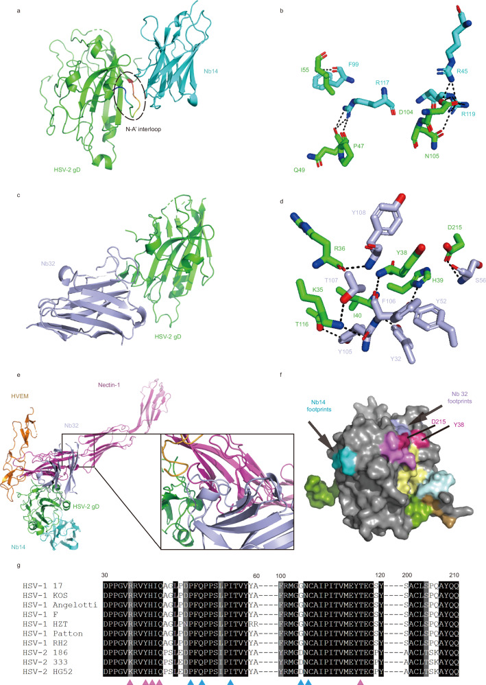
Herpes simplex virus (HSV) causes significant health burden worldwide. Currently used antiviral drugs are effective but resistance can occur. Here, we report two high-affinity neutralizing nanobodies, namely Nb14 and Nb32, that target non-overlapping epitopes in HSV gD. Nb14 binds a neutralization epitope located in the N-A' interloop, which prevents the interaction between gD and gH/gL during the second step of conformational changes during membrane fusion after virus attachment. The bispecific nanobody dimer (Nb14-32-Fc) exhibits high potency in vitro and in vivo. Mechanistically, Nb14-32-Fc neutralizes HSVs at both the pre-and post-attachment stages and prevents cell-to-cell spread in vitro. Administration of Nb14-32-Fc at low dosage of 1 mg/kg provides 100% protection in an HSV-1 infection male mouse model and an HSV-2 infection female mouse model. Our results demonstrate that Nb14-32-Fc could serve as a promising drug candidate for treatment of HSV infection, especially in the cases of antiviral drug resistance and severe herpes encephalitis.


Conflict of interest statement

Competing interests: The authors declare no competing interests.

Figures

Fig. 1
Fig. 1. Isolation and preliminary screening of anti-HSV-2 gD nanobodies.
a Enrichment of phages after panning on HSV-2 gD. b Monoclonal phage ELISA results for characterizing the binding of the isolated Nbs to HSV-2 gD. Each point represents a nanobody single colony. PBS group was used as a negative control. c, d Thirteen nanobodies (n = 13) at 1 μg/mL were tested for neutralization effects against HSV-1-GFP by flow cytometry. The intensity of fluorescence reflects the neutralization activity of antibody. c Representative images of Nb14-Fc group and control group (Fc alone). d Non-infection ratio of thirteen nanobodies. Nb14-Fc and Nb32-Fc could inhibit > 90% HSV-1 infection. Data and error bars are mean ± S.D, n = 3 biological independent experiments. Source data are provided as a Source Data file.
Fig. 2
Fig. 2. Identification of two neutralizing nanobodies (Nb14-Fc and Nb32-Fc) targeting two different epitopes on HSV gD.
a, b Multi-concentration ELISA-binding assay of Nb14-Fc (a) and Nb32-Fc (b) towards HSV gD. The OD450 emissions are depicted by curves. EC50 (nM) indicated median effect concentration and was calculated to assess the binding potency of Nbs. c, d The binding kinetics of Nb14-Fc (c) and Nb32-Fc (d) to HSV-1 gD were monitored by the Biacore 8 K system. e, f The binding kinetics of Nb14-Fc (e) and Nb32-Fc (f) to HSV-2 gD were monitored by the Biacore 8 K system. The actual responses (colored lines) and the data fitted to a 1:1 binding model (black dotted lines) are shown in (cf). KD, equilibrium dissociation constant; ka, association constant; kd, dissociation constant. g, h The neutralizing activities of Nb14-Fc (g) and Nb32-Fc (h) against HSV-1 and HSV-2. The half-maximal inhibitory concentration (IC50) values of the plaque reduction neutralization test (PRNT) were calculated by fitting the inhibition rates against antibody concentrations with a sigmoidal dose-response curve. i Competitive binding of Nb14-Fc and Nb32 to HSV-2 gD detected by ELISA. HSV-2 gD was coated on 96-well plates, Nb14-Fc was mixed with 4-fold serial dilutions of Nb32. The competition was determined by the reduction of the HRP-anti-IgG1 Fc induced chemiluminescence signal (OD450). The inhibition was calculated by comparing it to the Nb negative-control well. Data and error bars are mean ± S.D, n = 3 biological independent experiments in (a, b, and gi). Nb, Nanobody. Source data are provided as a Source Data file.
Fig. 3
Fig. 3. Structural basis of neutralization for Nb14 and Nb32.
a Overall structure of Nb14/HSV-2 gD complex shown in cartoon. HSV-2 gD is highlighted in green and Nb14 in cyan. The N-A’ interloop is highlighted with a black dotted line. b Detailed interactions between Nb14 and HSV-2 gD. Interacting residues are shown as sticks, and dotted lines indicate hydrogen bonds and salt bridges. c Overall structure of Nb32/HSV-2 gD complex shown in cartoon. HSV-2 gD is highlighted in green and Nb32 in light blue. d Detailed interactions between Nb32 and HSV-2 gD. Interacting residues are shown as sticks, and dotted lines indicate hydrogen bonds and salt bridges. e Structural alignment of Nb14/Nb32/HSV-2 gD and Nectin-1/HSV-2 gD complexes [PDB code: 4MYW] and HVEM/HSV-1 gD complexes [PDB code: 1JMA]. Steric clash between Nb32 and Nectin-1 is highlighted. f A grayscale surface representation of the HSV-1 gD with the epitopes for Nb14 (cyan), Nb32 (light blue), E317 (pale yellow), HSV8 (limon), CH42 (pale cyan), CH43 (sand), 4A3 (violet). Epitope overlap is colored hot pink. g Conservation analysis of binding epitope in different HSV strains. The binding epitope of Nb14 is displayed in blue, and the binding epitope of Nb32 is displayed in pink.
Fig. 4
Fig. 4. Nb32 interferes with gD binding to its cellular receptor Nectin-1 while Nb14 acts independent of Nectin-1 or HVEM.
a, b The competitive binding kinetics of Nb14-Fc (a) and Nb32-Fc (b) with receptors were identified by surface plasmon resonance (SPR). HSV-2 gD was immobilized onto CM5 sensor chip. Nbs and receptors were flowed over the ship successively. The real-time binding data are shown. c, d The inhibition of Nb14-Fc (c) and Nb32-Fc (d) activities were characterized by competitive ELISA, which demonstrated the ability of antibodies to inhibit the binding of HSV-2 gD to receptors expressed on CHO cells. e Neutralizing potency of Nb14-Fc and Nb32-Fc characterized in CHO-Nectin-1 or CHO-HVEM cells were measured by quantitative polymerase chain reaction (qPCR). The statistical significance was determined by comparing with the control group (no antibody group) at the same infection dose using Dunnett’s multiple comparison tests in a one-way ANOVA analysis, revealing a significant difference with ****p < 0.0001, ***p < 0.001, **p < 0.01, *p < 0.05, p > 0.05(ns). ns, no significant. Data and error bars are mean ± S.D, n = 3 biological independent experiments in c-e. Source data are provided as a Source Data file.
Fig. 5
Fig. 5. Nb14-32-Fc and its parent antibodies interfere with cell fusion and Nb14 interferes with gD binding to gH/gL.
a HEK-293T cells were co-transfected with glycoprotein expression plasmids (PLVX-gB-Zsgreen, PLVX-gD-Zsgreen, PLVX-gH-Zsgreen and PLVX-gL-Zsgreen) and cultured in medium containing 50 μg/ml antibodies at 37 °C for 48 h. Cells were stained with DAPI and observed under a fluorescence microscope. Scale bars, 500 μm. The relative Zsgreen area was quantified using ImageJ, with the fusion activity of the control set to 100%. b CHO cells which were co-transfected with glycoprotein expression plasmids and pG5-Luc were mixed with HEK-293T cells co-transfected with pACT-Myod and pBind-Id. Mixed cells were co-cultured in fresh media containing 50 μg/ml antibodies. After 48 h, cell-to-cell membrane fusion was evaluated using luciferase activity. The statistical significance in (a) and (b) was determined by comparing with the control group (no antibody group) using Dunnett’s multiple comparison tests in a one-way ANOVA analysis, revealing a significant difference with ****p < 0.0001, ***p < 0.001, **p < 0.01, *p < 0.05, p > 0.05(ns). ns, no significant. Data and error bars are mean ± S.D, n = 3 biological independent experiments. c Multi-concentration cell-based ELISA-binding assay of HSV-2 gD towards gH/gL expressed on CHO cells. The OD450 values are depicted by curves. d The inhibition of Nb14-Fc activities was characterized by competitive ELISA, which demonstrated the ability of Nb14-Fc to inhibit the binding of HSV-2 gD to gH/gL expressed on CHO cells. Data and error bars in (c) and (d) are mean ± S.D, n = 3 biological independent experiments. Source data are provided as a Source Data file.
Fig. 6
Fig. 6. Engineering of Nb-14-32-Fc with improved neutralizing potency.
a Diagram showing the modification of Nb-14-32-Fc. Nb14 and Nb32 are tandemly linked with a (GGGGS)5 linker and then fused with a human IgG1 Fc tag. HSV-2 gD is shown in gray surface. Nb14 and Nb32 are depicted in cartoon and highlighted in cyan and light blue, respectively. b The binding kinetics of Nb14-32-Fc to HSV gD were monitored by the Biacore 8 K system. The actual responses (colored lines) and the data fitted to a 1:1 binding model (black dotted lines) are shown. KD, equilibrium dissociation constant; ka, association constant; kd, dissociation constant. c, d The neutralizing activities of Nb-14-32-Fc and E317 against HSV-1(c) and HSV-2(d). The half-maximal inhibitory concentration (IC50) values of the plaque reduction neutralization test (PRNT) were calculated by fitting the inhibition rates against antibody concentrations with a sigmoidal dose-response curve. e, f Comparison of the neutralization efficacy of Nb14-32-Fc when serial dilutions of the antibody were added before (pre-attachment) or after (post-attachment) HSV-1(e) and HSV-2(f) virions interacted with Vero cells. Neutralization effects were determined after 72 h post-infection by the standard plaque assay. g, h Cell-to-cell spread of HSV-1 and HSV-2 in Vero cells was detected by infectious center assay. Vero cells were infected with HSV-1 and HSV-2 at an MOI of 5 in the presence of camel immune serum to neutralize any free virus released into the medium. After a total incubation of 5 h, the infected cells were detached with trypsin, plated onto uninfected Vero cells and then the antibodies were added. Cells were fixed and observed by fluorescence microscopy at 48 h post-infection. The infected cells without antibodies were used as a control. The statistical significance was determined by comparing with the control group using Dunnett’s multiple comparison tests in a one-way ANOVA analysis, revealing a significant difference with ****p < 0.0001, ***p < 0.001, **p < 0.01, *p < 0.05, p > 0.05(ns). ns, no significant. Scale bars, 200 μm. Data and error bars are mean ± S.D, n = 3 biological independent experiments in (ch). Source data are provided as a Source Data file.
Fig. 7
Fig. 7. Nb14-32-Fc and its parent nanobodies provide therapeutic protection against intracranial infection of HSV-1 F strain in mice.
a Animal experiment scheme. In comparison of treatment efficacy of Nb14-32-Fc and its parent nanobodies, mice were divided into four groups (n = 8 per group), including a control group administered vehicle (PBS) and three treatment groups. In comparison of treatment efficacy of Nb14-32-Fc and E317, mice were divided into three groups (n = 5 per group), including a control group administered vehicle (PBS) and two treatment groups. Mice were intracranially infected with the HSV-1 F strain;12 h later, mice were administered with antibodies or PBS via an intraperitoneal route (i.p.). The mice were then sacrificed on the 4th day of infection. b, c Viral RNA copies of HSV-1 in the brain and spinal cord of mice were measured with quantitative polymerase chain reaction (qPCR) on day 4 post infection. Each data point represents an individual mouse within the respective groups. d, e Weight loss of infected mice in the control group and treatment groups. Weight loss was monitored daily. d Nb32-Fc vs PBS(p = 0.0359), Nb14-Fc vs PBS(p = 0.0016), Nb14-32-Fc vs PBS(p < 0.0001) (n = 8 biologically independent animals per group). e E317 vs PBS(p = 0.3367), Nb14-32-Fc vs PBS(p = 0.0187) (n = 5 biologically independent animals per group). Error bars represent the mean ± SEM. Data in (be) were analyzed by the one-way ANOVA with Dunnett’s test in the comparison to PBS treated group, revealing a significant difference with ****p < 0.0001, ***p < 0.001, **p < 0.01, *p < 0.05, p > 0.05(ns). ns, no significant. f, g Survival curves of infected mice in the control group and treatment groups. Clinical symptoms were monitored daily. Data were analyzed by the two-sided log-rank (Mantel‒Cox) test. f Nb32-Fc vs PBS(p = 0.0048), Nb14-Fc vs PBS(p = 0.0072), Nb14-32-Fc vs PBS(p = 0.0002) (n = 8 biologically independent animals per group). g E317 vs PBS(p = 0.2019), Nb14-32-Fc vs PBS(p = 0.0048). (n = 5 biologically independent animals per group). Source data are provided as a Source Data file.
Fig. 8
Fig. 8. Nb14-32-Fc and its parent nanobodies provide therapeutic protection against vaginal infection of HSV-2 G strain in mice.
a Animal experiment scheme. Mice were divided into 5 groups (n = 8 per group), including a control group administered vehicle (PBS) and four treatment groups. b Viral RNA copies of HSV-2 in the brain and vagina of mice were measured with quantitative polymerase chain reaction (qPCR) on day 15 post infection. Each data point represents an individual mouse within the respective groups. The data (n = 8 biologically independent animals per group) were subjected to one-way ANOVA with Dunnett’s test in the comparison to PBS treated group. Error bars represent the mean ± SEM, revealing a significant difference with ****p <  0.0001, ***p < 0.001, **p < 0.01, *p < 0.05, p > 0.05(ns). ns, no significant. c Symptoms of genital disease in each group was scored daily after antibodies treatment. Error bars represent mean values with SEM (n = 8 biologically independent animals per group). d Weight loss of infected mice in the control group and treatment groups. Weight loss was monitored daily. Weight data (n = 8 biologically independent animals per group) represent mean ± SEM of mice remaining at each time point. The statistical significance of the 9th day was determined using Dunnett’s multiple comparison tests in a one-way ANOVA analysis. ****p < 0.0001, ***p < 0.001, **p < 0.01, *p < 0.05. Nb32-Fc vs PBS(p = 0.0019), Nb14-Fc vs PBS(p = 0.0132), Nb14-32-Fc vs PBS(p = 0.0036), E317 vs PBS(p = 0.0041). e Survival curves of infected mice in the control group and treatment groups. Clinical symptoms were monitored daily. Data (n = 8 biologically independent animals per group) were analyzed by the two-sided log-rank (Mantel‒Cox) test. Nb32-Fc vs PBS(p = 0.0002), Nb14-Fc vs PBS(p = 0.0039), Nb14-32-Fc vs PBS(p < 0.0001), E317 vs PBS(p = 0.0063). f The vaginal tissues of the control and treatment groups were examined by image taken and HE staining. Scale bars of image, 1 cm; Scale bars of HE stained section, 200 μm. Source data are provided as a Source Data file.

References

    1. Piret, J. & Boivin, G. Immunomodulatory strategies in herpes simplex virus encephalitis. Clin. Microbiol. Rev.33, e00105-19 (2020). - PMC - PubMed
    1. Rowe, A. M. et al. Herpes keratitis. Prog. Retinal Eye Res.32, 88–101 (2013). - PMC - PubMed
    1. Feng, E., Balint, E., Vahedi, F. & Ashkar, A. A. Immunoregulatory functions of interferons during genital HSV-2 infection. Front Immunol.12, 724618 (2021). - PMC - PubMed
    1. Arduino, P. G. & Porter, S. R. Herpes simplex virus type 1 infection: overview on relevant clinico-pathological features*. J. Oral. Pathol. Med.37, 107–121 (2008). - PubMed
    1. Qiao, H. et al. Herpes simplex virus type 1 infection leads to neurodevelopmental disorder-associated neuropathological changes. PLOS Pathog.16, e1008899 (2020). - PMC - PubMed

MeSH terms