Review
. 2025 May 7;10(1):146.
doi: 10.1038/s41392-025-02179-x.

Protein kinases in neurodegenerative diseases: current understandings and implications for drug discovery

Affiliations
Review

Protein kinases in neurodegenerative diseases: current understandings and implications for drug discovery

Xiaolei Wu et al. Signal Transduct Target Ther. .

Abstract

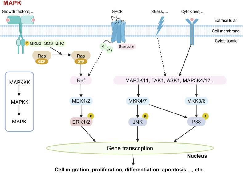
Neurodegenerative diseases (e.g., Alzheimer's, Parkinson's, Huntington's disease, and Amyotrophic Lateral Sclerosis) are major health threats for the aging population and their prevalences continue to rise with the increasing of life expectancy. Although progress has been made, there is still a lack of effective cures to date, and an in-depth understanding of the molecular and cellular mechanisms of these neurodegenerative diseases is imperative for drug development. Protein phosphorylation, regulated by protein kinases and protein phosphatases, participates in most cellular events, whereas aberrant phosphorylation manifests as a main cause of diseases. As evidenced by pharmacological and pathological studies, protein kinases are proven to be promising therapeutic targets for various diseases, such as cancers, central nervous system disorders, and cardiovascular diseases. The mechanisms of protein phosphatases in pathophysiology have been extensively reviewed, but a systematic summary of the role of protein kinases in the nervous system is lacking. Here, we focus on the involvement of protein kinases in neurodegenerative diseases, by summarizing the current knowledge on the major kinases and related regulatory signal transduction pathways implicated in diseases. We further discuss the role and complexity of kinase-kinase networks in the pathogenesis of neurodegenerative diseases, illustrate the advances of clinical applications of protein kinase inhibitors or novel kinase-targeted therapeutic strategies (such as antisense oligonucleotides and gene therapy) for effective prevention and early intervention.


Conflict of interest statement

Competing interests: P.L. serves on the editorial board of Signal Transduction and Targeted Therapy but was not involved in the handling of the manuscript. The other authors declare no competing interests.

Figures

Fig. 1
Fig. 1
Schematic diagram of the general working mechanism of kinases. Kinases catalyze the transfer of the γ-phosphate group of ATP to the specific substrate, making the specific amino acid of the substrate phosphorylated. This figure was created with BioRender.com
Fig. 2
Fig. 2
Kinase classification map. Based on the similarity of kinase domain sequences, 518 human protein kinases were divided into nine groups: tyrosine kinase group (TK group), tyrosine kinase-like group (TKL group), homologs of yeast Sterile 7, Sterile 11, Sterile 20 kinases group (STE group), containing PKA, PKG, PKC families group (AGC group), calcium/calmodulin-dependent protein kinase group (CAMK group), containing CDK, MAPK, GSK3, CLK families group (CMGC group), casein kinase 1 group (CK1 group), other group, atypical protein kinase group. Proteins of pathological significance for neurodegenerative diseases were highlighted in the figure using different colors as indicated in the figure. Figures generated using KinMap (http://kinhub.org/kinmap/index.html), and illustrations reproduced courtesy of Cell Signaling Technology, Inc. (www.cellsignal.com)
Fig. 3
Fig. 3
Typical RTKs-activated signaling cascades. Activation of the receptors (including growth factor receptors such as platelet-derived growth factor receptor (PDGFR), epidermal growth factor receptor (EGFR), vascular endothelial growth factor (VEGFR), and fibroblast growth factor receptor (FGFR), as well as tyrosine kinase receptor B (TrkB) and insulin-like growth factor 1 (IGF-1R)) by the corresponding cytokines, growth factors, and hormones induces autophosphorylation of their receptor tyrosine residues within the cell, thereby further amplifying the kinase activity, exposing the tyrosine phosphorylation docking sites, allowing them to be recognized by cytoplasmic proteins with Src homology 2 domain (SH2) or phosphotyrosine-binding (PTB) domains. Activated RTKs are able to recruit various signaling molecules and initiate downstream pathways. One major pathway involves the activation of phosphatidylinositol-3 kinase (PI3K), which converts PIP2 to PIP3, thereby activating AKT. Another pathway involves phospholipase C gamma (PLCγ), which, upon phosphorylation, stimulates protein kinase C (PKC) activity and mobilizes intracellular calcium (Ca2+). Concurrently, the RTK signaling cascade engages the Ras-Raf-MEK-ERK pathway via GRB2. Collectively, these pathways regulate diverse cellular processes, including cell cycle progression, proliferation, differentiation, survival, migration, metabolism, and other key functions. This figure was created with BioRender.com
Fig. 4
Fig. 4
Typical non-RTKs activated signaling cascades. In addition to the conserved kinase domain, non-RTKs also have a variable number of protein domains (e.g., SH2 or SH3 domains responsible for binding to other signaling molecules). Typical non-RTKs consist of the Src family (including Src, Fyn, Lyn, Lck), the Abelson tyrosine kinase (Abl) family (including Abl1, Abl2), and the Janus kinase (JAK) family (including JAK1, JAK2, JAK3, TYK2). The Src-mediated Ras-Raf-MEK pathway leads to transcriptional regulation in the nucleus, impacting cellular functions. Simultaneously, Fyn activates cyclin-dependent kinase 5 (CDK5), which modifies tau protein to facilitate microtubule remodeling. The activation of the JAK-STAT pathway allows STATs to translocate to the nucleus and directly regulate gene transcription. The regulation of non-RTK signal transduction pathways is closely related to synaptic function remodeling, neuronal excitability regulation, immune regulation, cell proliferation, etc. This figure was created with BioRender.com
Fig. 5
Fig. 5
CDK family and functions. In mammals, the CDK family can be divided into two categories according to their functions: cell cycle-related CDKs (such as CDK1, CDK4, and CDK5) and transcription-related CDKs (CDK7, CDK8, CDK9, CDK11, and CDK20). In the first category, CDK1, CDK2, and CDK4/6 are located in the cell nucleus and combine with CycA/E, CycA/B, and CycD, respectively, to regulate the transformation of different cell cycle stages; while CDK5 is located in the cytoplasm in cells, is mainly active in post-mitotic neurons, and participates in neuronal differentiation, migration, synaptic function, and memory consolidation. Unlike classical CDKs, CDK5 is not activated by cyclins. Instead, it is primarily activated by its neuron-specific cofactor, p35, a regulatory protein that binds to CDK5 and induces a conformational change, enabling its catalytic activity, while cleavage of p35 to p25 under pathological conditions such as oxidative stress, calcium dysregulation, or neurotoxic insults, results in the overactivation of CDK5, driving neurotoxic processes. This figure was created with BioRender.com
Fig. 6
Fig. 6
MAP3K-MAP2K-MAPK signaling pathway. In a typical MAPK cascade, MAP3K activates MAP2K (also known as MKK, MEK) by phosphorylating two conserved Ser/Thr residues in the activation loop, and the MAP2Ks directly phosphorylate MAPKs. The activation of MAPK cascades is initiated by various extracellular stimuli, including growth factors, G-protein-coupled receptor (GPCR) signaling, stress, and cytokines. Activated Ras recruits and activates Raf, which phosphorylates and activates downstream MEK1/2. MEK1/2 then phosphorylates ERK1/2, which translocates to the nucleus and regulates gene transcription. Stress signals (e.g., reactive oxygen species or osmotic stress) and cytokines activate distinct MAPKKKs, leading to the phosphorylation of different MAPKKs. MKK4/7 phosphorylates and activates JNK/p38 MAPK, while MKK3/6 phosphorylates p38 MAPK. The hierarchical organization of the MAPK pathways ensures signal specificity, playing a critical role in cell apoptosis, cell survival, and other cellular events. This figure was created with BioRender.com
Fig. 7
Fig. 7
Schematic description of kinase signaling pathways in Alzheimer’s disease. GSK3β, as one of the main kinases involved in tau phosphorylation, adds a phosphate group to the Thr231 site on tau. This process triggers tau oligomerization, NFTs formation, and participates in the regulation of the Nrf2-ARE pathway by phosphorylating Nrf2 Ser334-338 residues, thereby reducing the antioxidant capacity. The CDK5/p35 complex plays a critical role in maintaining synaptic function by modulating STAT3, synaptic components including PSD95 and DARPP32, ErbB3, BDNF/TrkB, or other regulators, while in AD, the CDK5/p35 is cleaved by Ca2+, Aβ, and calpain-1, giving rise to the abnormally hyperactive CDK5/p25 variant, promoting the pathway of cellular apoptosis, reentry into the cell cycle, and mitochondrial dysfunction. The phosphorylation of p38 MAPK exacerbates oxidative stress, decreases synaptic plasticity, and increases the release of inflammatory factors. Meanwhile, overactivated Fyn contributes to the phosphorylation of APP at Tyr682, leading to increased generation of intracellular Aβ. Fyn phosphorylation at Tyr416 causes cellular toxicity and imbalances in neural network function by modulating NMDAR, Pyk2, and eEF2. TTBK1 activates CDK5 and triggers downstream signaling, promoting NMDAR internalization and imbalanced degradation of the neural network. On the other hand, it triggers the phosphorylation of tau protein at Ser422 via calpain-1, exacerbating tau aggregation. In the progression of AD, the reduced activity of AMPK increases the phosphorylation level of mTOR. This, in turn, hinders autophagy processes while concurrently enhancing Aβ generation. The downregulation of PKA expression in AD pathology leads to decreased activation of both SIRT1 and CREB, increasing Aβ production and synaptic plasticity vulnerability. CK1 abnormalities not only regulate the transmission of their inherent signals but also wield regulatory influence over the downstream signaling pathways of crucial kinases such as GSK3β and CDK5. This intricate interplay between kinases forms an interconnected regulatory network that functions in AD. This figure was created with BioRender.com
Fig. 8
Fig. 8
Schematic description of kinase signaling pathways in Parkinson’s disease. PINK1 and LRRK2 are involved in mitochondrial and lysosomal function, and their mutations lead to mitochondrial dysfunction and autophagy defects. These mutations induce typical α-syn aggregation in the form of Lewy bodies and neurites, as well as neuronal loss. The accumulation of PINK1 in the mitochondrial membrane and subsequent recruitment of Parkin can trigger the initiation of mitophagy. LRRK2 mutations failed to phosphorylate AKT and promote cell survival via inhibition of FOXO1, whereas phosphorylated JNK and p38 MAPK, promoting cell death. Activated ASK1 can also phosphorylate MAPKK and subsequent MAPK to form a cascade amplification of signaling, promoting cell death by regulating the activation of JNK and p38. Phosphorylation of α-syn occurs at several sites involving kinases such as CK, PLK, c-Abl, and GRK, contributing to its aggregation and the formation of Lewy bodies, which alters the activity of numerous kinases and triggers neuroinflammation and increased ROS. This figure was created with BioRender.com
Fig. 9
Fig. 9
Schematic description of kinase signaling pathways in Huntington’s disease. mHTT promotes neuronal apoptosis and inflammatory responses by inhibiting ERK and activating the p38, JNK, and IKKβ signaling pathways. It establishes connections with HTT via the MEK/ERK and AKT signaling pathways, regulating neuronal autophagy, neuroprotection, and glucose uptake pathways. In addition to causing DNA damage by itself, mHTT’s aggregation also leads to pancreatic β-cells damage and activation of CDK5 through its interaction with IRS-2. The aberrant activation of CDK5 reduces microtubule stability, which, in turn, contributes to the exacerbation of mHTT pathology. In addition, the activation of the IKKβ signaling pathway by AKT promotes neuroinflammatory responses, while the inhibition of the JNK signaling cascade by AKT hinders cell apoptosis. The regulatory network formed by the mutual activation and inhibition of kinases plays distinct roles in different stages of the HD pathological process. This figure was created with BioRender.com
Fig. 10
Fig. 10
Schematic description of kinase signaling pathways in Amyotrophic lateral sclerosis. ALS genes encoding protein kinases, such as TBK1 and NEK1, affect the proteostasis process, autophagy, and DNA damage. TBK1 phosphorylates OPTN and p62, leading to autophagy clearance and thus ensuring efficient degradation of ubiquitinated mitochondria. TBK1 inactivates RIPK1 and loss of TBK1 boosts RIPK1 activation and promotes cell death. NEK1 mutations disable DNA damage response and its deficiency reduced the phosphorylation of VPS26B, leading to disruption of endosomal transport and further dysfunction of mitochondria and lysosome. Several kinases are involved in the phosphorylation of TDP-43 and others. The decrease of AKT activity in ALS leads to the upregulated GSK3β activity, contributing to TDP-43-induced axonal degeneration. c-Abl kinase mediates the accumulation of toxic FUS by phosphorylating FUS. Proteins abnormally aggregated alter the kinase signaling networks, eventually leading to cell death of motor neurons. This figure was created with BioRender.com

References

    1. Roskoski, R. Jr A historical overview of protein kinases and their targeted small molecule inhibitors. Pharm. Res.100, 1–23 (2015). - PubMed
    1. Attwood, M. M. et al. Trends in kinase drug discovery: targets, indications and inhibitor design. Nat. Rev. Drug Discov.20, 839–861 (2021). - PubMed
    1. Manning, G. et al. The protein kinase complement of the human genome. Science298, 1912–1934 (2002). - PubMed
    1. Zhang, H. et al. A subcellular map of the human kinome. Elife. 10, e64943 (2021). - PMC - PubMed
    1. Klaeger, S. et al. The target landscape of clinical kinase drugs. Science358, eaan4368 (2017). - PMC - PubMed

MeSH terms