. 2025 May 6;25(1):831.
doi: 10.1186/s12885-025-14220-x.

FLT3LG modulates the infiltration of immune cells and enhances the efficacy of anti-PD-1 therapy in lung adenocarcinoma

Affiliations

FLT3LG modulates the infiltration of immune cells and enhances the efficacy of anti-PD-1 therapy in lung adenocarcinoma

Fengyu Zhao et al. BMC Cancer. .

Abstract

Background: Immunotherapy, particularly anti-PD-1 therapy, has assumed a progressively significant position in the management of non-small cell lung cancer (NSCLC), especially in lung adenocarcinoma (LUAD). Nevertheless, a subset of patients exhibit resistance to anti-PD-1 therapy, and the exploration of biomarkers for evaluating the responsiveness to anti-PD-1 therapy necessitates further investigation. FLT3LG is regarded as being associated with tumor diagnosis and immunotherapy in a variety of tumor types, but its function in LUAD is uncertain.

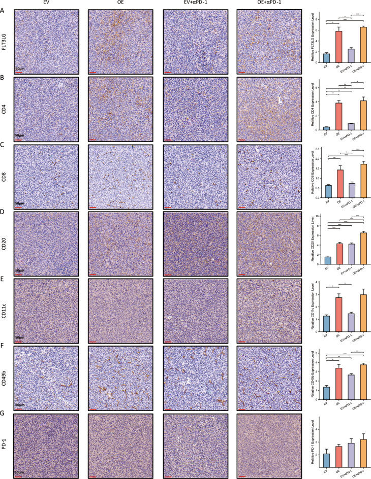
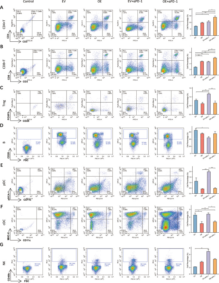
Methods: Bioinformatics analysis was conducted to evaluate the clinical value, functional enrichment, genetic correlation, and immune infiltration of FLT3LG in LUAD. We then used a mouse model to detect immune cell infiltration and relevant protein expression by flow cytometry and immunohistochemistry under anti-PD-1 treatment after overexpression of FLT3LG. The serum FLT3LG expression in LUAD patients was detected via ELISA, and PD-L1 expression in tumor samples was detected by immunohistochemistry.

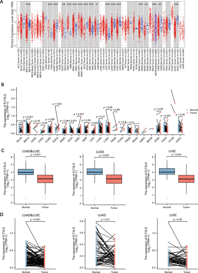
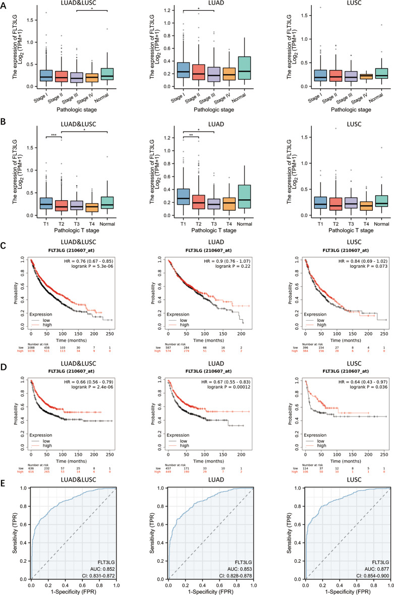
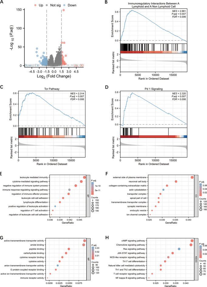
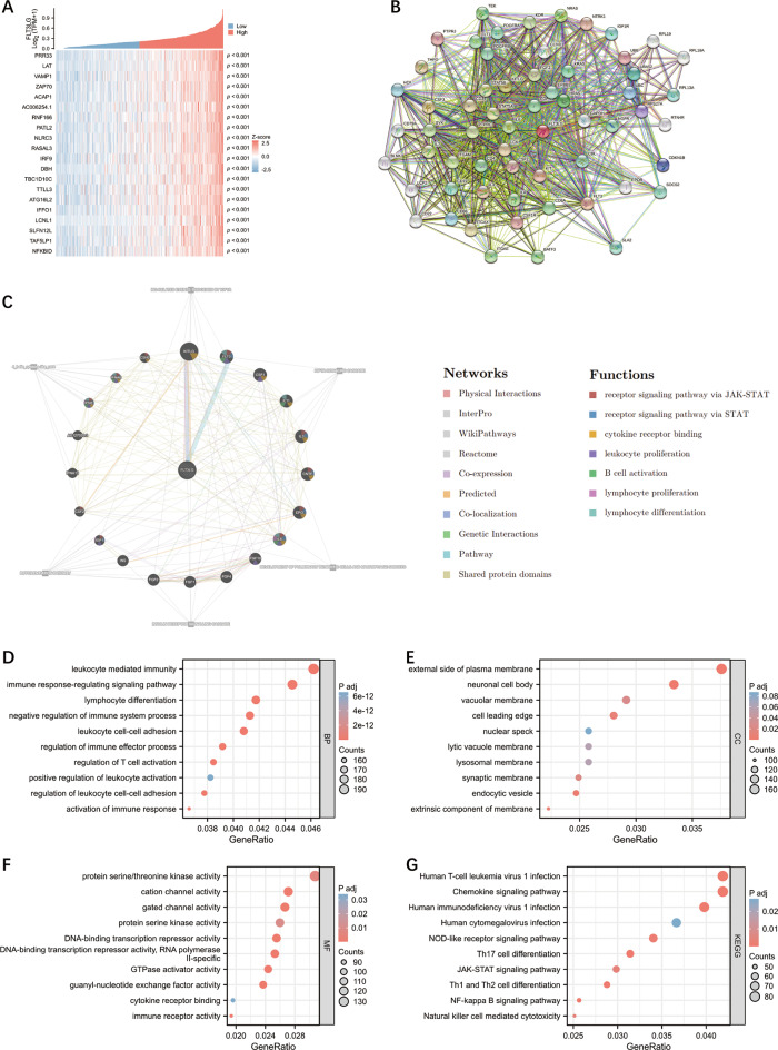
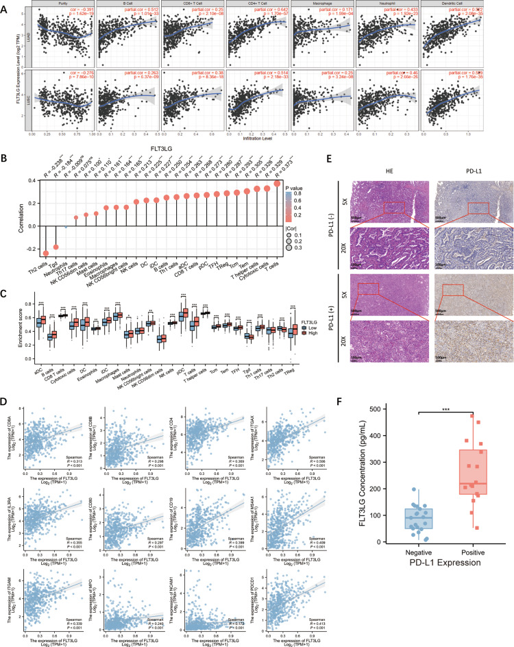
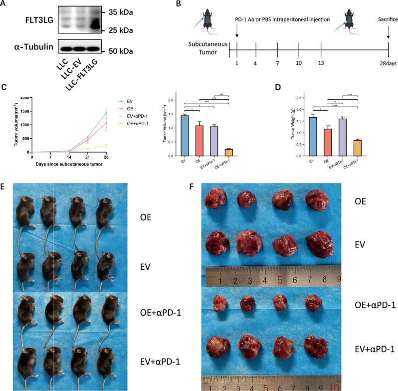
Results: In LUAD patients, a better prognosis is associated with elevated FLT3LG expression. Among the genes strongly associated with FLT3LG, the majority were involved in immune-related processes and were enriched predominantly in immune-related pathways. Moreover, high expression of FLT3LG was significantly positively correlated with increased infiltration of multiple immune cells, including T cells and natural killer (NK) cells, in lung adenocarcinomas, as well as the expression of several immune cell markers, such as CD4 and CD8a. In a mouse model, overexpression of FLT3LG in mice subjected to subcutaneous graft tumor elicited a pronounced immune response and could enhance the efficacy of anti-PD-1 therapy.

Conclusion: FLT3LG could be considered as a diagnostic and prognostic marker for LUAD and might play a role in enhancing the therapeutic response to immunotherapy in patients with LUAD.

Keywords: Biomarkers; Cancer immunotherapy; FLT3LG; Immune cell infiltration; Lung adenocarcinoma (LUAD).


Conflict of interest statement

Declarations. Ethics approval and consent to participate: All experiments involving human samples and animals were authorized by the Ethics Committee of the First Affiliated Hospital of Xi’an Jiaotong University. All of the ethics guidelines pertaining to animal research were followed in terms of both animal care and experimentation. The ethics approval number is NO. XJTU1AF2021LSK-334. Consent for publication: Not applicable. Competing interests: The authors declare no competing interests.

Figures

Fig. 1
Fig. 1
The expression of FLT3LG across cancers and in NSCLC. a. Expression of FLT3LG in tumor and normal tissues in the TCGA pan-cancer dataset. (*: p value < 0.05; **: p value < 0.01; ***: p value < 0.001) b. FLT3LG expression in tumor and paired normal tissues in the TCGA and GTEx pan-cancer datasets. c. FLT3LG expression in tumor and normal tissues from patients with NSCLC. d. FLT3LG expression in tumor and paired normal tissues from patients with NSCLC
Fig. 2
Fig. 2
Role of FLT3LG in the diagnosis and prognosis prediction of NSCLC. a. The expression of FLT3LG in different pathological stages of NSCLC. b. The expression of FLT3LG in patients with different T stages of NSCLC. c. K‒M analysis of OS in LUAD, LUSC, LUAD and LUSC. d. K‒M analysis of first progression (FP) in LUAD and LUSC tissues and LUAD and LUSC tissues. e. The predictive value of FLT3LG in patients with NSCLC. (LUAD and LUSC, AUC = 0.852, CI = 0.831–0.872; LUAD, AUC = 0.853, CI = 0.828–0.878; LUSC, AUC = 0.877, CI = 0.854–0.900). *: p < 0.05; **: p < 0.01; ***: p < 0.001
Fig. 3
Fig. 3
Differential gene analysis and differential gene enrichment analysis of FLT3LG in LUAD. a. Volcano plot of all differentially expressed genes (DEGs). b-d. Gene set enrichment analysis (GSEA) of FLT3LG based on DEGs. e-g. Gene Ontology (GO) analysis of the upregulated DEGs. (e. Biological process analysis; f. Cellular component analysis; g. Molecular function analysis). h. KEGG pathway analysis of upregulated DEGs
Fig. 4
Fig. 4
Correlated gene analysis of FLT3LG in LUAD. a. Heatmap of the top 20 genes correlated with FLT3LG. b. PPI networks constructed with STRING. c. PPI networks constructed in GeneMANIA. d-f. Gene Ontology (GO) analysis of the top 5000 correlated genes. (d. Biological process analysis; e. Cellular component analysis; f. Molecular function analysis) g. KEGG pathway analysis of the top 5000 correlated genes
Fig. 5
Fig. 5
Correlations between FLT3LG expression level and immune cell infiltration and the expression of immune signature. a. Correlation between the expression of FLT3LG and the infiltration of different immune cells according to TIMER. b. Correlation between the expression of FLT3LG and the infiltration of different immune cells in the TCGA cohort. c. Immune cell infiltration levels in patients with different expression levels of FLT3LG. d. Correlation between FLT3LG expression and immune cell marker expression. e. HE staining and PD-L1 immunohistochemical staining of tumor samples from LUAD patients. f. Serum FLT3LG concentrations in LUAD patients with negative or positive PD-L1 expression in tumor tissue samples. *: p < 0.05; **: p < 0.01; ***: p < 0.001
Fig. 6
Fig. 6
Effect of FLT3LG on anti-PD-1 therapy efficacy in a mouse LUAD model. a. Transfection of LLC cells. The blots were cut prior to hybridization with antibodies during blotting. b. Flowchart of the in vivo experiments. c. Growth curves and volume of LLC subcutaneous tumors. d. Weights of the LLC subcutaneous tumors. e. Mice bearing LLC subcutaneous tumors. f. LLC subcutaneous tumors. *: p < 0.05, **: p < 0.01, ***: p < 0.001
Fig. 7
Fig. 7
Overexpression of FLT3LG can promote the infiltration of diverse immune cells in the TME. Subcutaneously transplanted tumors from mice were excised for immunohistochemical staining to analyze the expression of multiple immune-related molecules. a. The expression of FLT3LG in the four groups. b-f. The expression of several common immune cell markers. (b. CD4+ T cells; c. CD8+ T cells; d. CD20+ B cells; e. CD11c+ DCs; f. CD49b+ NK cells.) g. PD-1 expression in the four groups. Scale bar, 50 μm. *: p < 0.05, **: p < 0.01, ***: p < 0.001
Fig. 8
Fig. 8
Flow cytometry analysis of immune cells from the spleen of a LUAD subcutaneous tumor mouse model. Lymphocytes isolated from the spleens of mice with subcutaneous LUAD tumors were evaluated via flow cytometry. a. CD4+ T cells (CD3+CD4+). b. CD8+ T cells (CD3+CD8+). c. Treg cells (CD4+CD25+FOXP3+). d. B cells (CD20+). e. pDC (CD11c+pDCA1+). f. cDC (CD11c+MHCII+). g. NK cells (CD49b+). *: p < 0.05, **: p < 0.01, ***: p < 0.001

Similar articles

References

    1. Bray F, Laversanne M, Sung H, Ferlay J, Siegel RL, Soerjomataram I, et al. Global cancer statistics 2022: GLOBOCAN estimates of incidence and mortality worldwide for 36 cancers in 185 countries. Cancer J Clin. 2024;74(3):229–63. 10.3322/caac.21834. - PubMed
    1. Ishida Y, Agata Y, Shibahara K, Honjo T. Induced expression of PD-1, a novel member of the Immunoglobulin gene superfamily, upon programmed cell death. EMBO J. 1992;11(11):3887–95. - PMC - PubMed
    1. Kim JW, Eder JP. Prospects for targeting PD-1 and PD-L1 in various tumor types. Oncol (Williston Park). 2014;28(Suppl 3):15–28. - PubMed
    1. Forde PM, Chaft JE, Smith KN, Anagnostou V, Cottrell TR, Hellmann MD, et al. Neoadjuvant PD-1 Blockade in resectable lung Cancer. N Engl J Med. 2018;378(21):1976–86. 10.1056/NEJMoa1716078. - PMC - PubMed
    1. Cascone T, William WN, Weissferdt A, Leung CH, Lin HY, Pataer A, et al. Neoadjuvant nivolumab or nivolumab plus ipilimumab in operable non-small cell lung cancer: the phase 2 randomized NEOSTAR trial. Nat Med. 2021;27(3):504–14. 10.1038/s41591-020-01224-2. - PMC - PubMed

MeSH terms