. 2025 May 6;23(1):333.
doi: 10.1186/s12951-025-03393-z.

Piezoelectric-immunomodulatory electrospun membrane for enhanced repair of refractory wounds

Affiliations

Piezoelectric-immunomodulatory electrospun membrane for enhanced repair of refractory wounds

Shu Liu et al. J Nanobiotechnology. .

Abstract

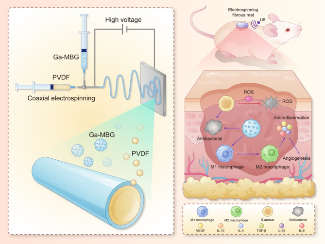
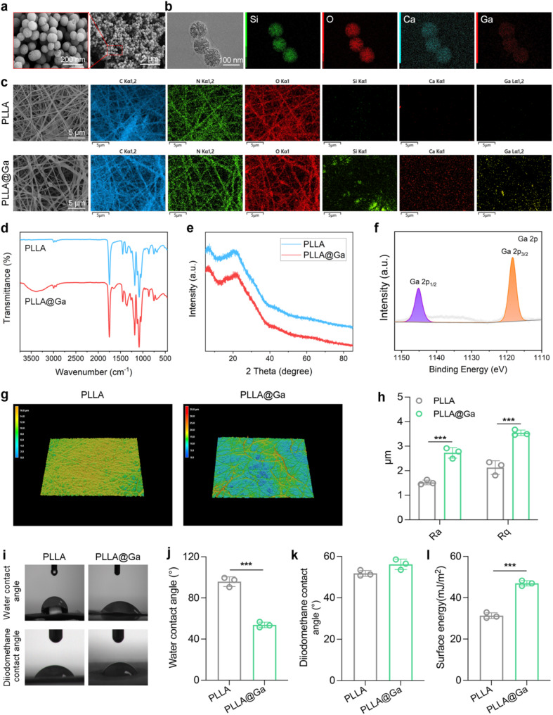
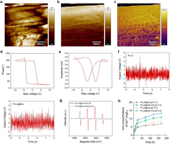
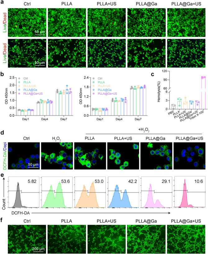
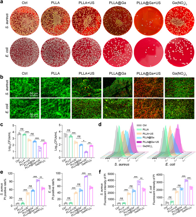
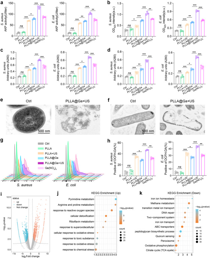
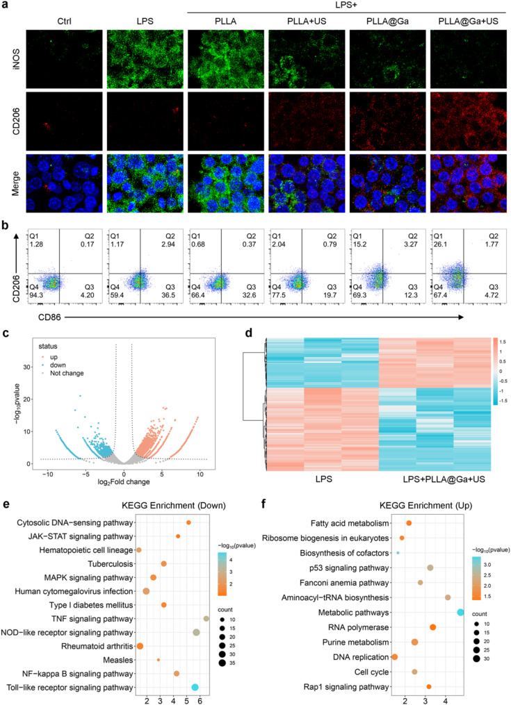
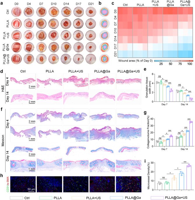
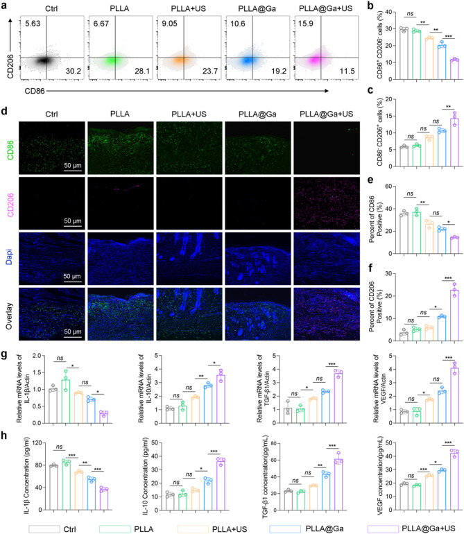
The microenvironment and healing process of diabetic wounds are highly complex, necessitating the development of wound dressings that combine excellent biocompatibility, superior antibacterial properties, and immune-regulating capabilities. However, achieving this goal remains a significant challenge. In this study, a multifunctional electrospun dressing (polylactic acid@Ga, PLLA@Ga) was designed and fabricated by integrating sonodynamic therapy with gallium-doped mesoporous bioactive glass (Ga-MBG). Compared to pure PLLA materials, PLLA@Ga exhibited remarkable antibacterial effects in vitro and demonstrated effective anti-infection properties in vivo. These effects are primarily attributed to the release of Ga ions, which competitively replace iron, thereby disrupting iron-dependent bacterial enzymes and ultimately leading to bacterial death. Additionally, in vitro experiments showed that PLLA@Ga could promote macrophage polarization from the M1 to M2 phenotype, effectively modulating the immune microenvironment of diabetic infected wounds. In vivo wound healing experiments further revealed that PLLA@Ga significantly enhanced collagen deposition and angiogenesis, accelerating the healing process of infected diabetic wounds. Thus, the multifunctional electrospun dressing developed in this study holds great potential as a promising candidate for the treatment of diabetic wounds.

Keywords: Antibacterial; Diabetic wounds; Electrospinning; Ga-MBG; Macrophage.


Conflict of interest statement

Declarations. Ethics approval and consent to participate: The procedures involving the animals and their care were conducted in conformity with national and international laws and policies. The animal experiments were approved by Changhai Hospital. Consent for publication: All authors agreed with the publisher to publish this work. Competing interests: The authors declare no competing interests.

Figures

Scheme 1
Scheme 1
Synthesis process of PLLA@Ga and its mechanism for promoting diabetic wound healing. The preparation of PLLA@Ga involves electrospinning a solution of PLLA and Ga-MBG. Under US stimulation, the PLLA@Ga membrane enhances the production of ROS, thereby increasing antibacterial activity. Furthermore, PLLA@Ga promotes the polarization of macrophages from the M1 phenotype to the M2 phenotype, thereby modulating the immune microenvironment and accelerating wound healing
Fig. 1
Fig. 1
Preparation and characterization of Ga-MBG and PLLA@Ga. (a) SEM images of Ga-MBG. (b) TEM and EDS images of Ga-MBG. (c) SEM and EDS mapping images of PLLA and PLLA@Ga. (d) FTIR of PLLA and PLLA@Ga. (e) XRD of PLLA and PLLA@Ga. (f) XPS of PLLA@Ga. (g) 3D-CLSM of PLLA and PLLA@Ga. (h) Surface roughness of PLLA and PLLA@Ga. (i) Water contact angle and diiodomethane contact angles of PLLA and PLLA@Ga. (j) Statistics on water contact angle of PLLA and PLLA@Ga. (k) Statistics on diiodomethane contact angles of PLLA and PLLA@Ga. (l) Surface energy of PLLA and PLLA@Ga
Fig. 2
Fig. 2
Characterization of PLLA@Ga. (a) PFM image of PLLA@Ga material surface. (b) Piezoelectric amplitude of PLLA@Ga. (c) Phase angle of PLLA@Ga. (d) Butterfly cycle curve of PLLA@Ga. (e) Piezoelectric flip curve of PLLA@Ga. (f) The output voltage of PLLA@Ga under US stimulation. (g) ESR of PLLA@Ga with US or without US. (h) Ga3+ concentration was determined by ICP-OES after immersion of PLLA@Ga in PBS at different pH (pH 7.4 and pH 5.5) for different times with or without US
Fig. 3
Fig. 3
Effects of PLLA@Ga + US in vitro. (a) Images of live/dead assays of HUVECs (upper panel) and RS-1 cells (lower panel). (b) CCK-8 assay of HUVECs (left panel) and RS-1 cells (right panel). (c) Hemolysis assay results of vatious materials exposure. (d) Fluorescence imaging was employed to assess the reduction of intracellular ROS in H2O2-stimulated Raw264.7 cells. (e) Analysis of intracellular ROS in H2O2-stimulated Raw264.7 cells after different treatments via flow cytometry. (f) Tube formation via coculturing HUVECs with various hydrogels
Fig. 4
Fig. 4
In vitro antibacterial activity of PLLA@Ga + US. (a) Representative plates of bacteria colonies after treatment with different samples. (b) The fluorescence images of live (green) and dead bacterial (red). (c) Results of bacteria treated with various materials that formed colonies after 24 h. (d-e) Representative flow cytometry plots and quantitative analysis of PI. (f) Detection of membrane damage in bacteria through monitoring PI influx. Data are presented as mean ± SD (n = 3). “ns” represent “no significant”; *P < 0.05; **P < 0.01; ***P < 0.001
Fig. 5
Fig. 5
In vitro antibacterial activity of PLLA@Ga + US. (a) The sensitivity to alkaline phosphatase activity. (b) The ONPG hydrolysis activity. (c-d) Leakage of cell contents after exposure to various treatments was investigated by monitoring the absorbance of extracellular material at 260 nm (A260) and 280 nm (A280) corresponding to nucleic acids and proteins, respectively. (e-f) Morphology of bacteria from different treatment groups as observed by TEM. (g) Assessment of ROS generation was conducted using DCFH-DA fluorescent probes by FCM. (h) Assessment of ROS generation was conducted using DCFH-DA fluorescent probes by microplate reader. (i) Volcano map of differentially expressed genes (DEGs). (j-k) KEGG pathway enrichment analysis. Data are presented as mean ± SD (n = 3). “ns” represent “no significant”; *P < 0.05; **P < 0.01; ***P < 0.001
Fig. 6
Fig. 6
Studies on the in vitro immunomodulatory of PLLA@Ga + US. (a) Expression of iNOS and CD206 following different hydrogel treatments. (b) Analysis of CD86 and CD206 in macrophages following various treatments via flow cytometry. (c) Volcano map of differentially expressed genes (DEGs). (d) Heat map of differentially expressed genes (DEGs). (e) KEGG enrichment for the down-regulated pathways. (f) KEGG enrichment for the up-regulated pathways
Fig. 7
Fig. 7
PLLA@Ga + US promotes diabetic wound healing. (a-b) Photographs of wounds at predetermined time points and superimposed images of wounds after various treatments. (c) Areas of unhealed wounds in each group at different time intervals. (d) H&E staining. (e) Corresponding quantitative analysis of the granulation tissue width. (f) Masson’s trichrome staining. (g) Statistical analysis of collagen deposition during the remolding phase. (h) Representative immunofluorescence staining of microvessel of wound. (i) Statistical analysis of microvessel density. Data are presented as mean ± SD (n = 3). “ns” represent “no significant”; *P < 0.05; ***P < 0.001
Fig. 8
Fig. 8
Impact of the PLLA@Ga + US on macrophage polarization in vivo. (a-c) Flow cytometry of M1and M2 macrophages retrieved from the wound tissue. (d) Immunofluorescence staining of tissue sections at the wound site for CD86 and CD206. (e-f) Statistical analysis concerning the proportion of CD86 (f) and CD206 (g) positive macrophages. (g) Gene expression levels of IL-1β, IL-10, VEGF, and TGF-β1 in wound tissues. (h) Secretion of IL-1β, IL-10, VEGF, and TGF-β1 in wound tissues assessed by Elisa. Data are presented as mean ± SD (n = 3). “ns” represent “no significant”; *P < 0.05; **P < 0.01; ***P < 0.001

Similar articles

References

    1. Chokshi T, Fickweiler W, Jangolla S, Park K, Wu IH, Shah H, Sun JK, Aiello LP, King GL. Reduced Aqueous Retinol-Binding Protein 3 Concentration Is Associated With Diabetic Macular Edema and Progression of Diabetic Retinopathy, Diabetes Care (2024). - PMC - PubMed
    1. Kuo S, Ye W, Wang D, McEwen LN, Villatoro Santos C, Herman WH. Cost-Effectiveness of the National Diabetes Prevention Program: A Real-World, 2-Year Prospective Study, Diabetes Care (2024). - PubMed
    1. Chu Z, Liu X, Zhao T, Jiang D, Zhao J, Dong X, Yeung KWK, Liu X, Liao Y, Ouyang L. Self-healing Ppy-hydrogel promotes diabetic skin wound healing through enhanced sterilization and macrophage orchestration triggered by NIR. Biomaterials. 2024;315:122964. - PubMed
    1. Zhang X, Ning F, Li Y, Lu J, He Y, Feng C, Dong CM. Pluripotent polysaccharide coordinated hydrogels remodel inflammation, neovascularization and reepithelization for efficient diabetic wound prohealing. J Control Release. 2024;377:37–53. - PubMed
    1. Pu C, Wang Y, Xiang H, He J, Sun Q, Yong Y, Chen L, Jiang K, Yang H, Li Y. Zinc-based polyoxometalate nanozyme functionalized hydrogels for optimizing the Hyperglycemic-Immune microenvironment to promote diabetic wound regeneration. J Nanobiotechnol. 2024;22(1):611. - PMC - PubMed

LinkOut - more resources