. 2024 May 9;12(4):101317.
doi: 10.1016/j.gendis.2024.101317. eCollection 2025 Jul.

The bidirectional effects of APPswe on the osteogenic differentiation of MSCs in bone homeostasis by regulating Notch signaling

Affiliations

The bidirectional effects of APPswe on the osteogenic differentiation of MSCs in bone homeostasis by regulating Notch signaling

Nan Wang et al. Genes Dis. .

Abstract

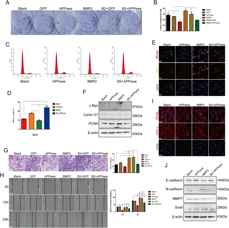
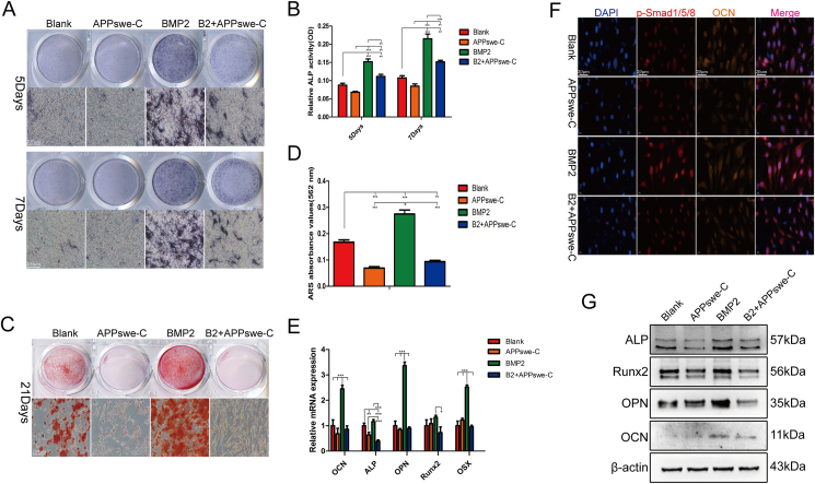
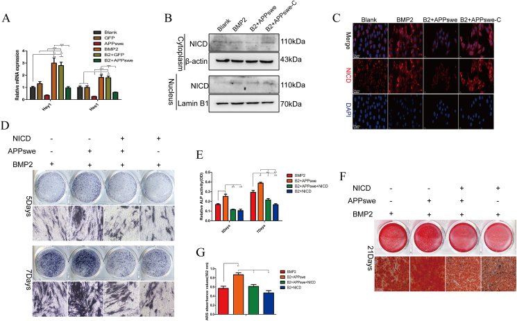
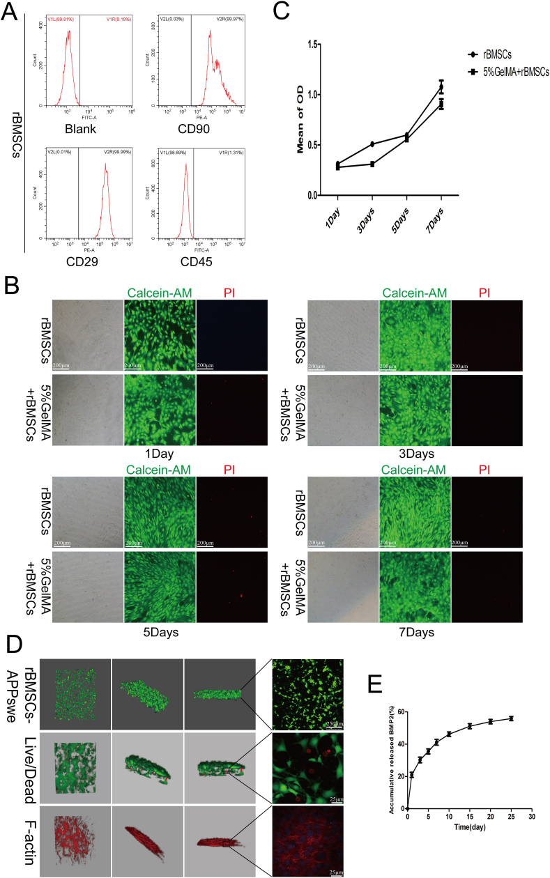
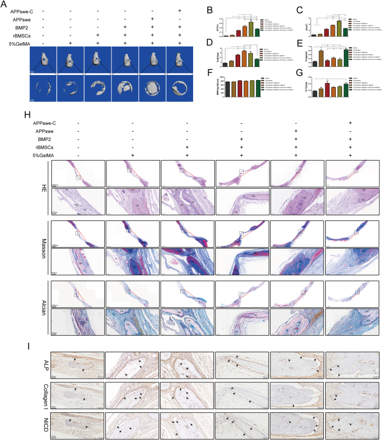
Amyloid precursor protein (APP), especially Swedish mutant APP (APPswe), is recognized as a significant pathogenic protein in Alzheimer's disease, but limited research has been conducted on the correlation between APPswe and the osteogenic differentiation of mesenchymal stem cells (MSCs). The effects of APPswe and its intracellular and extracellular segments on the osteogenic differentiation of bone morphogenetic protein 2 (BMP2)-induced MSCs were analyzed in this study. Our analysis of an existing database revealed that APP was positively correlated with the osteogenic differentiation of MSCs but negatively correlated with their proliferation and migration. Furthermore, APPswe promoted BMP2-induced osteogenic differentiation of MSCs, while APPswe-C (APPswe without an intracellular segment) had the opposite effect; thus, the intracellular domain of APPswe may be a key factor in promoting the osteogenic differentiation of MSCs. Additionally, both APPswe and APPswe-C inhibited the proliferation and migration of MSCs. Furthermore, the intracellular domain of APPswe inhibited the activity of the Notch pathway by regulating the expression of the Notch intracellular domain to promote the osteogenic differentiation of MSCs. Finally, APPswe-treated primary rat bone marrow MSCs exhibited the most favorable bone repair effect when a GelMA hydrogel loaded with BMP2 was used for in vivo experiments, while APPswe-C had the opposite effect. These findings demonstrate that APPswe promotes the osteogenic differentiation of MSCs by regulating the Notch pathway, but its extracellular segment blocks the self-renewal, proliferation, and migration of MSCs, ultimately leading to a gradual decrease in the storage capacity of MSCs and affecting long-term bone formation.

Keywords: Alzheimer's disease; Amyloid precursor protein; MSCs; Notch signaling; Osteogenic differentiation.


Conflict of interest statement

The authors declared no competing interests.

Figures

Fig. 1
Figure 1
Bioinformatics was used to preliminarily explore the molecular mechanism of APPswe in osteogenic transformation. (A) APP expression at different time points during the osteogenic differentiation of MSCs. (B) ALP expression at different time points during the osteogenic differentiation of MSCs. (C) ACP5 expression at different time points during the osteogenic differentiation of MSCs. (D) Analysis of the correlations between the expression of APP and those of ALP and ACP5. (E) Correlation analysis between APP and the proliferation genes PCNA and CCND1 and the migration genes MMP7 and MMP9. ∗p < 0.05, ∗∗p < 0.01, ∗∗∗p < 0.001; ns, no significant difference. APP, amyloid precursor protein; APPswe, Swedish mutant APP; MSCs, mesenchymal stem cells; ALP, alkaline phosphatase; ACP5, acid phosphatase 5; PCNA, proliferating cell nuclear antigen; CCND1, cyclin D1; MMP7/9, matrix metallopeptidase 7/9; OS, osteogenesis.
Fig. 2
Figure 2
Effect of APPswe on the osteogenic differentiation of MSCs induced by BMP2. (A, B) ALP staining and activity detection were performed after treating cells with APPswe or BMP2 for 5 days or 7 days. (C, D) ARS staining and semiquantitative analysis were used to detect calcium nodule formation after cells were treated with APPswe or BMP2 for 21 days. (E) The mRNA expression levels of genes related to osteogenic differentiation were measured by quantitative real-time PCR after cells were treated with APPswe or BMP2 for 48 h. (F) Western blot analysis was performed to detect the expression levels of osteogenic differentiation-related proteins after cells were treated with APPswe or BMP2 for 48 h. (G) The expression levels of the osteogenic differentiation proteins p-Smad1/5/8 and OCN in the different treatment groups were detected by cell immunofluorescence. MSCs were treated with APPswe for 24 h and then with BMP2 for 48 h to detect the expression of related proteins or mRNAs. n = 3; ∗p < 0.05, ∗∗p < 0.01, ∗∗∗p < 0.001. APPswe, Swedish mutant amyloid precursor protein; MSC, mesenchymal stem cell; ALP, alkaline phosphatase; BMP2, bone morphogenetic protein 2; OCN, osteocalcin.
Fig. 3
Figure 3
APPswe inhibits the self-renewal, proliferation, and migration of MSCs. (A, B) The effect of APPswe on the self-renewal ability of MSCs was detected by colony-forming unit-fibroblast assays. (C, D) Cell cycle changes in MSCs treated with APPswe were detected by flow cytometry. (E) The expression of the proliferation-related gene Ki67 in different groups was detected by cell immunofluorescence. (F) The expression levels of proliferation-related proteins in MSCs after treatment with APPswe were detected by western blotting. (G) Transwell assays were used to detect the effect of APPswe treatment on the cell migration ability of MSCs. (H) The effect of APPswe on MSC migration was detected by a wound healing test. (I) The expression levels of F-actin in the different treatment groups were detected by FITC-phalloidin staining. (J) Western blot analysis was performed to detect the expression levels of migration-related proteins in the different groups. n = 3; ∗p < 0.05; ∗∗p < 0.01, ∗∗∗p < 0.001. APPswe, Swedish mutant amyloid precursor protein; MSC, mesenchymal stem cell.
Fig. 4
Figure 4
AICD plays a crucial role in promoting the BMP2-induced osteogenic differentiation of MSCs. (A, B) ALP staining and activity detection were performed after treating cells with APPswe-C or BMP2 for 5 and 7 days. (C, D) ARS staining and semiquantitative analysis were used to detect calcium nodule formation in cells treated with APPswe-C or BMP2 for 21 days. (E) The mRNA expression levels of genes related to osteogenic differentiation were measured by quantitative real-time PCR after cells were treated with APPswe-C or BMP2 for 48 h. (F) The expression levels of the osteogenic differentiation proteins p-Smad1/5/8 and OCN in the different treatment groups were detected by cell immunofluorescence. (F) Western blot analysis was performed to detect the expression levels of osteogenic differentiation-related proteins after cells were treated with APPswe or BMP2 for 48 h. MSCs were treated with APPswe-C for 24 h and then with BMP2 for 48 h to detect the expression levels of related proteins or mRNAs. n = 3; ∗p < 0.05, ∗∗p < 0.01, ∗∗∗p < 0.001. APPswe, Swedish mutant amyloid precursor protein; AICD, the intracellular domain of APPswe; BMP2, bone morphogenetic protein 2; MSC, mesenchymal stem cell; ALP, alkaline phosphatase; APPswe-C, APPswe without an intracellular segment; OCN, osteocalcin.
Fig. 5
Figure 5
Effects of APPswe-C on the proliferation and migration of MSCs. (A, B) The effect of APPswe-C on the self-renewal of MSCs was detected by a colony-forming unit-fibroblast assay. (C, D) The effect of APPswe-C on the cell cycle was detected by flow cytometry. (E) The expression of the proliferation gene Ki67 was detected by cellular immunofluorescence. (F) The expression levels of proliferation-related proteins after APPswe-C treatment were detected by western blotting. (G, H) The cell migration of each group after APPswe-C treatment was detected by Transwell assay. (I, J) The effect of APPswe-C treatment on cell migration was detected by a wound healing test. (K) The expression of F-actin in the cells of each group after APPswe-C treatment was detected by FITC-phalloidin staining. (L) The expression levels of cell migration-related proteins were detected by western blot. MSCs were treated with APPswe-C for 24 h and then with BMP2 for 48 h to detect the expression levels of related proteins. n = 3; ∗p < 0.05, ∗∗p < 0.01, ∗∗∗p < 0.001. APPswe-C, Swedish mutant amyloid precursor protein without an intracellular segment; MSC, mesenchymal stem cell; BMP2, bone morphogenetic protein 2.
Fig. 6
Figure 6
APPswe affects the osteogenic differentiation and proliferation of MSCs by regulating the Notch signaling. (A) The mRNA expression levels of Hey1 and Hes1 in each treatment group were measured by quantitative real-time PCR. (B) Western blot analysis of NICD expression in the cytoplasm and nucleus in each treatment group. (C) The expression of NICD in each treatment group was detected by cellular immunofluorescence. (D, E) ALP staining and activity detection were performed in each treatment group. (F, G) The formation of calcium nodules in each treatment group was analyzed by ARS staining and semiquantitative analysis. MSCs were treated with APPswe or APPswe-C for 24 h and then with BMP2 for 48 h to detect the expression levels of related proteins. n = 3; ∗p < 0.05, ∗∗p < 0.01, ∗∗∗p < 0.001. APPswe, Swedish mutant amyloid precursor protein; APPswe-C, APPswe without an intracellular segment; MSC, mesenchymal stem cell; ALP, alkaline phosphatase; BMP2, bone morphogenetic protein 2; NICD, the Notch intracellular domain.
Fig. 7
Figure 7
Biocompatibility and protein release properties of the GelMA hydrogel. (A) Flow cytometry was used to detect rBMSCs extracted via the adherent method. (B) The rBMSCs cultured in GelMA hydrogel for 1, 3, 5, and 7 days were detected by live and dead staining. (C) MTT was used to detect the growth of rBMSCs cultured in GelMA hydrogels at different time points. (D) Confocal laser scanning was used to detect the specific cell morphology, growth, and extension of the rBMSCs in the GelMA hydrogel. (E) ELISA was used to measure the release of BMP2 in the GelMA hydrogel at different time points. APPswe-treated rBMSCs were cultured for 24 h and then collected. n = 3; ∗p < 0.05, ∗∗p < 0.01, ∗∗∗p < 0.001. APPswe, Swedish mutant amyloid precursor protein; rBMSC, rat bone mesenchymal stem cell; BMP2, bone morphogenetic protein 2; GelMA, gelatin methacryloyl.
Fig. 8
Figure 8
Effects of APPswe on BMP2-induced bone formation and repair in rBMSCs in vivo.(A) Six weeks after surgery, the rat skulls were recovered for micro-CT analysis, and representative images were shown. (BG) Histomorphometric analysis of structural bone parameters. Bone volume (BV, mm3), relative bone volume (BV/total volume, %), trabecular number (Tb.N, 1/mm), trabecular separation (Tb.Sp, mm), bone mineral density (BMD, mg HA/ccm), and trabecular thickness (Tb.Th, mm) were calculated based on the micro-CT scanning data. (H) Representative hematoxylin-eosin staining, Masson's trichrome staining, and Alcian blue staining of each group are shown. (I) The expression levels of ALP, collagen I, and NICD were confirmed by immunohistochemical staining. Representative images of each group are shown. The red line indicates the location and width of the skull defect in each group. The black box in the tissue staining image is the locally representative region of the lower-magnification image. The black arrows indicate positive protein expression in new bone. n = 3; ∗p < 0.05, ∗∗p < 0.01, ∗∗∗p < 0.001. APPswe, Swedish mutant amyloid precursor protein; rBMSC, rat bone mesenchymal stem cell; ALP, alkaline phosphatase; NICD, the Notch intracellular domain; NB, new bone; HB, host bone.
figs1
figs1

Similar articles

References

    1. Alzheimer’s disease facts and figures. Alzheimers Dement. 2022;18(4):700–789. - PubMed
    1. Frame G., Bretland K.A., Dengler-Crish C.M. Mechanistic complexities of bone loss in Alzheimer's disease: a review. Connect Tissue Res. 2020;61(1):4–18. - PubMed
    1. Chen Y.H., Lo R.Y. Alzheimer's disease and osteoporosis. Ci Ji Yi Xue Za Zhi. 2017;29(3):138–142. - PMC - PubMed
    1. Bonafede M., Shi N., Barron R., Li X., Crittenden D.B., Chandler D. Predicting imminent risk for fracture in patients aged 50 or older with osteoporosis using US claims data. Arch Osteoporosis. 2016;11(1):26. - PMC - PubMed
    1. Tiihonen M., Taipale H., Tanskanen A., Tiihonen J., Hartikainen S. Incidence and duration of cumulative bisphosphonate use among community-dwelling persons with or without Alzheimer's disease. J Alzheimers Dis. 2016;52(1):127–132. - PMC - PubMed

LinkOut - more resources