. 2025 May 6:29:0201.
doi: 10.34133/bmr.0201. eCollection 2025.

Impact of Culture Duration on the Properties and Functionality of Yeast-Derived Extracellular Vesicles

Affiliations

Impact of Culture Duration on the Properties and Functionality of Yeast-Derived Extracellular Vesicles

Gyeongchan Jeon et al. Biomater Res. .

Abstract

Extracellular vesicles (EVs), lipid bilayer nanovesicles secreted by cells, carry nucleic acids, proteins, and other bioactive molecules that influence recipient cells and modulate various biological processes. This study investigated how energy depletion and fermentation processes influence the characteristics and physiological functions of EVs secreted by Saccharomyces cerevisiae. Specifically, we analyzed EVs derived from 24-h cultures, representing the glucose utilization phase, and 72-h cultures, representing the starvation stage. Under energy-depleted conditions (72-h cultures), yeast secreted a higher number of EV particles, albeit with a smaller average particle size. In contrast, EVs from yeast cultured for 24 h, during the glucose utilization phase, were enriched in Pep12-rich endosome-derived vesicles and exhibited 71% higher cellular internalization efficiency. Proteomic and transcriptomic analyses revealed distinct protein and microRNA profiles between EVs from 24- and 72-h cultures, highlighting their potential roles in tissue regeneration, cell proliferation, and collagen synthesis. As a result, EVs derived from 24-h cultures exhibited a 15% greater effect in promoting collagen synthesis. The differential effects on collagen production may be attributed to the efficiency of endocytosis and the specific protein and microRNA cargo of the EVs. This study emphasizes the functional potential and unique properties of yeast-derived EVs while proposing strategies to modulate EV composition by adjusting the yeast culture duration and the energy source in the medium. Further research is needed to control yeast-produced EV components and to understand their mechanisms of action for effective therapeutic applications.


Conflict of interest statement

Competing interests: The authors declare that they have no competing interests.

Figures
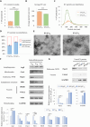
Fig. 1.
Fig. 1.
EV@Y24 and EV@Y72 were characterized, and the characteristics of yeast and yeast-derived extracellular vesicles (EVs) during each culture period were analyzed by western blotting. Particle size analysis was conducted by a ZetaView PMX-120 nanoparticle tracking analysis (NTA) instrument, and the instrument was calibrated with standard 100-nm polystyrene beads. (A) The contents of EV particles, (B) average particle size, and (C) size distribution were evaluated in EV samples. (D) The ratio of small particles (30 to 150 nm) and large particles (150 to 500 nm) was confirmed in each isolated EV sample. Data are presented as mean ± SD (n = 3). *P < 0.05, **P < 0.01, and ***P < 0.001. Yeast cells were cultured in YPD medium (1% yeast extract, 2% peptone, and 2% d-glucose) and harvested daily until 72 h. (E) EV@Y24 and EV@Y72 were imaged using a bio-transmission electron microscope. (F) In yeast cell samples, the expression of an autophagosome marker (Atg8), a nucleus marker (NSP1), a mitochondria marker (Cox4), vacuole markers (PHO8 and VMA2), a multivesicular body (MVB) marker (Pep12), and an endoplasmic reticulum (ER) marker (PDI1) was confirmed. (G) EVs were isolated from yeast-cultured medium and the expression of Pep12, VMA2, and glyceraldehyde-3-phosphate dehydrogenase (GAPDH) was confirmed. Y24, 24-h-cultured yeast; Y48, 48-h-cultured yeast; Y72, 72-h-cultured yeast; EV@Y24, EVs from 24-h-cultured medium; EV@Y48, EVs from 48-h-cultured medium; EV@Y72, EVs from 72-h-cultured medium. Atg8, autophagy-related protein 8; Cox4, cytochrome c oxidase subunit 4; NSP1, nuclear pore complex protein; PDI1, protein disulfide isomerase; Pep12, proteinase A vacuolar sorting protein 12; PHO8, alkaline phosphatase; VMA2, vacuolar membrane ATPase subunit B.
Fig. 2.
Fig. 2.
Fluorescence labeling and cellular uptake analysis of EV@Y24 and EV@Y72. DiO-labeled EVs were applied to Hs27 fibroblast cells at a concentration of 109 particles/ml. After 4 h, cellular uptake was assessed by flow cytometry. (A) The cellular uptake of EV@Y24 and EV@Y72 is presented as histogram plots, and (B) cellular uptake is quantified as a percentage relative to that of the EV@Y24-treated group. (C) Fluorescence imaging of the uptake of DiO-labeled EVs (yellow arrows) by Hs27 cells after 4-h incubation. (D) The number of EV particles per cell was counted and is visualized as a box plot (n = 10). The white dashed lines indicate cell boundaries. (E) To assess dye stability, DiO-labeled EVs were incubated at 37 °C for 4 h, and their fluorescence intensity was measured and compared before and after incubation. Data are presented as mean ± SD (n = 3). **P < 0.01. DIC, differential interference contrast image; DAPI, 4′,6-diamidino-2-phenylindole; DiO, 3,3′-dioctadecyloxacarbocyanine perchlorate.
Fig. 3.
Fig. 3.
Protein qualitative analysis of EV@Y24 and EV@Y72. (A) Venn diagram of identified proteins in EV@Y24 and EV@Y72. (B and E) Gene Ontology (GO) analysis of 29 proteins present only in EV@Y24. (C and F) GO analysis of 194 commonly existing proteins. (D and G) GO analysis of 164 proteins present only in EV@Y72. ATP, adenosine triphosphate.
Fig. 4.
Fig. 4.
Protein relative analysis of EV@Y24 and EV@Y72. The heat map of the shared proteins among EV@Y24 and EV@Y72 was visualized, and GO analysis was performed. Zone I contains a collection of proteins enriched in EV@Y24 samples (|Z score| > 0.9). Zone II is a collection of proteins whose abundances do not differ significantly in the 2 samples. Zone III contains a collection of proteins enriched in EV@Y72 samples (|Z score| > 0.9). GPI, glycosylphosphatidylinositol; mRNA, messenger RNA.
Fig. 5.
Fig. 5.
Small RNA sequencing in EV@Y24 and EV@Y72. (A) Small RNA length distribution of read counts from Illumina sequencing in EV samples. (B) A volcano plot was visualized, and 66 differentially expressed small RNAs (red spots) were selected based on a P value <0.05 and |log2(fold change)| > 2. (C) The heat map of the shared small RNAs among EV@Y24 and EV@Y72 is visualized. (D) The Kyoto Encyclopedia of Genes and Genomes (KEGG) analysis of the target genes of the identified small RNAs in EV@Y24 and EV@Y72. In the graph, a higher logFC indicates stronger suppression by EV@Y72, while a lower logFC suggests stronger suppression by EV@Y24. Circle size represents the number of mRNAs involved in each biological process targeted by microRNAs (miRNAs). Pathways with P values >0.05 were excluded. A lower P value indicates significant enrichment of the targeted genes in a biological process compared to random chance. Human mRNA target prediction of the identified yeast EV-derived miRNA sequences was performed using the psRNATarget tool. The GO terms and KEGG pathway of the target mRNA were categorized using DAVID Bioinformatics Resources. FC, fold change; MAPK, mitogen-activated protein kinase; NF-κB, nuclear factor-κB.
Fig. 6.
Fig. 6.
EV@Y24 and EV@Y72 promoted collagen synthesis in Hs27 fibroblast cells. Hs27 cells were grown until they reached 90% confluency and then switched to serum-free media containing retinol, EV@Y24, or EV@Y72. (A) After 24 h, the content of procollagen type 1 was confirmed in cultured medium using an enzyme-linked immunosorbent assay (ELISA) kit. (B) The expression of COL1A1, COL3A1, elastin, MMP1, and TIMP1 was evaluated by RT-PCR. (C) The expression of COL1A1, COL3A1, and MMP1 protein was evaluated by western blotting. Data are presented as mean ± SD (n = 3). *P < 0.05, **P < 0.01, and ***P < 0.001.

Similar articles

References

    1. Witwer KW, Théry C. Extracellular vesicles or exosomes? On primacy, precision, and popularity influencing a choice of nomenclature. J Extracell Vesicles. 2019;8(1):1648167. - PMC - PubMed
    1. Kalluri R, LeBleu VS. The biology, function, and biomedical applications of exosomes. Science. 2020;367(6478):eaau6977. - PMC - PubMed
    1. Yang BW, Lin YM, Huang YB, Zhu NX, Shen YQ. Extracellular vesicles modulate key signalling pathways in refractory wound healing. Burns Trauma. 2023;11:tkad039. - PMC - PubMed
    1. Casado-Díaz A, Quesada-Gómez JM, Dorado G. Extracellular vesicles derived from mesenchymal stem cells (MSC) in regenerative medicine: Applications in skin wound healing. Front Bioeng Biotechnol. 2020;8:146. - PMC - PubMed
    1. Ye Q, Li Z, Li Y, Li Y, Zhang Y, Gui R, Cui Y, Zhang Q, Qian L, Xiong Y, et al. . Exosome-derived microRNA: Implications in melanoma progression, diagnosis and treatment. Cancers. 2023;15(1):80. - PMC - PubMed

LinkOut - more resources