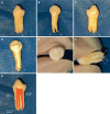
Mandibular left first premolar with three roots and three canals: A case report
- PMID: 40330286
- PMCID: PMC11736522
- DOI: 10.12998/wjcc.v13.i13.100822
Mandibular left first premolar with three roots and three canals: A case report
Abstract
Background: The numbers of mandibular first premolar roots and root canals vary, and the incidence of three roots and three canals is 0.09%.
Case summary: In this article, we review the root and root canal conditions for the mandibular first premolar and report the case of a mandibular left first premolar with three roots and three canals in a male patient, with suggestions for clinical diagnosis and treatment. The patient was referred by an orthodontist for the extraction of the tooth. Preoperative cone-beam computed tomography examination revealed that it had three roots. Under local anesthesia, the extraction socket was carefully expanded, and the tooth was successfully removed intact using forceps. The procedure was uneventful, with no root fractures, postoperative bleeding, or sensory abnormality observed.
Conclusion: The mandibular first premolar is characterized by multiple roots and canal variations that can increase the difficulty of treatment.
Keywords: Case report; Mandibular first premolar; Three root canals; Three roots; Tooth extraction.
©The Author(s) 2025. Published by Baishideng Publishing Group Inc. All rights reserved.
Conflict of interest statement
Conflict-of-interest statement: The authors declare that they have no competing interests.
Figures


Similar articles
-
Endodontic treatment of a two-rooted mandibular first premolar with four root canals: a case report.Front Dent Med. 2024 Dec 11;5:1498167. doi: 10.3389/fdmed.2024.1498167. eCollection 2024. Front Dent Med. 2024. PMID: 39917697 Free PMC article.
-
A cone-beam computed tomography study of root canal morphology of maxillary and mandibular premolars in a Turkish population.Acta Odontol Scand. 2014 Nov;72(8):701-6. doi: 10.3109/00016357.2014.898091. Epub 2014 May 15. Acta Odontol Scand. 2014. PMID: 24832561
-
Mandibular first premolar with five root canals: a case report.BMC Oral Health. 2020 Sep 10;20(1):253. doi: 10.1186/s12903-020-01241-0. BMC Oral Health. 2020. PMID: 32912222 Free PMC article.
-
The root and root canal morphology of the human mandibular second premolar: a literature review.J Endod. 2007 Sep;33(9):1031-7. doi: 10.1016/j.joen.2007.03.020. Epub 2007 Jun 5. J Endod. 2007. PMID: 17931927 Review.
-
Diagnosis and root canal treatment of a mandibular second premolar with three root canals.Int Endod J. 2003 Dec;36(12):912-9. doi: 10.1111/j.1365-2591.2003.00750.x. Int Endod J. 2003. PMID: 14689961 Review.
References
-
- Cleghorn BM, Christie WH, Dong CC. The root and root canal morphology of the human mandibular first premolar: a literature review. J Endod. 2007;33:509–516. - PubMed
-
- Zhang D, Chen JH, Lan GH, Xu Q, Yang JJ, Liu ZH, Wen XJ, Liu LC, Deng MJ, Stomatology DO. [Research on root and root canal morphology in mandibular first premolars of Chinese] Di-San Junyi Daxue Xuebao. 2016;38:1188–1194.
-
- Bürklein S, Heck R, Schäfer E. Evaluation of the Root Canal Anatomy of Maxillary and Mandibular Premolars in a Selected German Population Using Cone-beam Computed Tomographic Data. J Endod. 2017;43:1448–1452. - PubMed
-
- Yang H, Tian C, Li G, Yang L, Han X, Wang Y. A cone-beam computed tomography study of the root canal morphology of mandibular first premolars and the location of root canal orifices and apical foramina in a Chinese subpopulation. J Endod. 2013;39:435–438. - PubMed
Publication types
LinkOut - more resources
Full Text Sources

