

A Ring-Shaped Lateral Meniscus in an Anterior Cruciate Ligament Reconstruction Surgery
- PMID: 40330346
- PMCID: PMC12051721
- DOI: 10.7759/cureus.81729
A Ring-Shaped Lateral Meniscus in an Anterior Cruciate Ligament Reconstruction Surgery
Abstract
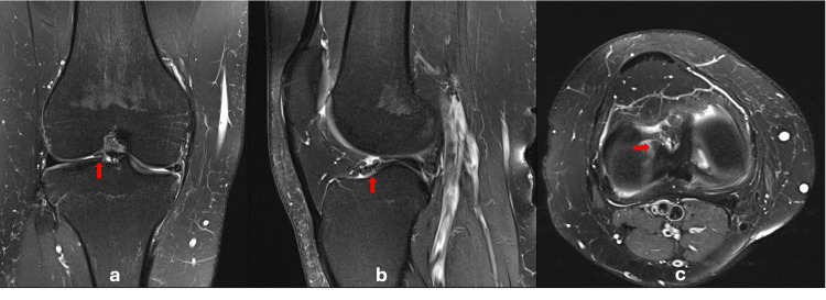
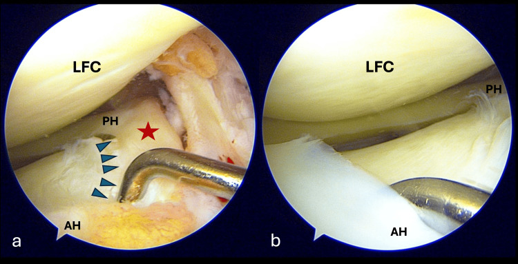
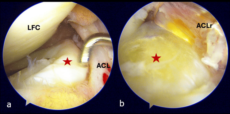
The ring-shaped meniscus is a rare variant of meniscus anomalies. We report a case of a young man who experienced right knee instability following a sports-related injury. Clinical examination suggested an anterior cruciate ligament (ACL) tear, which was confirmed through imaging studies. Intra-operatively, an incidental finding revealed the presence of a ring-shaped lateral meniscus. We discuss the diagnostic features of this uncommon abnormality.
Keywords: acl reconstruction; anatomical variant; discoid meniscus; lateral meniscus; ring-shaped meniscus.
Copyright © 2025, Mohd Miswan et al.
Conflict of interest statement
Human subjects: Consent for treatment and open access publication was obtained or waived by all participants in this study. Conflicts of interest: In compliance with the ICMJE uniform disclosure form, all authors declare the following: Payment/services info: All authors have declared that no financial support was received from any organization for the submitted work. Financial relationships: All authors have declared that they have no financial relationships at present or within the previous three years with any organizations that might have an interest in the submitted work. Other relationships: All authors have declared that there are no other relationships or activities that could appear to have influenced the submitted work.
Figures
Similar articles
-
Formation of ring-shaped lateral meniscus following anterior cruciate ligament reconstruction: A case report.Int J Surg Case Rep. 2017;31:229-232. doi: 10.1016/j.ijscr.2017.01.060. Epub 2017 Jan 30. Int J Surg Case Rep. 2017. PMID: 28189986 Free PMC article.
-
Regeneration of ring-shaped lateral meniscus after partial resection of discoid meniscus with anterior cruciate ligament reconstruction.Int J Surg Case Rep. 2013;4(12):1093-6. doi: 10.1016/j.ijscr.2013.08.023. Epub 2013 Sep 25. Int J Surg Case Rep. 2013. PMID: 24240076 Free PMC article.
-
Ring-shaped lateral meniscus with hypoplasic anterior cruciate ligament.Skeletal Radiol. 2015 Dec;44(12):1813-8. doi: 10.1007/s00256-015-2197-2. Epub 2015 Jul 16. Skeletal Radiol. 2015. PMID: 26178136
-
Symptomatic Complete Discoid Medial Meniscus Completely Coalesced with the Anterior Cruciate Ligament: A Case Report and Literature Review.Orthop Surg. 2022 Sep;14(9):2391-2395. doi: 10.1111/os.13377. Epub 2022 Aug 1. Orthop Surg. 2022. PMID: 35913195 Free PMC article. Review.
-
Early Operative Versus Delayed or Nonoperative Treatment of Anterior Cruciate Ligament Injuries in Pediatric Patients.J Athl Train. 2016 May;51(5):425-7. doi: 10.4085/1062-6050.51.5.11. Epub 2016 May 31. J Athl Train. 2016. PMID: 27244126 Free PMC article. Review.
References
-
- The ring-shaped meniscus: a case series demonstrating the variation of imaging appearances on MRI. Iqbal A, McLoughlin E, Botchu R, James SL. Skeletal Radiol. 2020;49:281–289. - PubMed
-
- Evaluation of the morphological variations of the meniscus: a cadaver study. Ryu K, Iriuchishima T, Oshida M, Saito A, Kato Y, Tokuhashi Y, Aizawa S. Knee Surg Sports Traumatol Arthrosc. 2015;23:15–19. - PubMed
-
- Watanabe M, Takeda S, Ikeuchi H. Tokyo: Igaku-Shoin. Tokyo, Japan: Igaku-Shoin; 1979. Atlas of arthroscopy; p. 156.
Publication types
LinkOut - more resources
Full Text Sources