. 2025 Apr 22:16:1571027.
doi: 10.3389/fimmu.2025.1571027. eCollection 2025.

Serum angiotensin type-1 receptor autoantibodies and neurofilament light chain as markers of neuroaxonal damage in post-COVID patients

Affiliations

Serum angiotensin type-1 receptor autoantibodies and neurofilament light chain as markers of neuroaxonal damage in post-COVID patients

Ana I Rodriguez-Perez et al. Front Immunol. .

Abstract

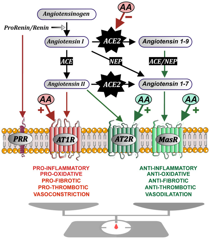
Introduction: Dysregulation of autoimmune responses and the presence of autoantibodies (AA), particularly those related to the renin-angiotensin system (RAS), have been implicated in the acute phase of COVID-19, and persistent dysregulation of brain RAS by RAS-related autoantibodies may also contribute to neurological symptoms of post-COVID.

Methods: We analyzed levels of serum and CSF RAS AA in post-COVID patients with neurological symptoms, individuals who have fully recovered from COVID-19 (after-COVID controls), and uninfected individuals, and their possible correlations with the serum marker of neuroaxonal damage neurofilament light chain (NfL) and the degrees of cognitive deficit.

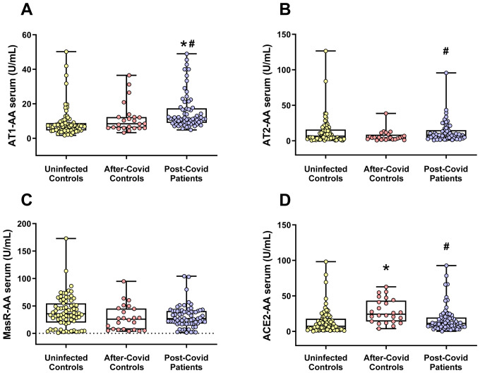
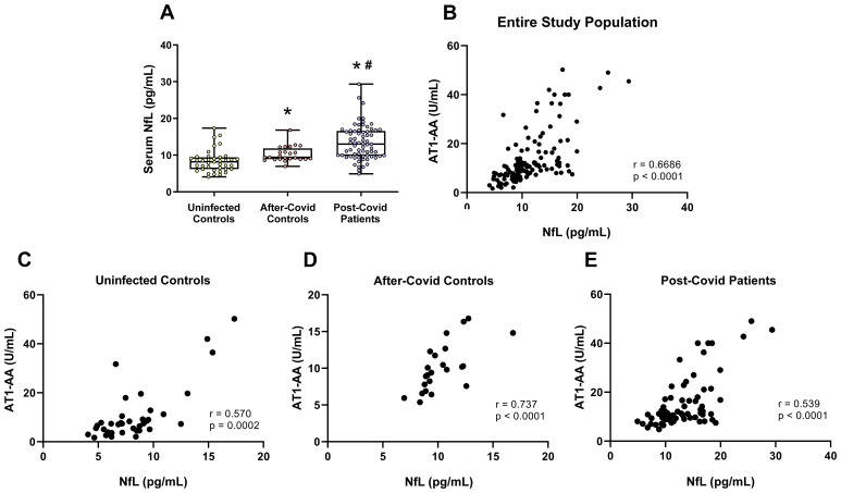
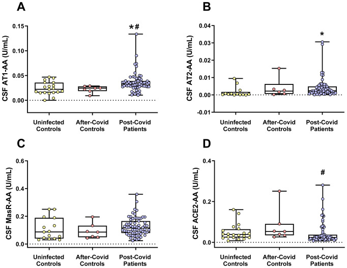
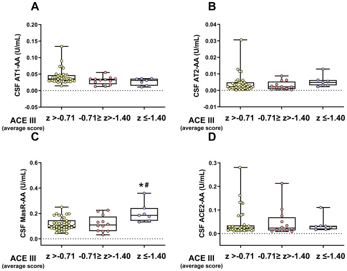
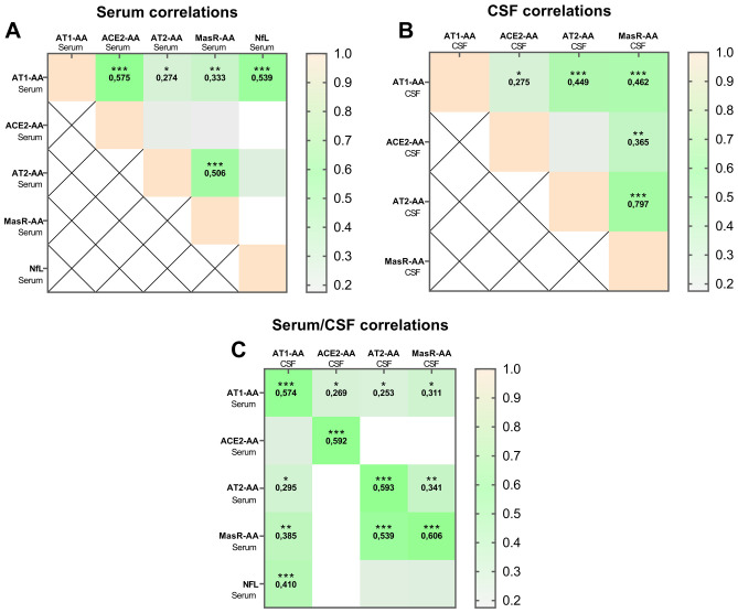
Results: Both in serum and CSF, levels of AA agonists of the pro-inflammatory angiotensin II type 1 receptors (AT1-AA) were significantly elevated in this cohort of neurological post-COVID patients compared to both uninfected and after-COVID controls and correlated with serum levels of NfL. Changes in serum and CSF levels of AA promoting the RAS anti-inflammatory axis (upregulation of AA agonists of AT2 and Mas receptors, downregulation of AA antagonists of ACE2) suggest upregulation of the RAS compensatory response in this cohort of neurological post-COVID patients. Post-COVID patients with more pronounced cognitive impairment exhibited significantly higher CSF levels of MasR-AA and a trend toward elevated AT2-AA. Persistent brain RAS dysregulation, particularly persistent increase in AT1-AA, and its correlation with neuroaxonal damage markers and cognitive impairment, may play a significant role in neurological symptoms associated with post-COVID. Serum levels of NfL and AT1-AA may be interesting biomarkers for the early identification of CNS involvement in patients with neurological symptoms and a history of COVID-19. However, post-COVID is a highly heterogeneous entity and may result from various underlying mechanisms. The present study includes a cohort, which may differ from other cohorts with different clinical profiles, which may show different results on NfLs and CSF RAS autoantibodies, particularly AT1-AA.

Conclusion: These findings highlight the potential of targeting AT1 receptors as a therapeutic strategy for mitigating cognitive deficits in post-COVID patients showing upregulated AT1-AA levels.

Keywords: AT1; COVID-19; autoantibody; autoimmunity; biomarkers; cognitive impairment; neurological long-COVID; post-acute sequelae of COVID-19 syndrome.


Conflict of interest statement

The authors declare that the research was conducted in the absence of any commercial or financial relationships that could be construed as a potential conflict of interest. The author(s) declared that they were an editorial board member of Frontiers, at the time of submission. This had no impact on the peer review process and the final decision.

Figures

Figure 1
Figure 1
Schematic organization of the renin-angiotensin system (RAS), which is formed by two opposite axes: a first axis (red arrows) mainly constituted by Angiotensin II acting on AT1 receptors (AT1R), and a compensatory axis (green arrows) constituted by Angiotensin II acting on AT2 receptors, and Angiotensin 1-7 acting on Mas receptors. Angiotensin II is produced by the enzyme prorenin/renin acting on the precursor protein angiotensinogen to produce Angiotensin I, which is converted by the angiotensin-converting enzyme (ACE) into Angiotensin II. Renin and its precursor prorenin (PR) can also act on specific PR receptors. Angiotensin-converting enzyme 2 (ACE2) plays a key role in the system balance because ACE2 (together with peptidases such as Neprilysin, NEP) transforms components of the pro-inflammatory axis (Angiotensin I and, particularly, Angiotensin II) into components of the compensatory axis (Angiotensin 1-9 and, particularly Angiotensin 1-7). Autoantibodies (AA) play an agonistic effect on the GPCR (G protein-coupled receptors: AT1, AT2, Mas) and antagonistic effect on ACE2. Figure modified with permission from: Drugs Modulating Renin-Angiotensin System in COVID-19 Treatment. J.L. Labandeira-Garcia et al., 2022, Biomedicines, 10 (DOI:10.3390/biomedicines10020502).
Figure 2
Figure 2
Serum levels of different RAS autoantibodies in the three studied groups. (A) AT1-AA serum levels were significantly higher in the post-COVID patient group. Changes in autoantibody levels for the RAS anti-inflammatory axis [i.e. AT2 (B) and MasR (C)] were less clear. (D) Serum levels of ACE2-AA were significantly higher in the after-COVID control group. Data distribution is shown using a box plot with boxes representing the IQR, i.e., the limits between the first quartile (Q1, 25%) and the third quartile (Q3, 75%). The whiskers extend up to maximum and minimum values. *p < 0.05 relative to uninfected control; # p<0.05 relative to after-COVID controls. Kruskal–Wallis One Way Analysis of Variance on Ranks with Dunn’s Method post hoc test. ACE2-AA, ACE2 Autoantibodies; AT1-AA, Autoantibodies for AT1 receptors; AT2-AA, Autoantibodies for AT2 receptors; IQR, Interquartile range; MasR-AA, Autoantibodies for Mas receptors.
Figure 3
Figure 3
NfL serum levels in the three studied groups. (A) NfL serum levels were significantly higher in patients who had suffered COVID-19 but did not present post-COVID symptoms (after-COVID controls) and post-COVID patients than in uninfected controls and NfL levels in post-COVID patients were significantly higher than in after-COVID controls. Furthermore, levels of serum NfL significantly correlated with serum levels of AT1-AA in the entire study population (B), uninfected controls (C); after-COVID controls (D), and post-COVID patients (E). In (A), data distribution is shown using a box plot with boxes representing the IQR, i.e., the limits between the first quartile (Q1, 25%) and the third quartile (Q3, 75%). The whiskers extend up to maximum and minimum values. *p < 0.05 relative to uninfected control; # p<0.05 relative to after-COVID controls. Kruskal–Wallis One Way Analysis of Variance on Ranks with Dunn’s Method post hoc test. In (B–D), correlations between NfL serum levels and AT1-AA serum levels were assessed using Spearman’s rank correlation coefficient. AT1-AA, Autoantibodies for AT1 receptors; IQR, Interquartile range; NfL, Neurofilament light chain.
Figure 4
Figure 4
CSF levels of different RAS autoantibodies in the three studied groups. (A) CSF AT1-AA levels were significantly higher in the post-COVID patients. (B) CSF levels of AT2-AA were significantly higher in post-COVID patients than in uninfected patients but not significantly different compared to after-COVID controls. (C) In the post-COVID patient group, CSF MasR-AA levels exhibited a trend toward being higher than in the other groups, although this difference did not reach statistical significance. (D) CSF ACE2-AA levels were significantly lower in the post-COVID patient group than in the after-COVID control group. Data distribution is shown using a box plot with boxes representing the IQR, i.e., the limits between the first quartile (Q1, 25%) and the third quartile (Q3, 75%). The whiskers extend up to maximum and minimum values. *p < 0.05 relative to uninfected controls; # p<0.05 relative to after-COVID controls. Kruskal–Wallis One Way Analysis of Variance on Ranks with Dunn’s Method post hoc test. ACE2-AA, ACE2 Autoantibodies; AT1-AA, Autoantibodies for AT1 receptors; AT2-AA, Autoantibodies for AT2 receptors; IQR, Interquartile range; MasR-AA, Autoantibodies for Mas receptors.
Figure 5
Figure 5
CSF levels of different RAS autoantibodies in post-COVID patients with different degrees of cognitive impairment. No significant differences in CSF AT1-AA or ACE2-AA levels were found between the three post-COVID subgroups: unimpaired overall CI (z > -0.71), mild overall CI (-0.71 ≥ z >-1.40), and severe overall CI (z ≤ -1.40), (A, D). However, the group with more marked CI exhibited significantly higher CSF MasR-AA levels (C) and a trend toward higher CSF AT2-AA levels (B), though the latter did not reach statistical significance. Data distribution is shown using a box plot with boxes representing the IQR, i.e., the limits between the first quartile (Q1, 25%) and the third quartile (Q3, 75%). The whiskers extend up to maximum and minimum values. *p < 0.05 relative to ACE III (Average score) of Z> -0.71; # p<0.05 relative to ACE III (Average score) of -071<Z>-1.40. Kruskal–Wallis One Way Analysis of Variance on Ranks with Dunn’s Method post hoc test. ACE2-AA, ACE2 Autoantibodies; AT1-AA, Autoantibodies for AT1 receptors; AT2-AA, Autoantibodies for AT2 receptors; CI, Cognitive Impairment; IQR, Interquartile range; MasR-AA, Autoantibodies for Mas receptors.
Figure 6
Figure 6
Autoantibody correlations in post-COVID patients. Results of Spearman’s correlation matrixes show variables (AT1-AA, ACE2-AA, AT2-AA, MasR-AA and NfL) correlating at varying levels of significance (*p < 0.05; **p < 0.01; ***p < 0.001) in serum (A), CSF (B) and between serum and CSF (C). AT1-AA, Autoantibodies for AT1 receptors; ACE2-AA, ACE2 Autoantibodies; AT2-AA, Autoantibodies for AT2 receptors; MasR-AA, Autoantibodies for Mas receptors; NfL, Neurofilament light chain.

Similar articles

Cited by

References

    1. Soriano JB, Murthy S, Marshall JC, Relan P, Diaz JV, Condition WHOCCDWGoP-C- . A clinical case definition of post-covid-19 condition by a delphi consensus. Lancet Infect Dis. (2022) 22:e102–e7. doi: 10.1016/S1473-3099(21)00703-9 - DOI - PMC - PubMed
    1. Ceban F, Ling S, Lui LMW, Lee Y, Gill H, Teopiz KM, et al. . Fatigue and cognitive impairment in post-covid-19 syndrome: A systematic review and meta-analysis. Brain Behav Immun. (2022) 101:93–135. doi: 10.1016/j.bbi.2021.12.020 - DOI - PMC - PubMed
    1. Monje M, Iwasaki A. The neurobiology of long covid. Neuron. (2022) 110:3484–96. doi: 10.1016/j.neuron.2022.10.006 - DOI - PMC - PubMed
    1. Soung AL, Vanderheiden A, Nordvig AS, Sissoko CA, Canoll P, Mariani MB, et al. . Covid-19 induces cns cytokine expression and loss of hippocampal neurogenesis. Brain. (2022) 145:4193–201. doi: 10.1093/brain/awac270 - DOI - PMC - PubMed
    1. Diez-Cirarda M, Yus-Fuertes M, Sanchez-Sanchez R, Gonzalez-Rosa JJ, Gonzalez-Escamilla G, Gil-Martinez L, et al. . Hippocampal subfield abnormalities and biomarkers of pathologic brain changes: from sars-cov-2 acute infection to post-covid syndrome. EBioMedicine. (2023) 94:104711. doi: 10.1016/j.ebiom.2023.104711 - DOI - PMC - PubMed