

WTAP-mediated N6-methyladenosine mRNA methylation regulates laser-induced macular neovascularization
- PMID: 40330498
- PMCID: PMC12055036
WTAP-mediated N6-methyladenosine mRNA methylation regulates laser-induced macular neovascularization
Abstract
Purpose: Neovascular age-related macular degeneration (nAMD) is now a major cause of central vision loss in older adults worldwide. The primary characteristic of nAMD is the formation of macular neovascularization (MNV), which is a pathologic form of angiogenesis. Epigenetics plays a role in multiple pathological physiologic processes. N6-methyladenosine (m6A) modification is the most common, abundant, and reversible modification in eukaryotic mRNAs, and it plays a role in various pathological angiogenesis processes. This study intends to reveal the expression and functions of m6A during the macular neovascularization (MNV) process.
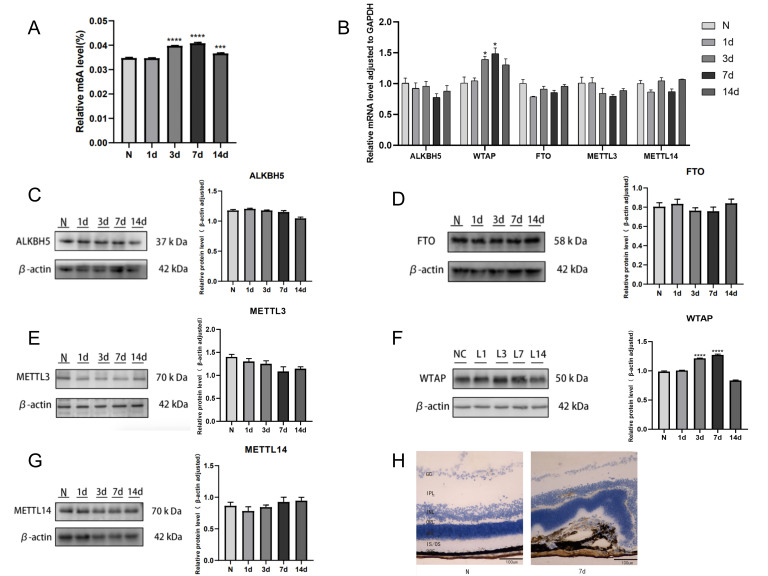
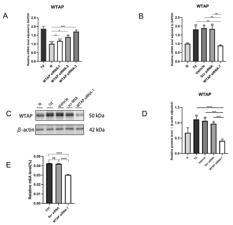
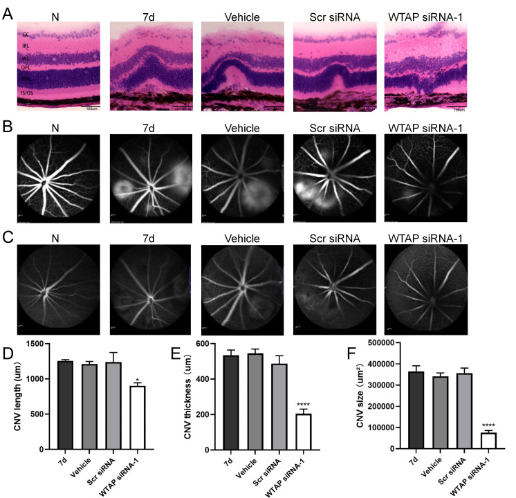
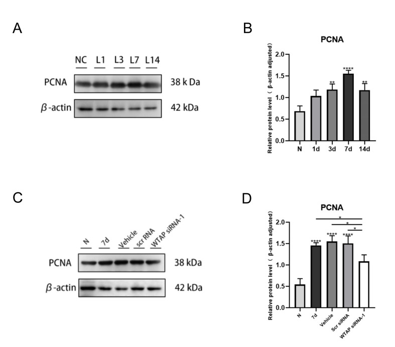
Methods: A laser-induced MNV mouse model was used in this study. m6A quantitative analysis was performed to detect the expression of m6A. Subsequently, the expression of various m6A writers and erasers was detected using quantitative real-time polymerase chain reaction (qRT-PCR) and western blot. Immunohistochemistry was used to detect Wilms' tumor 1-associating protein (WTAP) expression in the MNV lesions. Intravitreal injection of WTAP siRNA in MNV mice to silence the WTAP gene. Hematoxylin and eosin (H&E) were used to determine the thickness and length of the MNV. Fundus fluorescein angiography (FFA) and indocyanine green angiography (ICGA) were examined to measure the leakage area of the MNV. Proliferating cell nuclear antigen (PCNA) expression was detected with a western blot. The mRNA and protein levels of β-catenin were tested with qRT-PCR and western blot.
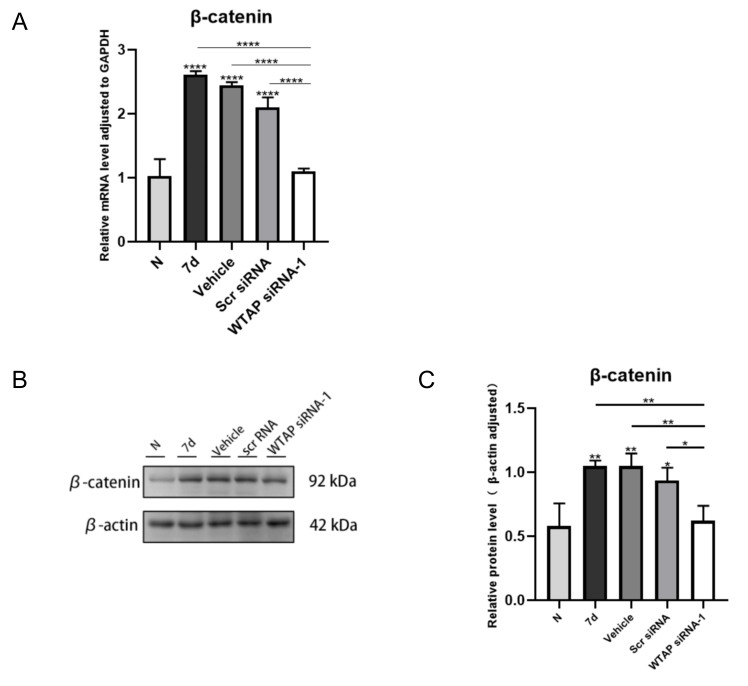
Results: We found increased m6A modification levels after laser induction compared with the normal control group. Subsequently, the expression of various m6A writers and erasers was detected. The results showed that WTAP increased in the MNV model in mice. After the injection of WTAP siRNA into the vitreous body, the expression of WTAP significantly decreased, subsequently decreasing the m6A modification levels. The width, breadth, and leakage area of MNV damage markedly decreased, and endothelial cell proliferation was inhibited. After laser-induced MNV, the expression of β-catenin increased, and that of β-catenin significantly decreased after WTAP knockout.
Conclusions: In conclusion, this study suggests that WTAP-mediated m6A methylation can regulate pathological angiogenesis during MNV and that WTAP may participate in the formation of MNV through the wingless-related integration site (Wnt) pathway. WTAP may be a potential target for MNV treatment.
Copyright © 2024 Molecular Vision.
Figures
Similar articles
-
Inhibition of YAP ameliorates choroidal neovascularization via inhibiting endothelial cell proliferation.Mol Vis. 2018 Jan 31;24:83-93. eCollection 2018. Mol Vis. 2018. PMID: 29422766 Free PMC article.
-
Fibronectin binds integrin α5β1 to regulate macular neovascularization through the Wnt/β-catenin signaling pathway.Exp Eye Res. 2024 May;242:109880. doi: 10.1016/j.exer.2024.109880. Epub 2024 Mar 27. Exp Eye Res. 2024. PMID: 38552713
-
WTAP-Mediated m6A Modification of TRAIL-DR4 Suppresses MH7A Cell Apoptosis.Int J Rheum Dis. 2025 Jan;28(1):e70065. doi: 10.1111/1756-185X.70065. Int J Rheum Dis. 2025. PMID: 39797510
-
Role of WTAP in Cancer: From Mechanisms to the Therapeutic Potential.Biomolecules. 2022 Sep 2;12(9):1224. doi: 10.3390/biom12091224. Biomolecules. 2022. PMID: 36139062 Free PMC article. Review.
-
Novel Insight of N6-Methyladenosine in Cardiovascular System.Medicina (Kaunas). 2025 Jan 26;61(2):222. doi: 10.3390/medicina61020222. Medicina (Kaunas). 2025. PMID: 40005339 Free PMC article. Review.
References
-
- Bouchon A, Dietrich J, Colonna M. Cutting edge: inflammatory responses can be triggered by TREM-1, a novel receptor expressed on neutrophils and monocytes. J Immunol. 2000;164:4991–5. - PubMed
-
- Ferrara N, Adamis AP. Ten years of anti-vascular endothelial growth factor therapy. Nat Rev Drug Discov. 2016;15:385–403. - PubMed
MeSH terms
Substances
LinkOut - more resources
Full Text Sources
Medical
Miscellaneous
