Case Reports
. 2025 Apr 22:15:1529976.
doi: 10.3389/fonc.2025.1529976. eCollection 2025.

DEB-TACE combined with ici for advanced squamous cell carcinoma of the skin: a case report

Affiliations
Case Reports

DEB-TACE combined with ici for advanced squamous cell carcinoma of the skin: a case report

Zhuo Wu et al. Front Oncol. .

Abstract

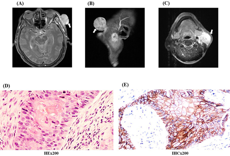
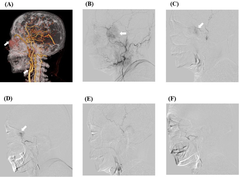
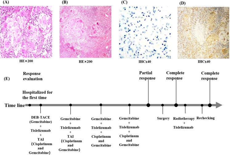
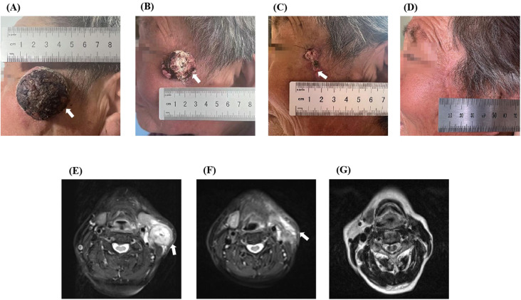
We report the case of an older female patient with squamous cell carcinoma of the skin, cTXN1M0, who initially presented with a mass of approximately 4.5 cm in diameter in the left temporal region, which had a tendency to break and bleed. Given the patient's poor general status and ECOG score of grade 2, it was considered that she could not tolerate systemic chemotherapy. Therefore, we applied drug-eluting beads transarterial chemoembolization combined with immune checkpoint inhibitors to treat this patient and unexpectedly found that this regimen resulted in complete classified remission and in partial pathological response without significant adverse events.

Keywords: CSCC; DEB-TACE; ICI; case report; complete remission.


Conflict of interest statement

The authors declare that the research was conducted in the absence of any commercial or financial relationships that could be construed as a potential conflict of interest.

Figures

Figure 1
Figure 1
The pre-treatment images of the patient and pathology images. (A–C) Enhanced MRI images of the face and neck show an abnormally enhancing shadow in the left temporal region with a maximum diameter of 3.2 cm × 2.1 cm and enlarged left cervical lymph nodes. (D) The results of pre-treatment cervical lymph node aspiration biopsy suggest infiltration by highly differentiated squamous cell carcinoma. (E) Immunohistochemical findings of P63 (+) and PDL-1 expression. (HEx200, Hematoxylin and Eosin staining observed at 200x magnification; IHCx200, Immunohistochemistry staining observed at 200x magnification).
Figure 2
Figure 2
The preoperative arteriography results of the patient and the digital subtraction angiography (DSA) images of transcarotid chemoembolization. (A) The preoperative computed tomography angiography results of the patient show that the mass was supplied by the external carotid artery branch. (B) DSA image of the patient’s frontotemporal mass before embolization. After anesthesia, imaging examination through the left distal radial artery approach revealed an abnormal vessel with a diameter of about 3.5 cm on the left side of the face. A microcatheter was inserted into the external carotid artery branch, and 0.3 mg of gemcitabine was administered. The microspheres were then embolized with polyvinyl alcohol (blue spheres 100–300 microns in diameter) containing 0.5 g (1000 mg/m2) gemcitabine. (C, D) Intraoperative superselective arterial DSA images. (E, F) DSA images of the patient after embolization show that the tumor is not visible.
Figure 3
Figure 3
Pathological findings of the patient at the time of surgery. (A) Pathological findings of the facial mass suggest only squamous epithelial cell hyperplasia and no tumor cells. (B) Pathological results of the cervical lymph nodes suggest no active tumor cells were seen. (C) shows immunohistochemistry of the facial mass, suggesting P40 (−); (D) Postoperative immunohistochemistry of the cervical lymph nodes suggests CD68 (+). (E) Flow chart of the patient during treatment. (HEx200, Hematoxylin and Eosin staining observed at 200x magnification; IHCx40, Immunohistochemistry staining observed at 40x magnification).
Figure 4
Figure 4
The comparison images of before and after treatment of the patient. Upon admission, a mass of about 3.5 cm × 3.5 cm was visible in the patient’s left temporal region, neck, and MRI showed abnormal signals in the shadow of the neck mass (A, E). After one cycle of treatment, the quality was significantly lower than before treatment, and repeated MRI showed that the shadow quality of the neck was lower than before treatment (B, F). (C) shows a visual view of the patient’s left temporal tumor after two cycles of treatment, leaving only postoperative scabs. (D) shows the visual observation of the patient after one year of treatment, leaving only postoperative scars, and (G) shows the MRI images of the patient’s neck after one year of treatment.

Similar articles

References

    1. Maubec E. Update of the management of cutaneous squamous-cell carcinoma. Acta Derm Venereol. (2020) 100:adv00143. doi: 10.2340/00015555-3498 - DOI - PMC - PubMed
    1. Ma VT, Haring CT, Warrier G, Swiecicki PL. Targeted therapy and traditional chemotherapy in melanoma and cutaneous squamous cell carcinoma. Facial Plast Surg. (2020) 36:186–93. doi: 10.1055/s-0040-1709126 - DOI - PubMed
    1. Bie Z, Li Y, Li B, Wang D, Li L, Li X. The efficacy of drug-eluting beads bronchial arterial chemoembolization loaded with gemcitabine for treatment of non-small cell lung cancer. Thorac Cancer. (2019) 10:1770–8. doi: 10.1111/1759-7714.13139 - DOI - PMC - PubMed
    1. Bi Y, Shi X, Zhang W, Lu H, Han X, Ren J. Drug-eluting embolics chemoembolization for the management of recurrent or advanced head and neck cancer. J Vasc Interv Radiol. (2022) 33:949–55. doi: 10.1016/j.jvir.2022.05.002 - DOI - PubMed
    1. Fania L, Didona D, Di Pietro FR, Verkhovskaia S, Morese R, Paolino G, et al. . Cutaneous squamous cell carcinoma: From pathophysiology to novel therapeutic approaches. Biomedicines. (2021) 9:171. doi: 10.3390/biomedicines9020171 - DOI - PMC - PubMed

Publication types

LinkOut - more resources