. 2025 Apr 22:16:1550879.
doi: 10.3389/fphar.2025.1550879. eCollection 2025.

GluN2B influences the progression of status epilepticus by modulating calcium ion homeostasis through its interaction with CaMKIIα

Affiliations

GluN2B influences the progression of status epilepticus by modulating calcium ion homeostasis through its interaction with CaMKIIα

Lin Zhang et al. Front Pharmacol. .

Abstract

Background: Status epilepticus (SE) is a neurological emergency characterized by prolonged, unresolved epileptic seizures, often resulting in adverse outcomes. Conventional pharmaceuticals are not universally effective in terminating epileptic seizures; therefore, identifying novel targets for seizure cessation and the prevention of SE is crucial. This study aimed to assess the expression levels and interactions of the N-methyl-D-aspartate receptor (NMDAR) subunit GluN2B and CaMKIIα following epileptic convulsions and to explore their potential mechanisms of action.

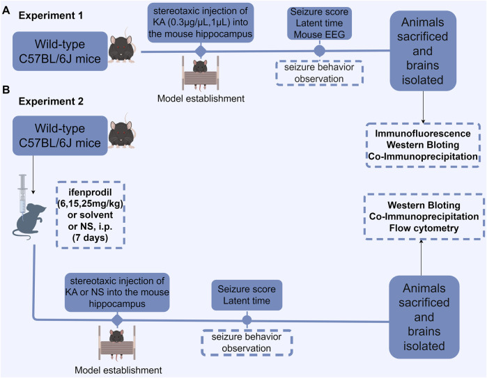
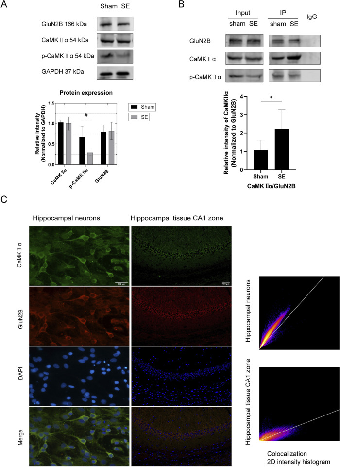
Methods: This study utilized Western blotting to evaluate the protein expression levels of CaMKIIα, p-CaMKIIα, and GluN2B in the hippocampus of mice subjected to kainic acid-induced SE. Immunofluorescence colocalization analysis and co-immunoprecipitation were utilized to investigate the interaction between GluN2B and CaMKIIα in the hippocampus. Additionally, flow cytometry was employed to measure intracellular calcium ion levels.

Results: Compared to the sham operation group, the intracellular calcium ion concentration in the hippocampus of SE mice was elevated, whereas the expression of p-CaMKIIα was markedly reduced. The levels of CaMKIIα and GluN2B remained unchanged, and the immune complex of GluN2B and CaMKIIα in the SE group exhibited a significant increase. The GluN2B inhibitor ifenprodil was found to prolong the latency of epileptic seizures, counteract calcium influx, and modulate the expression of p-CaMKIIα, as well as the immune complex levels of GluN2B and CaMKIIα. These findings suggest that the interaction between GluN2B and CaMKIIα may be critical in the pathophysiological processes of SE, influencing the levels of p-CaMKIIα and calcium ion homeostasis.

Conclusion: The reduction in CaMKIIα phosphorylation levels depends on the NMDAR pathway. When GluN2B binds to CaMKIIα, it may occupy the autophosphorylation site of CaMKIIα (T286 binding site), thereby affecting its autophosphorylation. This results in decreased phosphorylation levels, disruption of NMDAR-dependent calcium homeostasis, and alteration of the excitation/inhibition balance.

Keywords: Camkiiα; GluN2B; calcium homeostasis; ifenprodil; phosphorylation; status epilepticus.


Conflict of interest statement

The authors declare that the research was conducted in the absence of any commercial or financial relationships that could be construed as a potential conflict of interest.

Figures

None
After GluN2B binds with CaMKIIa, GluN2B occupies the CaMKIIa autophosphorylation site at threonine 286 (T286), affecting CaMKIIa autophosphorylation, reducing phosphorylation levels, and disrupting NMDAR-dependent calcium ion homeostasis, thereby disrupting the excitation-inhibition balance in the central nervous system and influencing the onset and progression of SE.
FIGURE 1
FIGURE 1
Flowchart depicting the experimental design. (A) Protein expression in KA-induced status epilepticus (SE) was measured. The latent period and seizure score of SE were measured and recorded following KA injection into the hippocampus, after which EEG recordings were obtained, and the brain was subsequently isolated for biochemical examination. (B) The effect of the GluN2B antagonist ifenprodil on KA-induced SE and its associated protein expression was evaluated. Mice were intraperitoneally injected with normal saline, ifenprodil (6, 15, 25 mg/kg), or solvent with normal saline daily for 7 consecutive days. On the seventh day, 30 min to 1 h after administration, stereotaxic injections of KA (0.3 μg/μL, 1 µL) or saline (1 µL) were administered into the hippocampus. After modeling, the mice underwent behavioral assessments, followed by brain extraction for biochemical and histological analyses. NS, normal saline; i. p., intraperitoneal injection; KA, kainic acid.
FIGURE 2
FIGURE 2
EEG results. (A) Sham group. (B,C) SE group before seizure after KA injection. (D) SE group when the seizure reached SE after KA injection. KA, kainic acid; SE, status epilepticus; EEG, electroencephalography.
FIGURE 3
FIGURE 3
Protein expression in hippocampal tissue and interaction between GluN2B and CaMKⅡα in mouse primary neurons and hippocampal tissue. (A) In comparison to the sham group, the expression of p-CaMKIIα in the SE group was diminished (n = 6 per group, #p < 0.05). Nonetheless, there was no notable alteration in the expression of GluN2B and total CaMKⅡα protein. (B) Quantitative analysis of protein immunoprecipitation via Co-IP (in vivo) revealed a substantial increase in the pull-down of CaMKⅡα in the SE group compared to the control group (n = 6 per group, *p < 0.05). (C) Immunofluorescence colocalization examination of primary neurons (microscope: Olympus BX 53; objective lens: neuron; exposure time: 50 ms; optical/digital magnification: ×60) revealed colocalization of GluN2B and CaMKⅡα in neurons (PCC = 0.91). Scale bar: 100 µm. Immunofluorescence colocalization study of hippocampus tissue (microscope: Olympus BX 53; objective lens: hippocampal tissue; exposure time: 200 ms; optical/digital magnification: ×200) revealed that GluN2B colocalizes with CaMKⅡα in hippocampal tissue (PCC = 0.82). Scale bar: 100 µm. All trials were conducted in triplicate. The independent t-test was employed to compare mean values. KA, kainic acid; SE, status epilepticus; CaMKⅡα, calcium/calmodulin-dependent protein kinase II α subunit; DAPI, 2-(4-amidinophenyl)-6-indolecarbamidine dihydrochloride.
FIGURE 4
FIGURE 4
The GluN2B inhibitor ifenprodil impedes GluN2B, alters the interaction between GluN2B and CaMKⅡα, diminishes the occupancy of CaMKⅡα phosphorylation sites, affects the phosphorylation level of CaMKⅡα, disrupts intracellular calcium ion homeostasis, and decreases both epileptic sensitivity and the severity of KA-induced status epilepticus. (A) The graphs illustrate (1) the latency of seizure onset and (2) the latency from seizure to Racine IV. In comparison to the SE group, the latency of seizure onset and the latency to Racine IV in the ifenprodil intervention group were significantly prolonged (n = 7 in each group, **p < 0.01, ***p < 0.001), with no substantial discrepancies observed among the various dose groups (p > 0.05). (B) In comparison to the sham operation group, p-CaMKⅡα levels in the SE group and the solvent group were markedly reduced (n = 7 in each group, **p < 0.01, ***p < 0.001). Following ifenprodil intervention, p-CaMKⅡα levels increased relative to the SE and solvent groups, with no substantial discrepancies observed among the various doses of ifenprodil (p > 0.05). Additionally, no substantial discrepancies in total CaMKⅡα protein levels were observed across the different groups (n = 5 in each group, p > 0.05). (C) In comparison to the sham operation group, the pull-down of CaMKⅡα in SE and solvent groups was significantly increased (n = 5 in each group, *p < 0.05, **p < 0.01, ***p < 0.001). However, following ifenprodil intervention, CaMKⅡα levels were significantly diminished relative to the SE and solvent groups, with no notable differences observed among the groups receiving varying doses of ifenprodil intervention (p > 0.05). (D) Compared to the sham operation group, the calcium ion concentration in the hippocampal tissue cells of the SE group and the solvent group was markedly elevated (n = 5 in each group, **p < 0.01, ***p < 0.001). Conversely, following ifenprodil intervention, the calcium ion concentration in the hippocampal tissue cells significantly decreased relative to the SE group and the solvent group (n = 5 in each group, *p < 0.05, **p < 0.01, ***p < 0.001), with no significant differences observed among the groups receiving varying doses of ifenprodil (p > 0.05). KA, kainic acid; SE: status epilepticus; CaMKⅡα: calcium/calmodulin-dependent protein kinase II α subunit.

Similar articles

References

    1. Abbasova K., Kubová H., Mareš P. (2018). Does status epilepticus Modify the effect of ifenprodil on cortical epileptic Afterdischarges in Immature rats? Pharmacol. Rep. 70 (1), 126–132. 10.1016/j.pharep.2017.08.012 - DOI - PubMed
    1. Alldredge B. K., Gelb A. M., Isaacs S. M., Corry M. D., Allen F., Ulrich S., et al. (2001). A comparison of lorazepam, diazepam, and Placebo for the treatment of out-of-Hospital status epilepticus. N. Engl. J. Med. 345 (9), 631–637. 10.1056/NEJMoa002141 - DOI - PubMed
    1. Ashpole N. M., Song W., Brustovetsky T., Engleman E. A., Brustovetsky N., Cummins T. R., et al. (2012). Calcium/calmodulin-dependent protein kinase Ii (Camkii) inhibition induces neurotoxicity via dysregulation of glutamate/calcium signaling and Hyperexcitability. J. Biol. Chem. 287 (11), 8495–8506. 10.1074/jbc.M111.323915 - DOI - PMC - PubMed
    1. Avalos-Fuentes A., Albarrán-Bravo S., Loya-Lopéz S., Cortés H., Recillas-Morales S., Magaña J. J., et al. (2015). Dopaminergic Denervation Switches Dopamine D3 receptor signaling and disrupts its Ca(2+) dependent modulation by Camkii and calmodulin in Striatonigral Projections of the rat. Neurobiol. Dis., 74336–74346. 10.1016/j.nbd.2014.12.008 - DOI - PubMed
    1. Barcomb K., Hell J. W., Benke T. A., Bayer K. U. (2016). The Camkii/Glun2b protein interaction Maintains synaptic strength. J. Biol. Chem. 291 (31), 16082–16089. 10.1074/jbc.M116.734822 - DOI - PMC - PubMed

LinkOut - more resources