Review
. 2025 May-Jun;20(7-9):573-587.
doi: 10.1080/17460913.2025.2501924. Epub 2025 May 7.

Antiviral defense against filovirus infections: targets and evasion mechanisms

Affiliations
Review

Antiviral defense against filovirus infections: targets and evasion mechanisms

Bianca S Bodmer et al. Future Microbiol. 2025 May-Jun.

Abstract

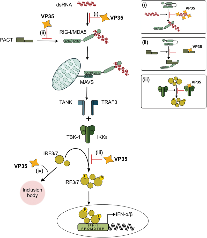
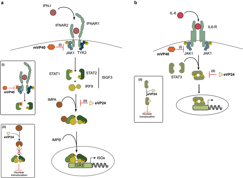
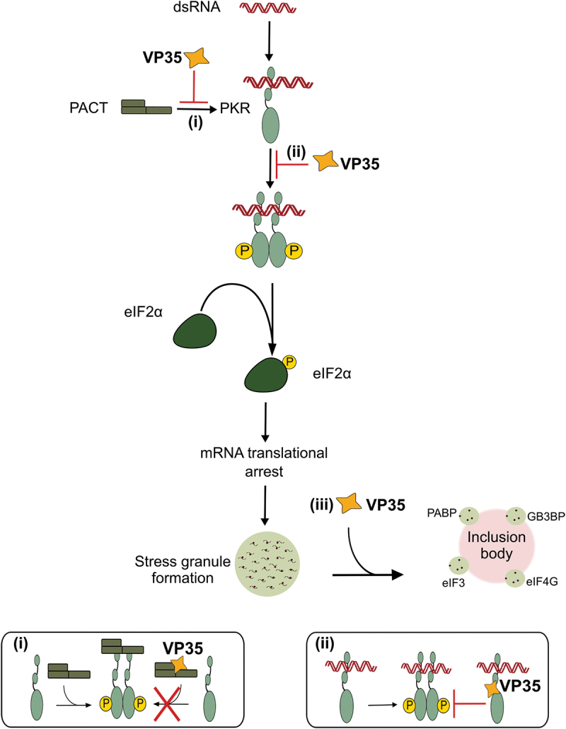
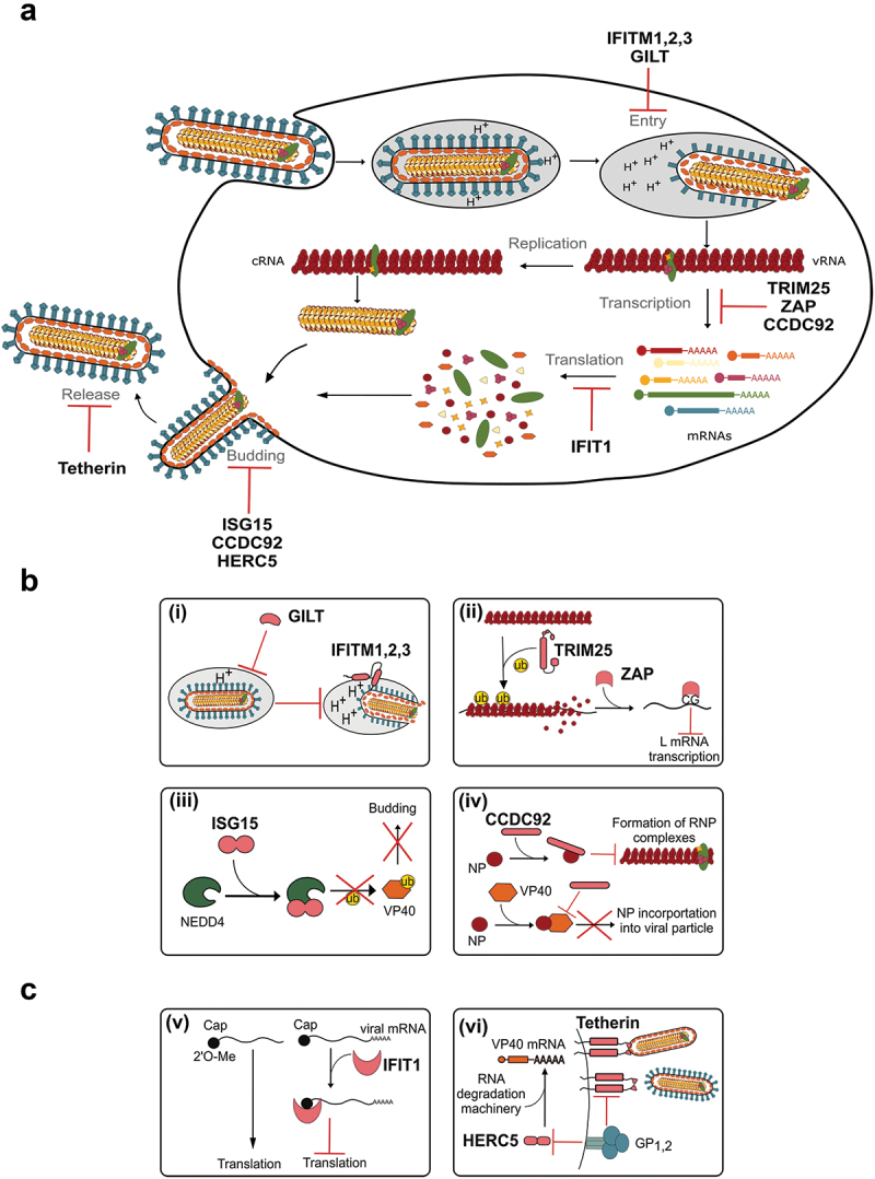
Filoviruses include a number of serious human pathogens, infections with which result in the development of hemorrhagic fevers with high case fatality rates. As for other RNA viruses, viral replication generates both protein and RNA species that can serve as danger signals, leading to the activation of antiviral defense pathways. However, in order to be able to efficiently infect humans these viruses have developed mechanisms that allow them to evade diverse host antiviral defense mechanisms. Consequently, in addition to their functions within the viral lifecycle many filovirus proteins have been shown to have accessory functions involved in the regulation of diverse host pathways. These include those of the type-I interferon response, other pathways involved in dsRNA-sensing, as well as the selective inhibition of interferon stimulated gene activities. Further, filoviruses have developed mechanisms to subvert recognition of infected cells and the generation of neutralizing antibodies. This review focuses on bringing together the evidence to date supporting the existence of diverse mechanisms aimed at regulating these pathways as well as providing details of the mechanisms involved.

Keywords: Ebola virus; Filoviruses; adaptive immunity; immune antagonism; immune evasion; innate immunity; virus-host cell interactions.

Plain language summary

Filoviruses like Ebola virus and Marburg virus can cause serious disease when they infect humans. During the infection these viruses generate danger signals that can be detected by the immune system to trigger defense pathways that can help combat these infections. Therefore, to be able to successfully infect humans, these viruses have needed to develop various ways to block these defense pathways. Many filovirus proteins have been shown to have these kinds of activities. Some of the mechanisms involved are common to many different filoviruses, but others are specific for certain filoviruses. Also, while different filoviruses may block the same pathway, they sometimes do so using different virus proteins or different mechanisms. This review summarizes our current knowledge about how filoviruses interact with host defense pathways to achieve successful infection and also how this can play a role in the development of disease.


Conflict of interest statement

The authors have no relevant affiliations or financial involvement with any organization or entity with a financial interest in or financial conflict with the subject matter or materials discussed in the manuscript. This includes employment, consultancies, honoraria, stock ownership or options, expert testimony, grants or patents received or pending, or royalties.

No writing assistance was utilized in the production of this manuscript.

Figures

Figure 1.
Figure 1.
Inhibition of type-I IFN production by VP35. Viral dsRNA is recognized by RIG-I and MDA5 leading to the production of IFN-I. VP35 antagonizes this pathway at different stages. (i) VP35 interacts with dsRNA to prevent its recognition by RIG-I and MDA5. (ii) VP35 binds to PACT, thus inhibiting its activation of RIG-I. (iii) VP35 inhibits phosphorylation of IRF3/7 and their nuclear translocation, resulting in the inhibition of IFN-I gene transcription. (iv) IRF3 is sequestered into inclusion bodies in a VP35-dependent manner.
Figure 2.
Figure 2.
Inhibition of the JAK-STAT pathway. (a) Inhibition of type-I IFN signaling. Filoviruses use different mechanisms to inhibit IFN signaling and subsequent ISG production. (i) MARV VP40 (mVP40) inhibits phosphorylation of JAK1, thereby blocking STAT1 phosphorylation. (ii) EBOV VP24 (eVP24) competes with phosphorylated STAT1 for binding to IMPA, thus blocking STAT1 nuclear translocation. (b) Inhibition of the IL-6 pathway. Filoviruses use different mechanisms to block activation of JAK/STAT signaling in response to IL-6. (i) MARV VP40 (mVP40) inhibits phosphorylation of JAK1, and thus also the downstream phosphorylation of STAT3. (ii) EBOV VP24 (eVP24) binds directly to STAT3, thereby blocking its nuclear translocation.
Figure 3.
Figure 3.
Inhibition of PKR-mediated translational arrest. VP35 is able to inhibit the PKR-mediated shut-off of translation by different mechanisms. (i) VP35 interacts with PACT, thus inhibiting PACT-mediated dimerization of PKR and its subsequent phosphorylation. (ii) VP35 binds directly to PKR, preventing its dimerization and phosphorylation. (iii) VP35 recruits stress granule marker proteins into viral inclusion body-associated granules, impairing the formation of stress granules.
Figure 4.
Figure 4.
Inhibition of the filovirus life cycle by ISGs and their antagonization by filoviral proteins. (a) Overview of the filovirus life cycle. Steps in the virus life cycle known to be targeted by ISGs are indicated. (b) Antiviral mechanisms of ISGs. (i) GILT as well as IFITMs 1, 2, and 3 inhibit virus entry at the level of the late endosome, likely by inhibition of cathepsin cleavage of GP1,2 and by interrupting the formation of fusion pores, respectively. (ii) TRIM25 ubiquitinates NP, blocking its interaction with viral ribonucleoprotein complexes. This allows ZAP to bind to the viral genomic RNA, resulting in decreased L mRNA transcription. (iii) ISG15 interacts with NEDD4 and blocks its ubiquitination of VP40, thereby inhibiting the budding of newly formed virions. (iv) CCDC92 interacts with NP, disrupting the formation of viral transcription complexes, and thus inhibiting viral mRNA transcription. Further, CCDC92 impairs the interaction between NP and VP40, which reduces the incorporation of NP into viral particles. (c) Inhibition of ISG activities by filoviral proteins. (v) IFIT1 inhibits viral translation by binding to viral mRNAs that are not 2’O-cap-methylated. Orthoebolavirus L prevents this through its 2’O-methyltransferase activity. (vi) Tetherin tethers newly formed viral particles to the cell membrane to prevent their release. GP1,2 prevents this by blocking the interaction of tetherin with VP40. Further, GP1,2 is also able to reverse the effect of HERC5, which appears to recruit the RNA degradation machinery to VP40 mRNAs.
Figure 5.
Figure 5.
Glycoprotein expression strategies and their roles in the evasion of immune cell functions. (a) Schematic of glycoprotein expression in orthoebolaviruses. The major mRNA transcript of the glycoprotein gene encodes pre-sGP, which is cleaved by furin to generate Δ-peptide and sGP, which in its mature form is a homodimer. If an additional adenosine is added during mRNA transcription of the glycoprotein gene, an mRNA encoding pre-GP is generated. Pre-GP is then cleaved by furin into the subunits GP1 and GP2, which heterotrimerize to form the mature GP1,2. The membrane-anchored GP1,2 can be further cleaved by TACE to generate GP1,2ΔTM. If two adenosines are added (or one is omitted) during transcriptional editing, ssGP is formed. sGP, Δ-peptide, GP1,2ΔTM and ssGP are all secreted from infected cells, whereas GP1,2 is anchored in the viral membrane. (b) Roles of the different forms of GP in immune evasion. (i) GP1,2ΔTM and sGP act as decoys for GP1,2 specific antibodies. (ii) The mucin-like domain of GP1,2 and GP1,2ΔTM can be recognized by TLR4, leading to activation of the NF-κB pathway, and resulting in cytotoxicity in non-infected immune cells. (iii) GP1,2 is able to sterically mask MHC-I present at the cell surface, thereby blocking the recognition of MHC-I-presented viral antigen by T cells.

Similar articles

References

    1. Burk R, Bollinger L, Johnson JC, et al. Neglected filoviruses. FEMS Microbiol Rev. 2016;40(4):494–519. doi: 10.1093/femsre/fuw010 - DOI - PMC - PubMed
    1. Hood G, Carroll M.. Host-pathogen interactions of emerging zoonotic viruses: bats, humans and filoviruses. Curr Opin Virol. 2024;68–69:101436. doi: 10.1016/j.coviro.2024.101436 - DOI - PubMed
    1. Penas JA, Miranda ME, de Los Reyes VC, et al. Risk assessment of Ebola Reston virus in humans in the Philippines. Western Pac Surveill Response J. 2019;10(3):1–8. doi: 10.5365/wpsar.2017.3.004 - DOI - PMC - PubMed
    1. Bodmer BS, Breithaupt A, Heung M, et al. In vivo characterization of the novel ebolavirus Bombali virus suggests a low pathogenic potential for humans. Emerg Microbes Infect. 2023;12(1):2164216. doi: 10.1080/22221751.2022.2164216 - DOI - PMC - PubMed
    2. • First assessment of a novel filovirus regarding its pathogenic potential.

    1. Messaoudi I, Basler CF. Immunological features underlying viral hemorrhagic fevers. Curr Opin Immunol. 2015;36:38–46. doi: 10.1016/j.coi.2015.06.003 - DOI - PMC - PubMed

LinkOut - more resources