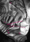
The influence of sacrocolporectopexy on pelvic anatomy assessed in an upright position using MRI
- PMID: 40331245
- PMCID: PMC12056576
- DOI: 10.1111/codi.70114
The influence of sacrocolporectopexy on pelvic anatomy assessed in an upright position using MRI
Abstract
Aim: Rectopexy with concomitant sacrocolpopexy (sacrocolporectopexy) is the favoured technique for treating combined pelvic organ prolapse and internal or external rectal prolapse, despite limited functional improvement. Previous studies have assessed anatomical change after standalone rectopexy or sacrocolpopexy, based on supine MRI defaecography. Since a supine position can underestimate the extent of pelvic organ prolapse, it might also incorrectly assess the anatomical effect of sacrocolporectopexy. The aim of this study was to assess the effect of sacrocolporectopexy on the pelvic anatomy in an upright position.
Method: Twenty one female patients undergoing sacrocolporectopexy from December 2022 to June 2024 were included. All patients underwent physical examination and MRI defaecography preoperatively and postoperatively. The descent of the bladder, vaginal vault and anorectal junction and the size of the rectocele and enterocele were assessed on the MRI defaecography images during maximum straining. Significance was tested using a paired t-test and an improvement of ≥10 mm was considered clinically relevant. The results were compared with previous studies, which used supine assessment.
Results: Postoperative improvement was found for the bladder, vaginal vault, anorectal junction, rectocele and enterocele with 14, 44, 5, 16 and 54 mm respectively. The bladder, vaginal vault, rectocele and enterocele showed clinically relevant improvement. Compared with supine results, upright assessments revealed a larger organ lift for the vaginal vault as well as a higher, overall, position of the anorectal junction.
Conclusion: Upright assessment of sacrocolporectopexy differs from supine assessment, with statistical and clinically relevant lift for the pelvic organs.
Keywords: Internal rectal prolapse; pelvic anatomy; pelvic organ prolapse; sacrocolporectopexy; upright magnetic resonance defaecography.
© 2025 The Author(s). Colorectal Disease published by John Wiley & Sons Ltd on behalf of Association of Coloproctology of Great Britain and Ireland.
Conflict of interest statement
None of the authors report a conflict of interest or a relevant financial relationship.
Figures


References
-
- Wijffels NAT, Consten E, van der Hagen SJ. Dutch Guidelines Rectal Prolapse . 2017. - PubMed
MeSH terms
Grants and funding
LinkOut - more resources
Full Text Sources
Medical

