Review
. 2025 Jul 28;12(15):5570-5593.
doi: 10.1039/d5mh00096c.

Poly(3-hexylthiophene) as a versatile semiconducting polymer for cutting-edge bioelectronics

Affiliations
Review

Poly(3-hexylthiophene) as a versatile semiconducting polymer for cutting-edge bioelectronics

Ilaria Abdel Aziz et al. Mater Horiz. .

Abstract

Semiconducting polymers (SPs), widely used in organic optoelectronics, are gaining interest in bioelectronics owing to their intrinsic optical properties, conductivity, biocompatibility, flexibility, and chemical tunability. Among them, poly(3-hexylthiophene) (P3HT) has attracted great attention as a versatile SP, being both optically active and conductive, for the fabrication of smart materials (e.g., films and nanoparticles), allowing the modulation of their performance and final biomedical applications. This review article provides an overview of the design of different kinds of P3HT-based materials, from chemical properties to structural engineering, to be used as key opto-electronic components in the development of opto-transducers for the modulation of cell fate, as well as biosensors such as organic electrochemical transistors (OECTs) and organic field effect transistors (OEFTs). Finally, their foremost applications in the biomedical field ranging from tissue engineering to biosensing will be discussed, including the future perspectives of P3HT derivatives towards cutting-edge applications for bioelectronics, in which optoceutics plays a key role.


Conflict of interest statement

There are no conflicts to declare.

Figures
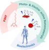
Fig. 1
Fig. 1. Schematic representation of the employment of P3HT for the synthesis of photo- and electro-active nanomaterials to be used in bioelectronic applications.
Fig. 2
Fig. 2. (A) Chemical structure and (B) energy band gap of P3HT, including its photocatalytic activity to produce H2O2.
Fig. 3
Fig. 3. (A) Schematic representation of the spin-coating methodology used to prepare P3HT thin films. (B) AFM topography image of a representative P3HT film. Reprinted with permission. Copyright 2016 The Royal Society of Chemistry. (C) Optical absorption spectrum of P3HT thin films. Reprinted with permission. Copyright 2017 Springer Nature. (D) Cyclic voltammetry at 100 mV s−1 of P3HT films. Reprinted with permission. Copyright 2016 The Royal Society of Chemistry. (E) Bode plots of P3HT films deposited on ITO, in the dark and upon photoexcitation (λ = 470 nm, 2.73 mW mm−2), including the equivalent circuits used to model experimental data. Reprinted with permission. Elsevier B.V. (F) Schematic representation of the detection strategy of H2O2 production upon P3HT film photoexcitation, and (G) fluorescence image of the illuminated region of the film (top), and SECM scan lines of H2O2 production (bottom) measured as H2O2 oxidation current at the black platinum probe. Reprinted with permission. Copyright 2020 Cell Press.
Fig. 4
Fig. 4. (A) Schematic representation of the P3HT micropillar fabrication process by moulding, and (B) SEM image of P3HT pillar structure. Reprinted with permission. Copyright 2019 American Chemical Society. (C) Schematic representation of the fabrication of porous thin films by PLA hydrolysis; (D) topographical AFM image of a representative porous P3HT film after PLA hydrolysis of P3HT60-g-PLA40 copolymer film; and (E) photocurrent curves of non-porous P3HT films with different thicknesses and porous films made of P3HTx-g-PLAy copolymers when irradiated with a LED (530 nm, 110 mW cm−2). Reprinted with permission. Copyright 2023 American Chemical Society. (F) SEM images of porous P3HT/SEBS films, and (G) transfer curves and corresponding gm evolutions for OECTs based on multilayers of P3HT. Reprinted with permission. Copyright 2023 Wiley-VCH GmbH. (H) AFM images of porous P3HT films obtained from phase separation P3HT/PS followed by dissolution of PS by washing with acetone, and (I) CV scans (100 mV s−1) of the O2 plasma-treated porous P3HT films. Reprinted with permission. Copyright 2020 The Royal Society of Chemistry.
Fig. 5
Fig. 5. (A) Schematic representation of the precipitation method used for the preparation of P3HT nanoparticles (NPs), and (B) representative SEM image of P3HT NPs. Reprinted with permission. Copyright 2017 The Royal Society of Chemistry. (C) Optical absorption and fluorescence (PL) spectra of P3HT NPs in aqueous dispersion. (D) Experimental set-up used for photocurrent experiments and (E) Photocurrent spectra with and without molecular oxygen in solution at V = OCP–300 mV: ambient conditions with oxygen (black solid line), upon removal of oxygen in a nitrogen atmosphere (blue solid line), and upon partial re-oxygenation (black dashed line). The yellow shaded area represents the temporal window corresponding to optical excitation. Adapted and reprinted with permission. Copyright 2018 Frontiers.
Fig. 6
Fig. 6. (A) Schematic representation of the formation of core–shell P3HT@PTDO nanoparticles, and surface photovoltage values corresponding to different nanoparticles (red circle, P3HT-NPs; black circles, P3HT@PTDO-NPs). Reprinted with permission. Copyright 2017, American Chemical Society. (B) Chemical structure of the P3HT-b-PEG block copolymer and AFM image of P3HT-b-PEG micelles formed in aqueous solution with an illustration of the aggregated P3HT core and the disordered PEG corona; UV/vis absorption spectra of the P3HT-b-PEG block copolymer and a P3HT homopolymer dissolved in the “good” solvent chloroform (black solid and dashed line), as well as of P3HT-b-PEG micelles in aqueous solution (red). Reprinted with permission. Copyright 2021 American Chemical Society. (C) Schematic representation of the synthesis of P3HT porous nanoparticles (PSPNs) and TEM-EDX images of a representative PSPN. (D) Photocurrent curves of SPNs and PSPNs upon their irradiation with a LED (λ = 530 nm; 6 mW cm−2). (E) Representative confocal fluorescence image of HUVECs treated with P3HT-PSPNs: nuclei (blue), actin filaments (green), CD31 (red), and PSPNs with intrinsic fluorescence represented in yellow for better visualization. Reprinted with permission. Copyright 2024 American Chemical Society.
Fig. 7
Fig. 7. (A) Sketch reporting the illumination conditions for the living cells in the home-made setup. The cells were illuminated from the top (400 μW mm−2) with two different illumination patterns (left), and average variation of Ca2+ peaks under different experimental conditions is shown, after the 20/200 ms stimulation protocol. The statistical significance is reported for p < 0.001 (***), all other differences are not significantly different (right). Reprinted with permission. Copyright 2022 The Royal Society of Chemistry. (B) Schematic representation of the biological pathways possibly induced by photostimulation of semiconducting polymer NPs. Reprinted with permission. Copyright 2022 The Authors. Advanced Healthcare Materials published by Wiley-VCH GmbH. (C) SEM image of a neuronal soma suspended over two P3HT pillars. (D) Cortical neurons cultured on P3HT micropillars after 3 DIV, where neurons were fixed and fluorescently labeled for β-III-tubulin (green) and Tau-1 (red). (E) Average neurite length for 257 neurons on glass, 225 neurons on flat P3HT, and 229 neurons on pillar P3HT (data were compared using the non-parametric Mann–Whitney U-test with Bonferroni–Holm multiple comparison correction (0.05 significance level). ***p < 0.001, ns—not significant). Reprinted with permission. Copyright 2021 American Chemical Society.
Fig. 8
Fig. 8. (A) Optical microscope image of an EGOFET fabricated by ink-jet printing and standard photolithography; (B) picture of the device after a polystyrene cell is glued around the transistor channel; (C) percentage variation of ID max after the acquisition of the first 64 transfer curves till the 200th curve measured on an EGOFET comprising a spin-coated (blue circles) or an ink-jet printed (black circles) P3HT film; (D) P3HT based EGOFETs’ threshold voltage (VT). Reprinted with permission. Copyright 2020 The Royal Society of Chemistry. (E) Schematic diagram of the device based on a P3HT/PVA/ITO multilayer and operated in an aqueous solution, with the Ag/AgCl electrode placed in the electrolyte; (F) schematic diagram for the voltage-controlled permeability and switchable molecule release of a P3HT film; and (G) the concentration of released FSA (left axis) as a function of time under the application of bias voltage (right axis). Reprinted with permission. Copyright 2017 Wiley-VCH Verlag GmbH & Co.
Fig. 9
Fig. 9. (A) Chemical structure of terthiophene (thiophene trimer). (B) Chemical structures of ETE-N and ETE-S trimers polymerized in vivo along the roots of living plants due to the presence of native peroxidase enzymes, and cross-sectional (top) and lateral views (bottom) of the bean roots (scale bar = 100 μm). Adapted and reprinted with permission. Copyright 2020 American Chemical Society.
None
Ilaria Abdel Aziz
None
Gabriele Tullii
None
Maria Rosa Antognazza
None
Miryam Criado-Gonzalez

Similar articles

Cited by

References

    1. Garg S. Goel N. J. Phys. Chem. C. 2022;126:9313–9323. doi: 10.1021/acs.jpcc.2c02938. - DOI
    1. Ding B. Le V. Yu H. Wu G. Marsh A. V. Gutiérrez-Fernández E. Ramos N. Rimmele M. Martín J. Nelson J. Paterson A. F. Heeney M. Adv. Electron. Mater. 2024;10:2300580. doi: 10.1002/aelm.202300580. - DOI
    1. Criado-Gonzalez M. Dominguez-Alfaro A. Lopez-Larrea N. Alegret N. Mecerreyes D. ACS Appl. Polym. Mater. 2021;3:2865–2883. doi: 10.1021/acsapm.1c00252. - DOI - PMC - PubMed
    1. Jiang Y. Pu K. Acc. Chem. Res. 2018;51:1840–1849. doi: 10.1021/acs.accounts.8b00242. - DOI - PubMed
    1. Gao D. Parida K. Lee P. S. Adv. Funct. Mater. 2020;30:1907184. doi: 10.1002/adfm.201907184. - DOI

LinkOut - more resources