Review
. 2025 Sep;94(3):913-936.
doi: 10.1002/mrm.30510. Epub 2025 May 7.

Engineering clinical translation of OGSE diffusion MRI

Affiliations
Review

Engineering clinical translation of OGSE diffusion MRI

Ante Zhu et al. Magn Reson Med. 2025 Sep.

Abstract

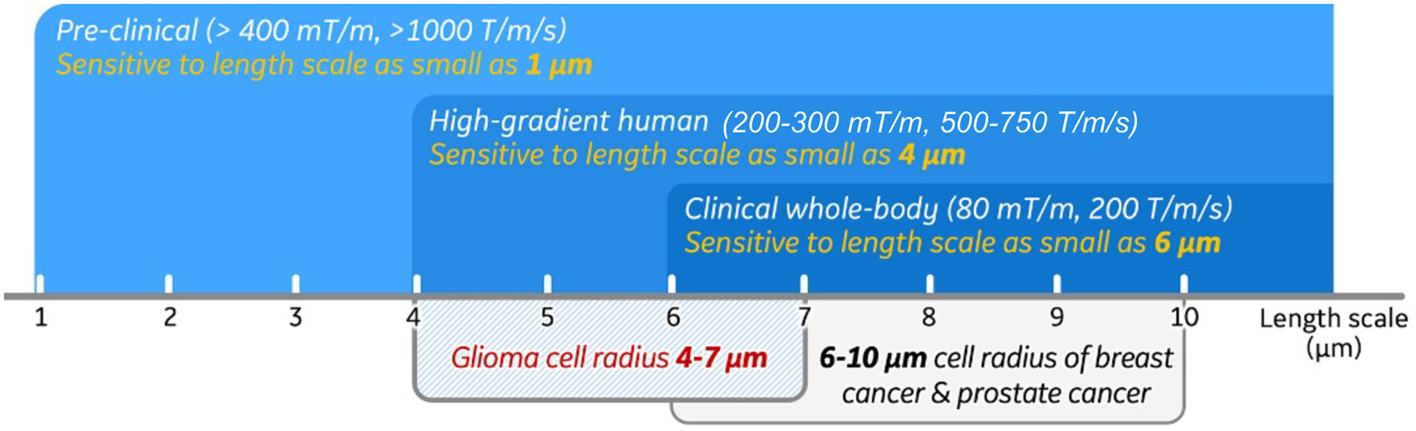
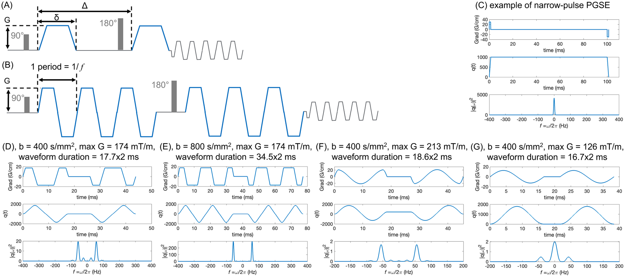
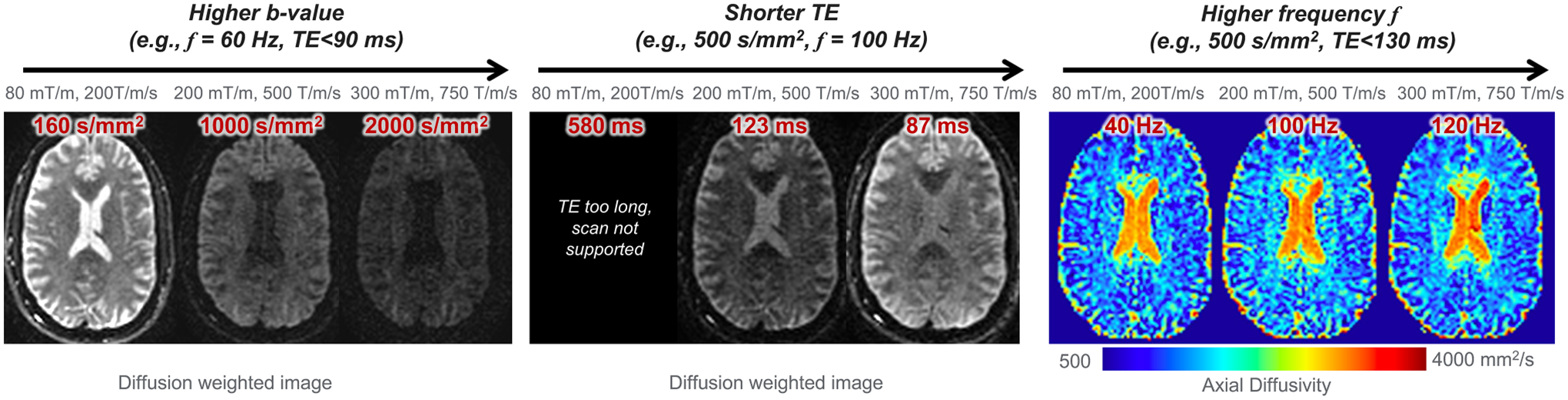
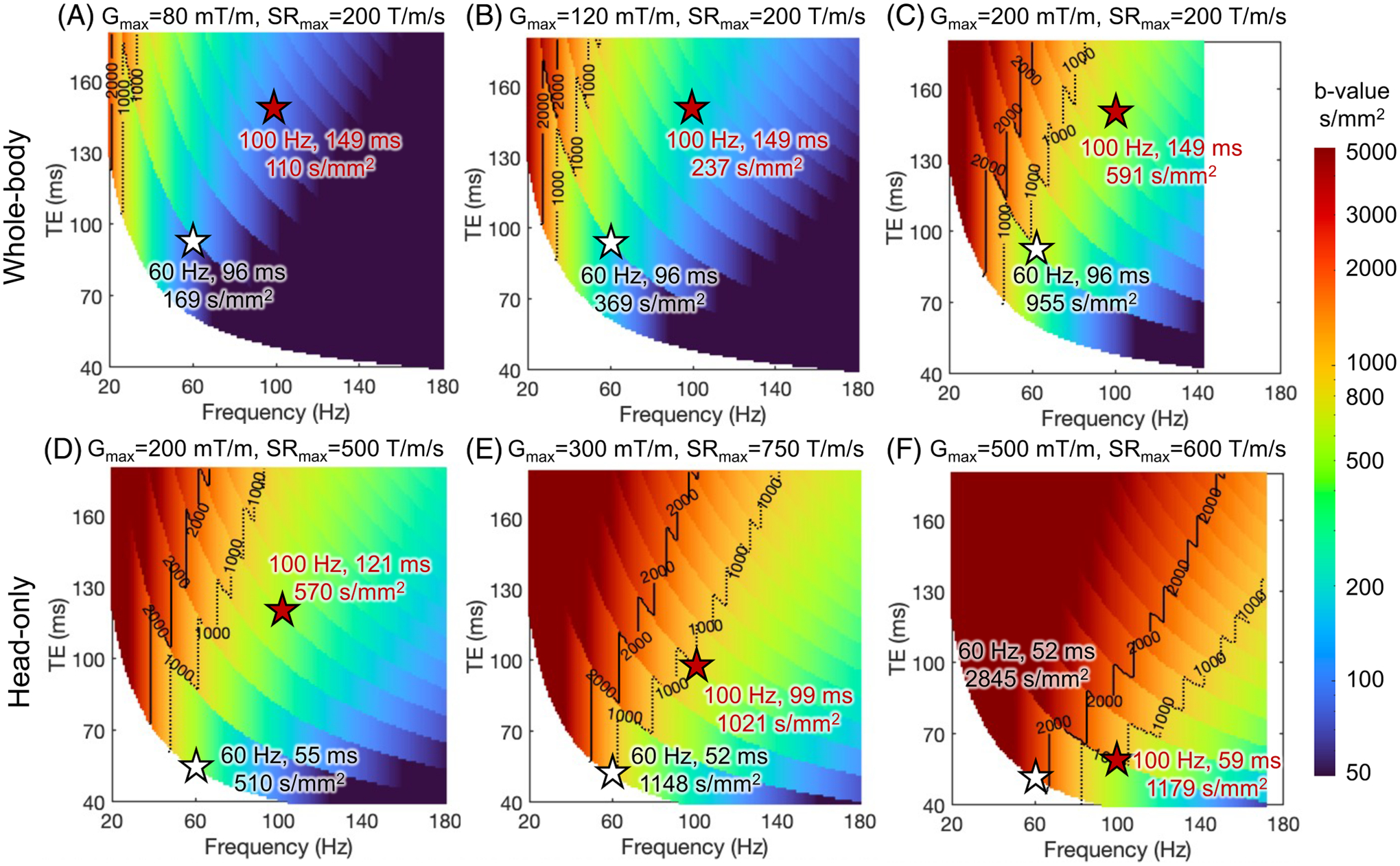
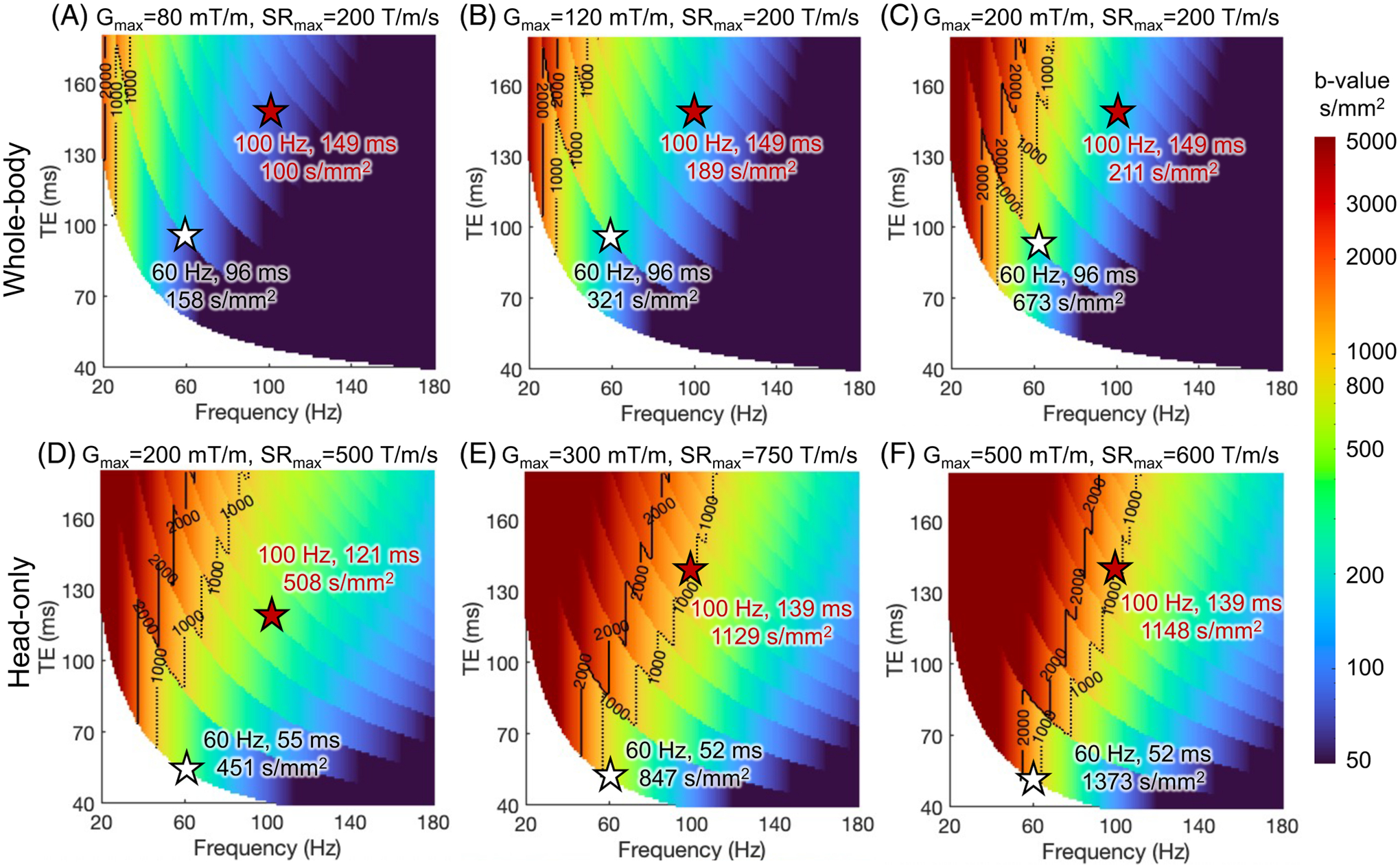
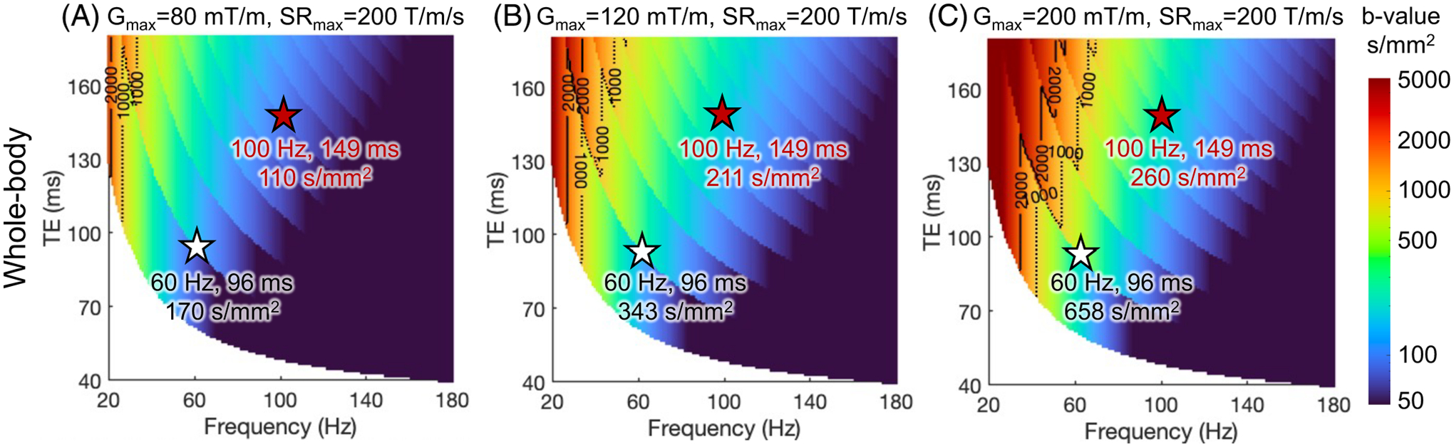
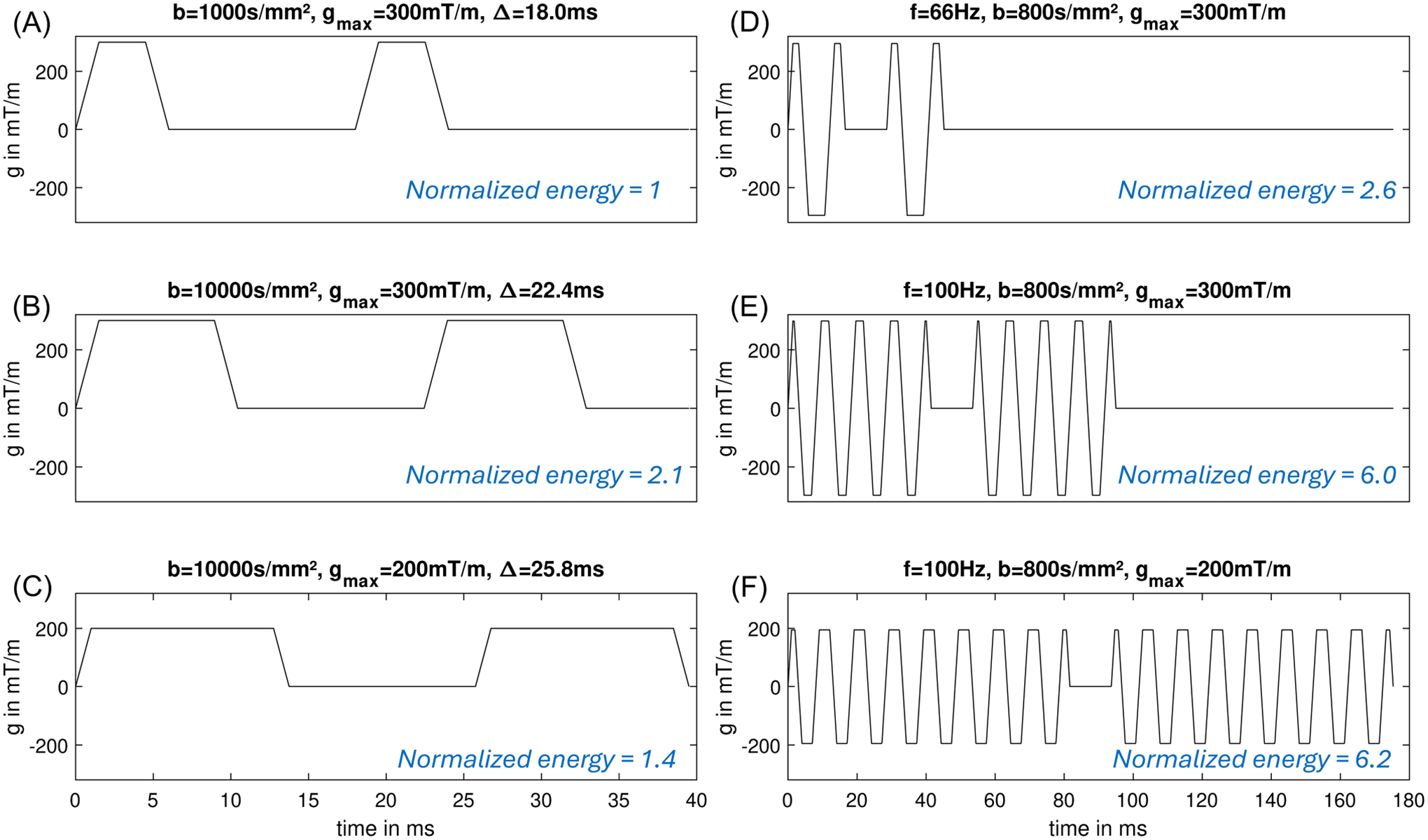
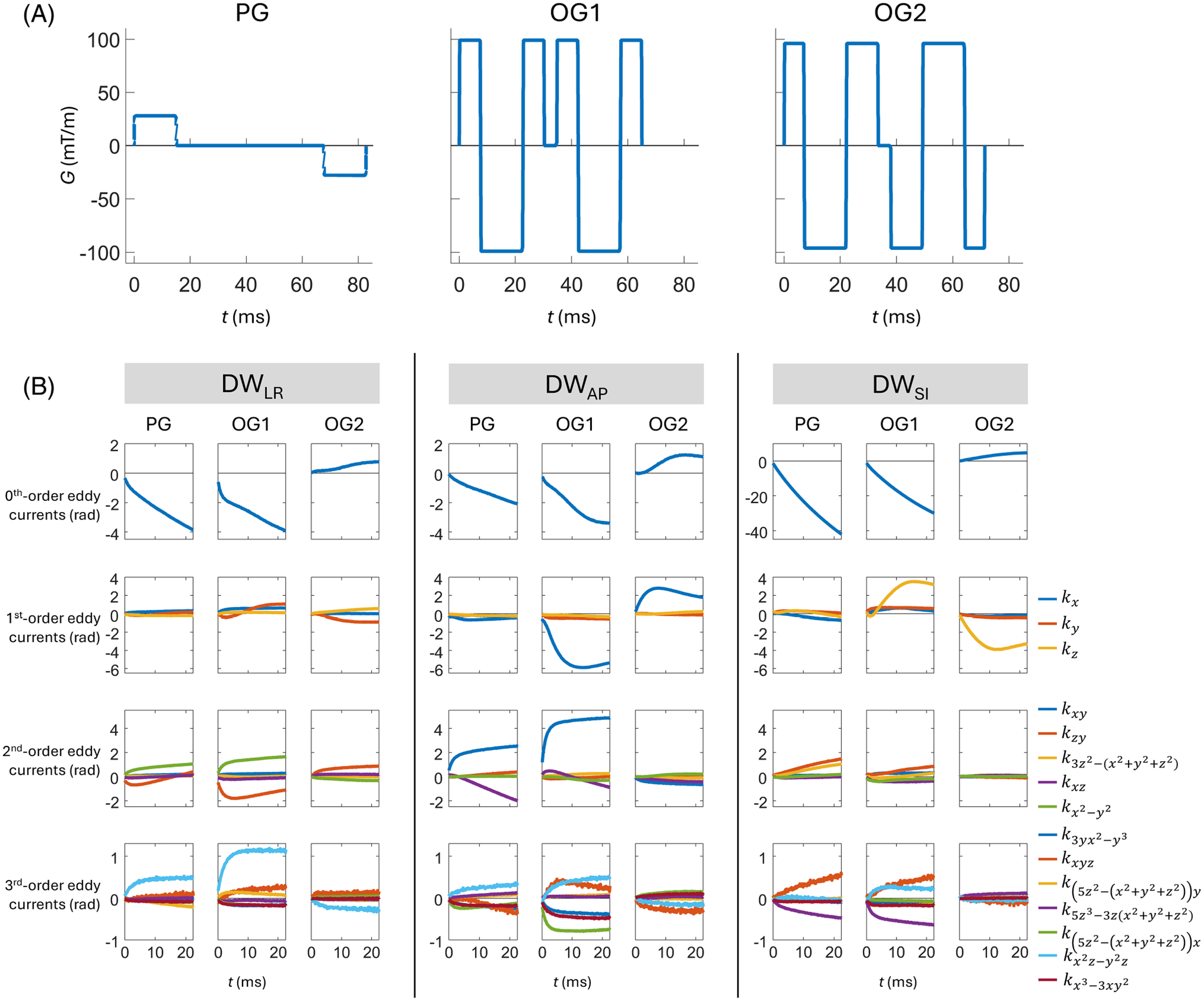
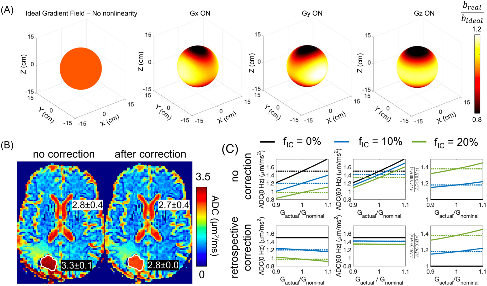
Oscillating gradient spin echo (OGSE) diffusion MRI (dMRI) can probe the diffusive dynamics on short time scales ≲10 ms, which translates into the sensitivity to tissue microstructure at the short length scales 10 μ $$ \lesssim 10\kern0.3em \upmu $$ m. OGSE-based tissue microstructure imaging techniques able to characterize the cell diameter and cellular density have been established in pre-clinical studies. The unique image contrast of OGSE dMRI has been shown to differentiate tumor types and malignancies, enable early diagnosis of treatment effectiveness, and reveal different pathophysiology of lesions in stroke and neurological diseases. Recent innovations in high-performance gradient human MRI systems provide an opportunity to translate OGSE research findings in pre-clinical studies to human research and the clinic. The implementation of OGSE dMRI in human studies has the promise to advance our understanding of human brain microstructure and improve patient care. Compared to the clinical standard (pulsed gradient spin echo), engineering OGSE diffusion encoding for human imaging is more challenging. This review summarizes the impact of hardware and human biophysical safety considerations on the waveform design, imaging parameter space, and image quality of OGSE dMRI. Here we discuss the effects of the gradient amplitude, slew rate, peripheral nerve stimulation, cardiac stimulation, gradient driver, acoustic noise and mechanical vibration, eddy currents, gradient nonlinearity, concomitant gradient, motion and flow, and signal-to-noise ratio. We believe that targeted engineering for safe, high-quality, and reproducible imaging will enable the translation of OGSE dMRI techniques into the clinic.

Keywords: MRI system engineering; clinical translation; high‐performance gradient; microstructure; oscillating gradient; time‐dependent diffusion.


Conflict of interest statement

CONFLICT OF INTEREST STATEMENT

Dr. Zhu, Dr. Li, Dr. Sprenger, Dr. Hua, Dr. Lee, Dr. Yeo, and Dr. Foo are employees of GE HealthCare.

Figures

FIGURE 1
FIGURE 1
The length scales that pre-clinical, human high-performance gradient and clinical whole-body diffusion MRI are sensitive to, compared with the cell radii of different human tumors.
FIGURE 2
FIGURE 2
(A) and (B) show the schematic of PGSE and trapezoidal-modulated OGSE waveforms, respectively. (C–G) show PGSE waveform and different OGSE waveforms with the polarity of post-180 diffusion pulse reversed, the anti-derivative q(t) of the gradient, and its spectrum q(ω). OGSE waveforms can achieve higher b-values with multiple oscillating periods (E vs. D) while maintaining the same diffusion time (or OGSE frequency), for example, at f=60Hz. (F) and (G) show the cosine and sine OGSE waveforms, respectively, at the same b-value and frequency ranges.
FIGURE 3
FIGURE 3
Example of human brain OGSE dMRI at different gradient configurations, including Gmax of 80 mT/m and SRmax of 200 T/m/s; Gmax of 200 mT/m and SRmax of 500 T/m/s; and Gmax of 300 mT/m and SRmax of 750 T/m/s. Images were acquired within peripheral nerve stimulation and cardiac stimulation limits in a head-only high-performance gradient MRI system.
FIGURE 4
FIGURE 4
The maximum b-value of trapezoid-cosine OGSE achieved at varying TE’s and frequencies, using simultaneous Gmax and SRmax of whole-body MRI systems (A–C) and head-only MRI systems (D–F). The calculation is detailed in Supplementary Material S1. At a fixed frequency, the maximum b-value shows discontinuity at certain TE, which is due to the use of different numbers of OGSE waveform periods, as shown in Supplementary Figure S2. Other system constraints are not imposed.
FIGURE 5
FIGURE 5
The maximum b-value of trapezoid-cosine OGSE achieved at TE’s and frequencies using the gradient and slew rate constrained by both MRI system limits in Figure 4 and PNS threshold of gradient coils. As an example in this review, the head-only gradient coils are characterized with ΔGmin=39.5mT/m and SRmin=185.6T/m/s, in the linear gradient field threshold curve of bipolar gradients. As an example in this review, the whole-body gradient coil is characterized with ΔGmin=19.7mT/m and SRmin=84.8T/m/s.
FIGURE 6
FIGURE 6
The maximum b-value of trapezoid-cosine OGSE achieved at TE’s and frequencies using the gradient and slew rate constrained by both MRI system limits in Figure 4 and CS threshold of whole-body gradient coils. As an example, the CS threshold of the whole-body gradient coil is characterized with ΔGmin=113mT/m and SRmin=62.5T/m/s.
FIGURE 7
FIGURE 7
Energy over the diffusion encoding waveforms of OGSE and PGSE.
FIGURE 8
FIGURE 8
(A) Pulsed (PG), trapezoid-cosine oscillating (OG1 at 33 Hz), and a modified oscillating (OG2 at 35 Hz) diffusion waveforms, and (B) the eddy currents resulting from them for different axes of diffusion encoding measured by a field camera in a 3T high-performance head-only gradient MRI system. In (B), all subplots show the temporally evolving phase accrual (in radians) during a subsequent EPI readout, resulting from the application of diffusion gradients. These evolutions are given in terms of coefficients of spherical harmonic basis functions of different orders (denoted in legend), with scaling that reflects the maximum phase accrual on a sphere of radius 100 mm. The contributions resulting from diffusion encoding were isolated by taking differences between the coefficients of dMRI acquisitions and corresponding b=0s/mm2 acquisitions. Note that vertical scaling is consistent within rows except for the first row (zeroth-order eddy currents), for which the vertical scale is much larger for diffusion encoding along the SI axis than for diffusion encoding along the LR and AP axes. LR: Left-right; AP: Anterior-posterior; SI: Superior-inferior.
FIGURE 9
FIGURE 9
(A) The nonlinear gradient field results in spatially varying b-value when the diffusion encoding gradient is played along the physical X,Y, and Z axis, for example, in the 10-cm diameter sphere. (B) ADC is incorrectly estimated without correcting the gradient nonlinearity effect on the b-value. For example, in the fluid-filled resected cavity of a glioblastoma patient after surgery, ADC was overestimated, compared to the ADC of the cerebrospinal cord. After correction, the ADC of the fluid-filled resected cavity was close to the ADC of the cerebrospinal cord. (C) Simulation of the effect of gradient nonlinearity on non-Gaussian diffusion measurements, for example, ADC(60Hz)ADC(0Hz), in a tumor model with a cell radius of 5 μm and different intra-tumoral volume fraction. The retrospective gradient nonlinearity correction largely reduces the bias on ADC(60 Hz) and ADC(0 Hz). However, the bias on ADC(60Hz)ADC(0Hz) cannot be corrected.

Similar articles

References

    1. Hanahan D, Weinberg RA. Hallmarks of cancer: The next generation. Cell. 2011;144:646–674. - PubMed
    1. Louis DN, Perry A, Wesseling P, et al. The 2021 WHO classification of tumors of the central nervous system: A summary. Neuro-Oncology. 2021;23:1231–1251. doi: 10.1093/neuonc/noab106 - DOI - PMC - PubMed
    1. Senkus E, Kyriakides S, Ohno S, et al. Primary breast cancer: ESMO clinical practice guidelines for diagnosis, treatment and follow-up. Ann Oncol. 2015;26:v8–v30. - PubMed
    1. Leenders GJLH, Kwast TH, Grignon DJ, et al. The 2019 International Society of Urological Pathology (ISUP) consensus conference on grading of prostatic carcinoma. Am J Surg Pathol. 2020;44:e87–e99. - PMC - PubMed
    1. Jiang X, Dudzinski S, Beckermann KE, et al. MRI of tumor T cell infiltration in response to checkpoint inhibitor therapy. J Immunother Cancer. 2020;8:e000328. doi: 10.1136/jitc-2019-000328 - DOI - PMC - PubMed

MeSH terms