. 2025 Jun 1;328(6):L832-L843.
doi: 10.1152/ajplung.00287.2024. Epub 2025 May 7.

Lumacaftor inhibits channel activity of rescued F508del cystic fibrosis transmembrane conductance regulator

Affiliations

Lumacaftor inhibits channel activity of rescued F508del cystic fibrosis transmembrane conductance regulator

Adam D Ambrosetti et al. Am J Physiol Lung Cell Mol Physiol. .

Abstract

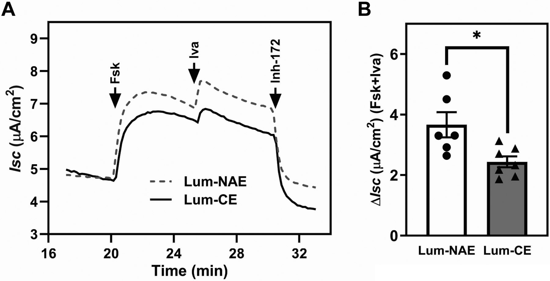
Lumacaftor, the corrector of Orkambi, enhances the processing of F508del cystic fibrosis transmembrane conductance regulator (CFTR), but its impact on the channel activity of rescued F508del CFTR (rF508del) is unclear. Using an electrode-based, real-time iodide efflux assay performed at room temperature, acute exposure to lumacaftor was shown to increase the processing of F508del CFTR without a proportional increase in channel activity in a CFBE41o-cell line stably expressing F508del CFTR (CFBE-DF). A similar effect was not observed on wild-type CFTR in a HEK293 cell line. At 37°C, rF508del channel activity is significantly inhibited in CFBE-DF cells by acute exposure to 5 µM lumacaftor, but not to 5 µM tezacaftor or 1 µM elexacaftor, the two correctors of Trikafta. Lumacaftor's inhibitory effect was characterized by a major left shift of the peak channel activity relative to the peak CFTR processing in the dose-response chart, which is absent for tezacaftor or elexacaftor. Ussing chamber analysis on polarized CFBE-DF cells reveals an inhibitory effect for lumacaftor on the forskolin- and ivacaftor-induced change in short-circuit current. Single channel patch clamp on HEK-DF cells shows that acute application of cytosolic lumacaftor significantly decreases rF508del channel open probability. Taken together, despite its strong corrector activity, lumacaftor inhibits rF508del channel activity, compromising the degree of functional rescue. This effect may contribute to the limited clinical efficacy of Orkambi.NEW & NOTEWORTHY Small-molecule correctors bind to F508del cystic fibrosis transmembrane conductance regulator (CFTR) and restore its trafficking to the plasma membrane to function as an anion channel. Despite its high efficacy as a corrector, lumacaftor inhibits the channel opening of rescued F508del CFTR, making it a weak CFTR modulator. The current work highlights the impact of CFTR correctors on the channel activity of rescued F508del CFTR as an important variable in the efficacy of modulator therapy.

Keywords: cystic fibrosis; cystic fibrosis transmembrane conductance regulator (CFTR); iodide efflux assay; ion channel; lumacaftor.


Figures

Figure 1.
Figure 1.
Comparison of sequential wash and real-time iodide efflux assays. A. Shown is the setup for real-time measurement. B. Correlation between iodide concentrations of iodide standard solutions and the measured voltages using an iodide selective electrode. The mean and standard errors of the mean (SEMs) of the R2 of five independent experiments are shown. C. For the sequential wash method, HEK and HEK-WT cells were loaded with iodide at 37°C and washed six times with an iodide-free efflux buffer at room temperature. Then the cells were sequentially washed 8 times with 2.5 ml each of efflux buffer containing 10 μM forskolin at room temperature for 1 minute each wash. The washes were collected, and their iodide concentrations were measured with an iodide-selective electrode. Shown are the data from 2 experiments for HEK and 3 for HEK-WT. D. HEK and HEK-WT cells were loaded with loading buffer at 37°C and washed six times with efflux buffer at room temperature as described above. The washed cells were overlaid with 2.5 ml of efflux buffer and an iodide-selective electrode was inserted into the efflux buffer above the cells. The extracellular iodide concentration was monitored at room temperature for 5 minutes to establish a base line. Then the cells were stimulated with forskolin (black arrowhead) and the extracellular iodide concentration was monitored for another 10 minutes at room temperature. Finally, a 10% Triton X-100 (TX-100) solution was added to a final concentration of 0.5% to permeabilize the plasma membrane (grey arrowhead) and the extracellular iodide concentration monitored for another 3 minutes. Shown are representative traces for the two cell lines. The iodide efflux rates were calculated by determining the slope of the baseline trace immediately before stimulation (red line) and the maximal slope of the trace after stimulation (blue line). E. The relative efflux rates (RERs) were calculated by dividing the stimulated rates by the corresponding baseline rates. The means and SEMs of 4 experiments for HEK and 6 for HEK-WT are shown.
Figure 2.
Figure 2.
Acute exposure to lumacaftor increases the mean processing of F508del CFTR but not forskolin-dependent iodide efflux rate in HEK-DF cells at room temperature. HEK-DF cells were treated with increasing concentrations of lumacaftor (Lum) at 37°C for at least 24 hours before electrode-based real-time iodide efflux assay was performed. Iodide loading was performed in the presence of Lum at the indicated concentrations or vehicle control DMSO. The cells were washed with the iodide-free efflux buffer in the absence of Lum at room temperature before baseline and forskolin-stimulated recording. The baseline and forskolin-stimulated recordings were conducted in either the absence (no acute exposure, or NAE) or presence (acute exposure, or AE) of Lum at the indicated concentrations. The cells were stimulated with either forskolin alone (A&B) or in combination with 50 μM genistein (C&D). The cells were lysed after the assay, and the lysates were subjected to quantitative immunoblotting for CFTR. A&C. Top, representative real-time traces of extracellular iodide concentration. Bottom, RERs were normalized to that of the DMSO vehicle control. The means and SDs of two experiments are shown. B&D. Top, representative immunoblots for CFTR and representative images of Ponceau S-stained membrane before immunoblotting (Ponc), serving as the loading control; Bottom, the means and SDs of CFTR levels in band C normalized to Ponc and then to that of DMSO vehicle control from two experiments.
Figure 3.
Figure 3.
Lumacaftor acute exposure increases F508del processing without proportional increase in forskolin-induced iodide efflux in CFBE-DF cells at room temperature. CFBE-DF cells were treated with 5 μM Lumacaftor (Lum) at 37°C for at least 24 hours before real-time iodide efflux assay. DMSO was used as a vehicle control. Iodide loading was done at 37°C, and the subsequent wash and recording were performed at room temperature. The cells were washed once with efflux buffer without Lum. The subsequent wash, baseline recording, and stimulation were performed in the absence (NAE) or presence (AE) of 5 μM Lumacaftor. The cells were stimulated with forskolin and genistein, and the extracellular iodide concentration was monitored for at least 10 minutes. After the assay, the cells were lysed, and the cell lysates were immunoblotted for CFTR. A. Top, representative traces. Bottom, the means and SEMs of 4 NAE and 3 AE experiments. B. Top, representative CFTR immunoblots of the cell lysates from the assays and corresponding Ponceau S staining of the nitrocellulose membranes (Ponc). Bottom, the means and SEMs of the levels of CFTR band C normalized to Ponc, and then to DMSO.
Figure 4.
Figure 4.
Lumacaftor acute exposure does not alter the forskolin-induced iodide efflux rate or processing in HEK-WT cells at room temperature. Real-time iodide efflux assay was performed on unpretreated HEK-WT cells at room temperature. The assay was performed in either the absence (none) or presence (Lum AE) of 1 μM lumacaftor during baseline and forskolin-stimulated recordings. After the assay, cells were lysed, and the cell lysates were subjected to quantitative immunoblotting for CFTR. A. Top, the means and SEMs of three pairs of real-time iodide efflux traces. Bottom, the means and SEMs of the RERs. B. Top, representative images of the CFTR immunoblot and protein staining of the membrane prior to immunoblotting (Ponc). Bottom, the means and SEMs of the levels of CFTR band C normalized to Ponceau S staining.
Figure 5.
Figure 5.
Lumacaftor inhibits forskolin-induced iodide efflux from CFBE-DF cells at 37°C. CFBE-DF cells were treated with 5 μM lumacaftor (Lum) for at least 24 hours before real-time iodide efflux assay was performed. DMSO was used as vehicle control. The iodide loading, washing, and recording were all done at 37°C. After loading, the cells were washed three times before recording. The washing and baseline recording were performed in the absence (NAE) or continuous presence (CE) of 5 μM lumacaftor, after which the cells were stimulated with 10 μM forskolin in combination with either 50 μM genistein or 10 μM ivacaftor, and the extracellular iodide concentration was recorded for at least 10 minutes. After the assay, the cells were lysed, and the cell lysates were immunoblotted for CFTR. A. Top, representative traces of extracellular iodide concentration. Bottom, the means and SEMs of RER normalized to DMSO from 3 experiments. Unpaired, two-tailed t-tests yielded p values of 0.04 and 0.02 respectively. B. Top, representative CFTR immunoblots of the cell lysates from the assays and the Ponceau S staining of the nitrocellulose membrane prior to immunoblotting (Ponc). Bottom, the means and SEMs of the levels of CFTR band C normalized to Ponc, and then to DMSO.
Figure 6.
Figure 6.
Acute exposure to either tezacaftor or elexacaftor fails to inhibit forskolin-induced iodide efflux in the presence of genistein from CFBE-DF cells at 37°C. CFBE-DF cells were treated with 5 μM tezacaftor (A&B) or 1 μM elexacaftor (C&D) for at least 24 hours before real-time iodide efflux assay was performed. The iodide loading, washing, and recording were all done at 37°C. After loading, the cells were washed three times before recording. The washing and baseline recording were performed in the absence (NAE) or continuous presence (CE) of 5 μM tezacaftor (TZ) or 1 μM elexacaftor (ELX), after which the cells were stimulated with forskolin and genistein, and the extracellular iodide concentration was recorded for at least 10 minutes. After the assay, the cells were lysed, and the cell lysates were immunoblotted for CFTR. A&C. Top, representative traces of extracellular iodide concentration. Bottom, the means and SEMs of RERs normalized to DMSO from 5 TZ NAE, 4 TZ CE, 3 ELX NAE, and 5 ELX CE experiments. Unpaired, two-tailed t-tests yielded p values of 0.77 and 0.12 for TZ and ELX, respectively. B&D. Top, representative CFTR immunoblots and Ponceau staining images (Ponc). Bottom, the means and SEMs of the levels of CFTR band C normalized to Ponc, and then to DMSO.
Figure 7.
Figure 7.
Relative dose responses between iodide efflux rate and F508del processing of lumacaftor, tezacaftor, and elexacaftor. CFBE-DF cells were treated with increasing concentrations of lumacaftor (A) tezacaftor (B), and elexacaftor (C) for at least 24 hours before real-time iodide efflux assay was performed. DMSO was used as vehicle control. The iodide loading, washing, and recording were performed in the continuous presence of each corrector at 37°C. CFTR channel was stimulated with forskolin and genistein. Cell lysates were immunoblotted for CFTR. Shown are the means and SEMs of relative efflux rate (RER) and level of CFTR band C (CFTR-C), both normalized to the values of DMSO.
Figure 8.
Figure 8.
Lumacaftor acute exposure reduces the cAMP-dependent transepithelial current across polarized CFBE-DF monolayer. CFBE-DF cells were pretreated with 5μM lumacaftor in complete culture media without puromycin at 37°C for 48 hours before Ussing chamber analyses were performed in the absence (Lum-NAE) or presence (Lum-CE) of 5 μM lumacaftor at 37°C. A. Representative traces of short-circuit transepithelial current before and after the sequential addition of 10 μM forskolin (Fsk), 10 μM ivacaftor (Iva), and 10 μM CFTR Inhibitor-172 (Inh-172). B. The means and SEMs of the increases in short-circuit transepithelial current (ΔIsc) induced by forskolin and ivacaftor from 6 Lum-NAE and 7 Lum-CE assays.
Figure 9.
Figure 9.
Lumacaftor acute exposure reduces single channel open probability of rF508del CFTR in HEK-DF cells. A. Representative single-channel recording of CFTR activity in excised inside-out membrane from HEK-DF cells pretreated with 3 μM Lumacaftor for 24 hr. Mg2+-ATP (1 mM) and PKA (125 U/ml) were continuously present in the intracellular solution with symmetrical 140 mM Cl across the membrane at a holding potential of −50 mV (Vm=+50 mV). Data were recorded in the presence of vehicle control DMSO (Vehicle, Lum-NAE), 1 μM lumacaftor (VX-809, Lum-CE), or 1 μM lumacaftor plus 10 μM CFTR inhibitor 172 (1 μM VX-809 + 10 μM CFTR (inh)172). The dotted line represents closed state and upward deflections of the traces reflect channel openings. Scale bars are the same for each trace. Data recorded in the presence of lumacaftor (1 μM VX-809) are discontinuous. B. Shown are the means and SEMs of Po from five individual patches.

References

    1. Riordan JR, Rommens JM, Kerem B, Alon N, Rozmahel R, Grzelczak Z, Zielenski J, Lok S, Plavsic N, Chou JL, Drumm ML, Iannuzzi MC, Collins FS, Tsui L-C. Identification of the cystic fibrosis gene: cloning and characterization of complementary DNA. Science 245: 1066–73, 1989. - PubMed
    1. Kartner N, Hanrahan JW, Jensen TJ, Naismith AL, Sun SZ, Ackerley CA, Reyes EF, Tsui LC, Rommens JM, Bear CE. Expression of the cystic fibrosis gene in non-epithelial invertebrate cells produces a regulated anion conductance. Cell 64: 681–691, 1991. doi: 10.1016/0092-8674(91)90498-n. - DOI - PubMed
    1. Cheng SH, Gregory RJ, Marshall J, Paul S, Souza DW, White GA, O’Riordan CR, Smith AE. Defective intracellular transport and processing of CFTR is the molecular basis of most cystic fibrosis. Cell 63: 827–34, 1990. - PubMed
    1. Du K, Sharma M, Lukacs GL. The DeltaF508 cystic fibrosis mutation impairs domain-domain interactions and arrests post-translational folding of CFTR. Nat Struct Mol Biol 12: 17–25, 2005. - PubMed
    1. Dalemans W, Barbry P, Champigny G, Jallat S, Dott K, Dreyer D, Crystal RG, Pavirani A, Lecocq JP, Lazdunski M. Altered chloride ion channel kinetics associated with the delta F508 cystic fibrosis mutation. Nature 354: 526–8, 1991. - PubMed

MeSH terms