. 2025 Apr 22;53(8):gkaf324.
doi: 10.1093/nar/gkaf324.

The structure of the Vibrio natriegens 70S ribosome in complex with the proline-rich antimicrobial peptide Bac5(1-17)

Affiliations

The structure of the Vibrio natriegens 70S ribosome in complex with the proline-rich antimicrobial peptide Bac5(1-17)

Karoline Raulf et al. Nucleic Acids Res. .

Abstract

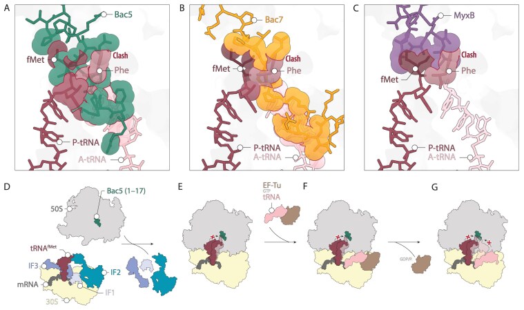
Proline-rich antimicrobial peptides (PrAMPs) are produced as part of the innate immune response of animals, insects, and plants. The well-characterized mammalian PrAMP bactenecin-5 (Bac5) has been shown to help fight bacterial infection by binding to the bacterial ribosome and inhibiting protein synthesis. In the absence of Bac5-ribosome structures, the binding mode of Bac5 and exact mechanism of action has remained unclear. Here, we present a cryo-electron microscopy structure of Bac5 in complex with the 70S ribosome from the Gram-negative marine bacterium Vibrio natriegens. The structure shows that, despite sequence similarity to Bac7 and other type I PrAMPs, Bac5 displays a completely distinct mode of interaction with the ribosomal exit tunnel. Bac5 overlaps with the binding site of both A- and P-site transfer RNAs bound at the peptidyltransferase center, suggesting that this type I PrAMP can interfere with late stages of translation initiation as well as early stages of elongation. Collectively, our study presents a ribosome structure from V. natriegens, a fast-growing bacterium that has interesting biotechnological and synthetic biology applications, as well as providing additional insights into the diverse binding modes that type I PrAMPs can utilize to inhibit protein synthesis.


Conflict of interest statement

None declared.

Figures

Graphical Abstract
Graphical Abstract
Figure 1.
Figure 1.
Summary of the origin, nature and mode of action of the most commonly known PrAMPs. (A) Sequence alignment of naturally occurring and synthetically produced PrAMPs of types I and II. The consensus sequence is indicated by a dashed box, and the conserved PRP motif is highlighted in bold. (B) Superimposition of the type I peptides Onc112 [6], Pyr [8], Met-1 (1–10) [8], and Bac7 (1–16) [8]. The alignments are based on the ribosomal RNA (rRNA) structure of the respective model organism. Additionally, the alignment of the initiator tRNA in the P-site (fMet) and the A-site tRNA (Val) [36] illustrates the mechanism of action of type I PrAMPs. This mode of action is further visualized in panel (C) (Elongating ribosome with EF-Tu*GTP). (D) Illustration of the mode of action of type II PrAMPs (RF1) and P-site tRNAIle. (E) Superimposition of type II PrAMPs Api137 [12] and Dro [15]. The alignment with RF1 and the P-site tRNAIle highlights the previously described mechanism. This figure was adapted from reference [2].
Figure 2.
Figure 2.
Growth assays and inhibition of translation by Bac5(1–17) and Bac7(1–16). (A) Growth assay showing the residual growth of wildtype V. natriegens (lacking SbmA, pAL0167_ColE1_sfGFP) after 6 h at 37°C when incubated with 0, 1, 10, 25, or 100 μM of Bac5(1–17) (green bars) or Bac7(1–16) (yellow bars), or 25 μM of the positive control antibiotic erythromycin (Ery). (B) Growth assay showing the residual growth of E. coli wildtype strain BW251113 (left) or BW25113 ΔsbmA (right) after 6 h at 37°C when incubated with 0, 1, 10, 25, or 100 μM of Bac5(1–17) (green bars), or 25 μM of the positive control antibiotic erythromycin (Ery). (C) Growth assay showing the residual growth of V. natriegens expressing heterologous SbmA (+SbmA, pAL0155_ColE1_SbmA) after 6 h at 37°C when incubated with 0, 1, 10, 25, or 100 μM of Bac5(1–17) (green bars) or Bac7(1–16) (yellow bars), or 25 μM of the positive control antibiotic erythromycin (Ery). For panels (A)–(C), all experiments were performed in triplicate (n = 3) and the bars represent the mean. (D) In vitro translation of firefly luciferase on V. natriegens ribosomes in the presence of increasing concentrations of Bac5(1–17). The luminescence in the absence of Bac5(1–17) was normalized as 100% and all bars represent the normalized mean of triplicate experiments (n = 3). Each replica is individually plotted as a white dot.
Figure 3.
Figure 3.
Cryo-EM structure of the V. natriegens Bac5-50S and SCM-30S complex. (A, B) Molecular model of the 30S ribosomal subunit with the individual ribosomal proteins and the 16S rRNA highlighted. The second view of the 30S is rotated 180° around the y axis. (C, D) Molecular model of the 50S ribosomal subunit with the individual ribosomal proteins, 5S and 23S rRNA highlighted. The 50S is shown in the crown view; the second panel shows this view rotated by 180°. (E) Isolated cryo-EM density map (mesh) with the molecular model of spectinomycin (SCM) and surrounding water molecules (spheres). The 16S nucleotide labels follow V. natriegens numbering, and interacting atoms are color-coded by element. SCM binds between the head and body of the 30S subunit. Dashed light blue lines indicate potential hydrogen bonds, while rose-colored dashed lines represent water-mediated interactions. (F) Isolated cryo-EM map density (mesh) of Bac5(1–17) and waters with the respective molecular models. Bac5(1–17) binds with its N-terminus in the PTC, while the C-terminus extends deeper into the polypeptide tunnel.
Figure 4.
Figure 4.
Interactions of Bac5(1–17) with nucleotides of the V. natriegens 50S subunit. Interactions between Bac5(1–17) and its binding site are highlighted with a purple background for residues from the N-terminal region and a light green background for those from the C-terminal region (A) Direct hydrogen bond interactions (blue dashed line) between the backbone nitrogen atoms and carbonyl oxygens of the N-terminal end of Bac5(1–17) and nucleotides of the 23S rRNA (A1–A5). (B) Direct hydrogen bond interactions involving N-terminal residues of Bac5(1–17) (B1–B2). (C) Stacking interactions (arrows) mediated by Arg8 in Bac5(1–17) (C1). (D) Water-mediated hydrogen bonds (dashed lines) of residues from the N-terminus (D1–D4). (E) Direct hydrogen bonds between C2596 towards the side chains of Arg12 from the C-terminal part of Bac5(1–17) (E1). (F) Stacking interactions of the C-terminal part of Bac5(1–17) (F1–F3). (G) Water-mediated hydrogen bonds between the backbone nitrogen atoms and carbonyl oxygens of the C-terminus (G1–G3).
Figure 5.
Figure 5.
Comparison of the binding modes of Bac5(1–17) and other type I PrAMPs, such as Bac7(1–16). (A) Overlay of Type I PrAMPs, including Bac7(1–16) [8], Pyr [8], Onc112 [6], and Tur1A [10], with the structure of Bac5 (1–17). (B) The top panel (B1) highlights a close-up of the consensus sequence of PrAMPs from (A), with residues numbered 1–9 and the aligned sequences shown below. The consensus sequence is outlined with dashed lines, while the conserved PRP-P motif is marked in bold. The lower panel (B2) displays the same overlay, now focusing on Bac5(1–17), with its corresponding sequence alignment shown below. (C) Comparison of the binding sites of Bac7(1–16) and Bac5(1–17) within ribosomes from T. thermophilus and V. natriegens, respectively. (D) Pro11 of Bac7 forms a stacking interaction with U2597 (equivalent to EcU2585) in the 23S rRNA of T. thermophilus. (E) The corresponding nucleotide in V. natriegens U2571 (EcU2585) adopts a different conformation. (F) In T. thermophilus, A2084 (EcA2062) extends into the tunnel lumen, stacking with Arg16 of Bac7. (G) In V. natriegens, the corresponding nucleotide (A2048) lies flat against the tunnel wall, stacking with Pro9 of Bac5. (H) Arg9 of Bac7(1–16) stacks with U2516 (EcU2504), a similar interaction to Bac5(1–17), where Arg7 stacks with U2490 (EcU2504). (I) Both Bac5(1–17) and Bac7(1–16) exhibit a conserved stacking interaction via Arg12 with EcC2610, differing only in the positioning of their side chains.
Figure 6.
Figure 6.
Model for the inhibition mechanism of Bac5(1–17). (A) The N-terminal region of Bac5(1–17) (teal green) causes steric problems with both the aminoacylated fMet-tRNA (dark red) in the P-site and the aminoacylated Phe-tRNA in the A-site (pale pink) [54], highlighted by red lines indicating the clashing atoms. (B) When aligned with Bac7(1–16) [8], similar steric interactions can be observed with the P- and A-site tRNAs. However, the clash with the A-site tRNA is more extensive compared to the one caused by Bac5(1–17). (C) Myxovalargin B (MyxB, purple) [53] also induces steric clashes with tRNAs in both the P- and A-sites, primarily due to interactions with the amino acids of the tRNAs. (DG) Based on structural alignments with tRNAs, a potential mechanism for Bac5-mediated translation inhibition is proposed. Bac5 binds to the 50S ribosomal subunit prior to the assembly of the 70S translating ribosome (D). This binding likely hinders translation initiation by disrupting the proper positioning of fMet-tRNA in the P-site (E). Additionally, the placement of the A-site tRNA by EF-Tu is blocked due to steric interference between Bac5 and both the amino acid and the CCA-end of the A-site tRNA (F-G). The illustration of the mechanism was inspired by [2].

References

    1. Scocchi M, Tossi A, Gennaro R Proline-rich antimicrobial peptides: converging to a non-lytic mechanism of action. Cell Mol Life Sci. 2011; 68:2317–30.10.1007/s00018-011-0721-7. - DOI - PMC - PubMed
    1. Graf M, Mardirossian M, Nguyen F et al. . Proline-rich antimicrobial peptides targeting protein synthesis. Nat Prod Rep. 2017; 34:702–11.10.1039/C7NP00020K. - DOI - PubMed
    1. Graf M, Wilson DN Intracellular antimicrobial peptides targeting the protein synthesis machinery. Adv Exp Med Biol. 2019; 1117:73–89.10.1007/978-981-13-3588-4_6. - DOI - PubMed
    1. Sola R, Mardirossian M, Beckert B et al. . Characterization of cetacean proline-rich antimicrobial peptides displaying activity against ESKAPE pathogens. Int J Mol Sci. 2020; 21:7367.10.3390/ijms21197367. - DOI - PMC - PubMed
    1. Panteleev PV, Pichkur EB, Kruglikov RN et al. . Rumicidins are a family of mammalian host-defense peptides plugging the 70S ribosome exit tunnel. Nat Commun. 2024; 15:8925.10.1038/s41467-024-53309-y. - DOI - PMC - PubMed

Supplementary concepts