. 2025 Apr 8;26(8):3466.
doi: 10.3390/ijms26083466.

Apoptotic Caspases-3 and -7 Cleave Extracellular Domains of Membrane-Bound Proteins from MDA-MB-231 Breast Cancer Cells

Affiliations

Apoptotic Caspases-3 and -7 Cleave Extracellular Domains of Membrane-Bound Proteins from MDA-MB-231 Breast Cancer Cells

Eva Vidak et al. Int J Mol Sci. .

Abstract

Apoptotic executioner caspases-3 and -7 are the main proteases responsible for the execution of apoptosis. Apoptosis is the main form of programmed cell death involved in organism development and maintenance of homeostasis and is commonly impaired in various pathologies. Predominately an immunologically silent form of cell death, it can become immunogenic upon loss of membrane integrity during progression to secondary necrosis, which mostly occurs when apoptotic bodies are not efficiently cleared by efferocytosis. In cancer, the efferocytic capacity can be overwhelmed following chemotherapeutic treatment, thereby providing an opportunity for the potential extracellular functions of executioner apoptotic caspases in the tumor microenvironment. By triggering apoptosis in Jurkat E6.1 acute T cell leukemia cells, we demonstrated that during progression to secondary necrosis, executioner caspases-3 and -7 can be found in the extracellular space. Furthermore, we showed that extracellularly active caspases-3 and -7 can cleave extracellular domains of membrane-bound proteins from MDA-MB-231 breast cancer cells, a function generally executed in the tumor microenvironment by several extracellular proteases from metalloprotease and cathepsin families. As such, this study provides the evidence for the potential involvement of apoptotic caspases-3 and -7 in extracellular proteolytic networks. Presented mass spectrometry data are available via ProteomeXchange with identifier PXD061399.

Keywords: apoptotic caspases; cancer cells; ectodomain shedding; extracellular activity; membrane-bound proteins.


Conflict of interest statement

The authors declare no conflicts of interest.

Figures

Figure 1
Figure 1
Detection of intracellular and extracellular caspase activity and flow cytometry analysis of STS-triggered apoptosis in Jurkat E6.1 cells. For all time-course experiments, Jurkat E6.1 cells were treated with 0.5 µM staurosporine (STS) or additionally with 25 µM zVAD-FMK. (A) DEVDase activity in lysates and cell media following STS treatment. Results are shown for 4 repeated experiments (n = 4) in technical triplicates (n = 3). (B) Statistical analysis of flow cytometry measurements depicting the percent of either viable (Annexin-PE−/PI−), early apoptotic (Annexin-PE+/PI−), late apoptotic or secondary necrotic (Annexin-PE+/PI+) or necrotic cells (Annexin-PE−/PI+) detected during four separate experiments (n = 4) and density diagrams of Annexin-PE positive cells (FL1-Height) against PI-positive cells (FL3-Height) present at the representative time points—viable cells (0 h), early apoptosis (6 h), secondary necrosis (20 h) and at the end of the experiment (24 h). (C) Cumulative percentages of detected dying cells (either Annexin-PE+/PI−, Annexin-PE−/PI+, or Annexin-PE+/PI+) and analysis of detected stained cells at representative time points for early (6 h) and late apoptosis (18 h) after treatment (n = 2). The data are shown as means ± SD.
Figure 2
Figure 2
Enzymatic activity and immunological detection of caspases-3 and -7 in lysates and cell media of apoptotic Jurkat E6.1 cells. For all time-course experiments, Jurkat E6.1 cells were treated with 0.5 µM staurosporine (STS). (A) DEVDase activity in lysates and corresponding Western blot detection of cleaved caspases-3 and -7. For Western blot detection, β-actin was used as a loading control. (B) DEVDase activity in cell media and the corresponding Western blot detection of cleaved caspases-3 and -7. (C) Quantification of detected caspases in cell lysates presented as relative intensity of bands normalized to loading control. Three (n = 3) independent repetitions were used to calculate the relative intensity of bands. Kinetic measurements were performed in triplicates (n = 3). Data are presented as means ± SD.
Figure 3
Figure 3
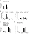
Enzymatic activity of caspases-3 and -7 in a normal (pH 7.4) and acidic (pH 6.0) environment. For DEVDase activity detection, either recombinant caspases (10 nM) were used (A,C) or apoptosis was induced with STS in Jurkat E6.1 cells and activity was detected in lysates (B) and media (D). All activity measurements were performed using 20 μM Ac-DEVD-AFC as a substrate (2000:1 ratio) when recombinant caspases were used (A,C). Jurkat E6.1 cells were treated with 0.5 µM STS at indicated time points. All experiments were performed as technical triplicates (n = 3) and data are presented as means ± SD. (A) DEVDase activity of recombinant caspases-3 and -7 in standard (HEPES buffer, pH 7.4) and low-pH (MES buffer, pH 6.0) activity buffer. (B) Detection of DEVDase activity in lysates and cell media of STS-treated Jurkat E6.1 cells in normal (pH 7.4) and acidic (pH 6.0) conditions. (C) Analysis of DEVDase activity of recombinant caspases-3 and -7 in different buffers (DPBS, HEPES, MES) under normal (pH 7.4) and acidic pH (pH 6.0). (D) Detected DEVDase activity in concentrated media from untreated (0 h STS) and secondary necrotic (20 h STS) Jurkat E6.1 cells under shedding conditions.
Figure 4
Figure 4
Identification of potential extracellular targets of caspases-3 and -7 and validation of neuropilin-1 cleavage. (A) Percentage of identified proteins with significantly increased presence (SCR ≥ 3) of MDA-MB-231 cells in sheddomes upon treatment with both caspases. (B) Localization of identified proteins with SCR ≥ 3. (C) Division of identified substrates among unique substrates of each investigated caspase and shared substrates, and further division according to the molecular function of identified substrates. (D) Validation of caspase-mediated cleavage of the extracellular domain of NRP-1 by caspases-3 and -7 using immunological detection. The experiment was performed in triplicate, but only one experiment is shown.
Figure 5
Figure 5
Immunological detection of caspase-mediated cleavages of extracellular proteins in normal (pH 7.4) and acidic (pH 6.0) environments. For the detection of extracellular cleavages, MDA-MB-231 cells were used. Red arrows indicate bands representing cleaved fragments, while black arrows indicate full-length proteins present in total cell lysates. (A) Detected caspase-3 or -7 extracellular cleavages of the selected targets (NRP-1, CD44, CSPG4) in an acidic (pH 6.0) environment in either DPBS or MES buffer. (B) Detection of caspase extracellular cleavages in a normal (pH 7.4) environment using either DPBS or HEPES buffer. NRP-1, neuropilin-1; CD44, CD44 antigen; CSPG4, chondroitin sulfate proteoglycan 4.
Figure 6
Figure 6
Western blot detection of extracellular cleavages of selected targets following treatment by cathepsins L and S and caspases-3 and -7. For all experiments, MDA-MB-231 cells were used. Cleavages were detected in DPBS buffer with pH 6.0 (A) and pH 7.4 (B). Red arrows indicate bands representing cleaved fragments produced by caspase-mediated cleavage, black arrows indicate full-length proteins as present in total cell lysates, and blue arrows indicate fragments specific for cathepsin cleavages. Additionally, the gray arrow indicates a non-specific band likely belonging to active cathepsin S.

Similar articles

References

    1. Lamkanfi M., Declercq W., Kalai M., Saelens X., Vandenabeele P. Alice in caspase land. A phylogenetic analysis of caspases from worm to man. Cell Death Differ. 2002;9:358–361. doi: 10.1038/sj.cdd.4400989. - DOI - PubMed
    1. Van Opdenbosch N., Lamkanfi M. Caspases in Cell Death, Inflammation, and Disease. Immunity. 2019;50:1352–1364. doi: 10.1016/j.immuni.2019.05.020. - DOI - PMC - PubMed
    1. Ramirez M.L.G., Salvesen G.S. A primer on caspase mechanisms. Semin. Cell Dev. Biol. 2018;82:79–85. doi: 10.1016/j.semcdb.2018.01.002. - DOI - PMC - PubMed
    1. Boice A., Bouchier-Hayes L. Targeting apoptotic caspases in cancer. Biochim. Biophys. Acta Mol. Cell Res. 2020;1867:118688. doi: 10.1016/j.bbamcr.2020.118688. - DOI - PMC - PubMed
    1. Dabrowska C., Li M., Fan Y. Apoptotic Caspases in Promoting Cancer: Implications from Their Roles in Development and Tissue Homeostasis. Adv. Exp. Med. Biol. 2016;930:89–112. doi: 10.1007/978-3-319-39406-0_4. - DOI - PubMed