. 2025 Apr 9;26(8):3502.
doi: 10.3390/ijms26083502.

Phenotypic and Genotypic Bacterial Virulence and Resistance Profiles in Hidradenitis Suppurativa

Affiliations

Phenotypic and Genotypic Bacterial Virulence and Resistance Profiles in Hidradenitis Suppurativa

Corina Ioana Cucu et al. Int J Mol Sci. .

Abstract

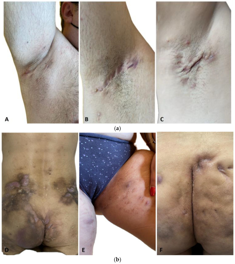
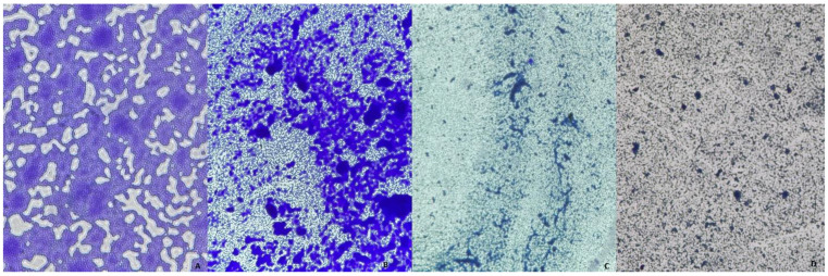
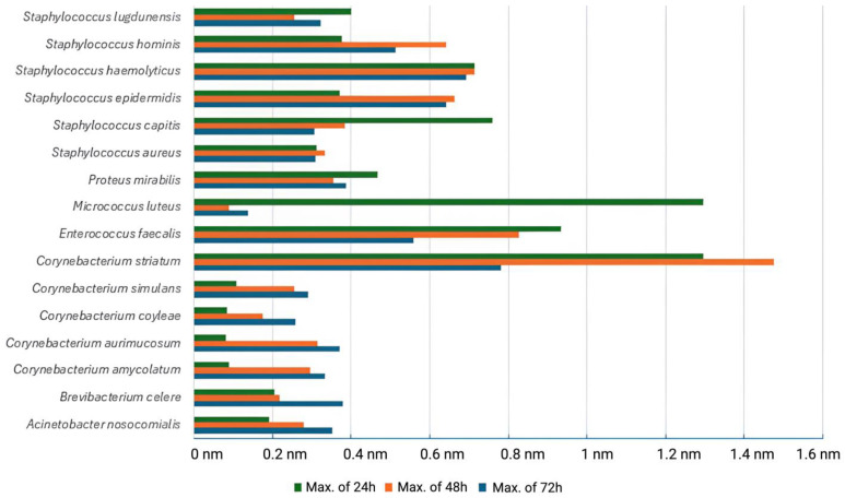
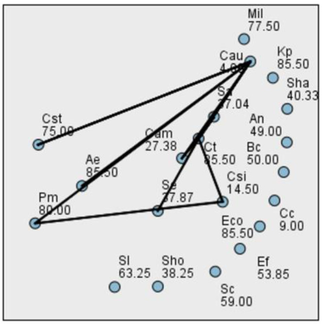
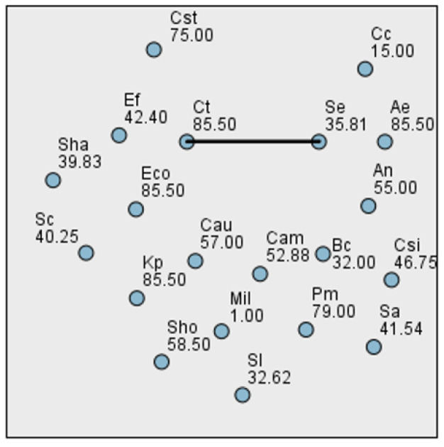
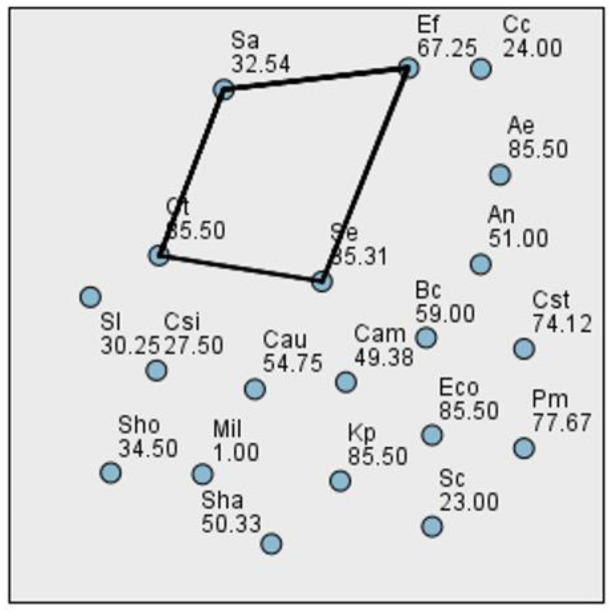
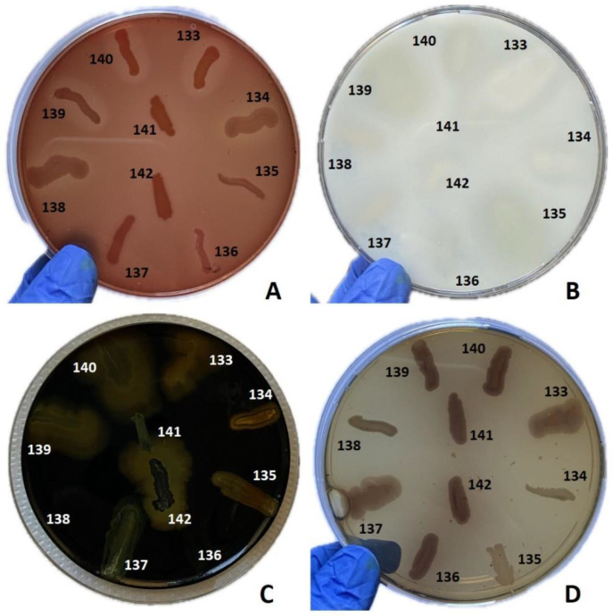
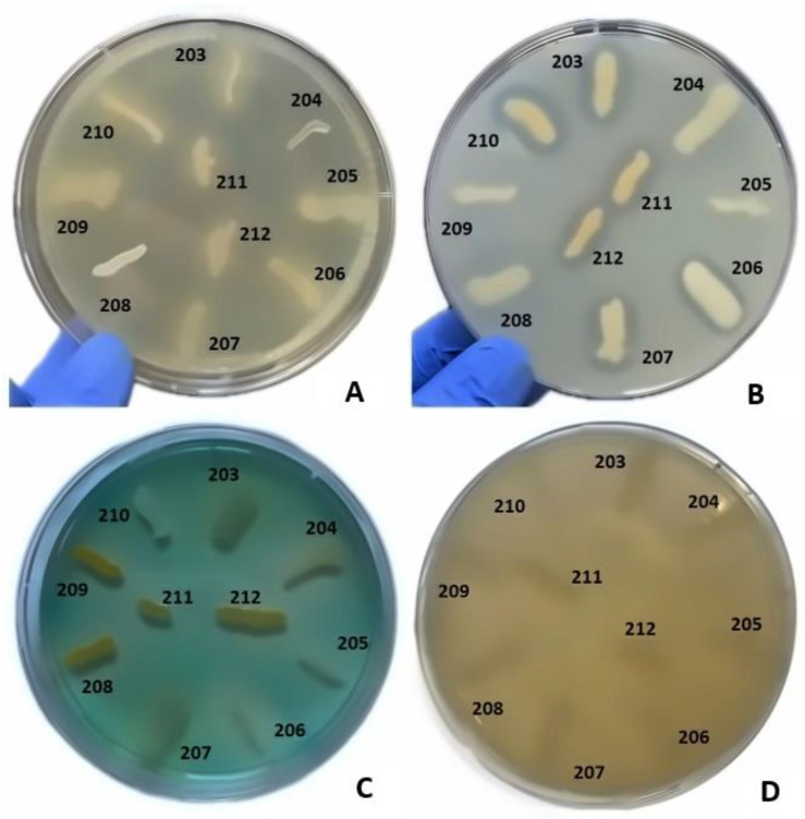
Hidradenitis suppurativa (HS) is a chronic inflammatory skin condition, primarily affecting young individuals, with a significant impact on their quality of life due to recurrent, painful nodules, abscesses, and oozing sinus tracts, primarily affecting intertriginous areas. The pathogenesis of HS is multifactorial, involving a complex interplay between genetic predisposition, immune dysregulation, microbial, and environmental factors. While it is known that cutaneous and gut microbiome contribute to innate immune dysregulation in HS, their precise involvement in disease pathogenesis remains unclear. Despite several studies investigating the microbiome of HS lesions, either by culture-dependent or independent methods, there is no data available on the interplay between bacterial virulence profiles, clinical manifestations, and the host immune response. This study aimed to explore the phenotypic and genotypic resistance and virulence profiles of microorganisms isolated from HS lesions (including the expression of soluble virulence factors and the ability to develop biofilms), with a special focus on Staphylococcus aureus (S. aureus), one of the most frequent infectious agents of HS. A total of 92 bacterial strains, belonging to 20 different bacterial species, were isolated from the HS lesions of 23 patients. The strains of Staphylococcus, Corynebacterium, and Enterococcus expressed the highest levels of soluble virulence factors, such as hemolysins, lecithinase, and lipase, which are involved in bacterial persistence, local invasivity, and tissue damage. Moreover, a significant variation among bacterial species was noted regarding the capacity to develop biofilms, with a potential impact on disease chronicization, bacterial tolerance to antibiotics, and immune defense mechanisms. The genetic characterization of methicillin-resistant staphylococci revealed the presence of adhesins, hemolysin and enterotoxin genes as well as methicillin and macrolides resistance genes. Our findings highlight the critical role of virulence determinants, including bacterial biofilms, in HS pathogenesis, emphasizing the need for targeted therapeutic strategies to disrupt biofilms and mitigate infection severity.

Keywords: Staphylococcus aureus; biofilm; hidradenitis suppurativa; skin microbiome; virulence factors.


Conflict of interest statement

The authors declare no conflicts of interest.

Figures

Figure 1
Figure 1
Axillary HS in different male patients included in this study: (a) (A). HS, Hurley stage I—inflammatory nodules without skin tunnels or scarring; (B). HS, Hurley stage II—recurrent abscesses with skin tunnels and scarring, single or multiple widely separated lesions; (C). HS Hurley stage III—diffuse or almost diffuse involvement, or multiple interconnected skin tunnels and abscesses across the entire affected area (b) (D). Severe gluteal HS with multiple fistulous tracts in a male patient (E). Inguinal HS in a female patient (F). Gluteal HS with several inflammatory nodules and scars in a male patient.
Figure 2
Figure 2
Biofilms of (A) Proteus mirabilis; (B) Alcaligenes faecalis; (C) Staphylococcus lugdunensis; (D) Micrococcus luteus at 24 h under the optical microscope at 20× magnification.
Figure 3
Figure 3
Biofilm forming capacity of isolated bacterial species at 24, 48, and 72 h.
Figure 4
Figure 4
Pairwise comparison between bacterial species biofilm production at 24 h.
Figure 5
Figure 5
Pairwise comparison between bacterial species biofilm production at 48 h.
Figure 6
Figure 6
Pairwise comparison between bacterial species biofilm production at 72 h.
Figure 7
Figure 7
Assessment of the production of different virulence factors in 10 bacterial strains analyzed. (A) Virulence factor: hemolysins, medium: agar supplemented with 5% sheep blood; (B) virulence factor: caseinase, medium: agar with addition of 15% casein; (C) virulence factor: amylase, medium: agar with 1% starch added, flooded with Lugol solution; (D) virulence factor: esculinase, medium: agar supplemented with 1% esculin and iron citrate. Bacterial strains tested: no. 133 Staphylococcus epidermidis (S. epidermidis), no. 134 S. epidermidis, no. 135 S. epidermidis, no. 136 S. epidermidis, no. 137 S. epidermidis, no. 138 Staphylococcus aureus, no. 139 S. epidermidis, no. 140 Enterococcus faecalis, no. 141 Corynebacterium amycolatum, no. 142 Corynebacterium aurimucosum.
Figure 8
Figure 8
Assessment of the production of different virulence factors in 10 bacterial strains analyzed. (A) Virulence factor: lipase, medium: agar supplemented with 1% Tween 80; (B) virulence factor: lecithinase, medium: agar with the addition of 2.5% egg yolk; (C) virulence factor: DNase, medium: DNase; (D) virulence factor: gelatinase; medium: agar with the addition of 3% gelatin. Bacterial strains tested: no. 203 Staphylococcus aureus (S. aureus), no. 204 S. aureus, no. 205 S. aureus, no. 206 S. haemolyticus, no. 207 Corynebacterium striatum (C. striatum), no. 208 C. striatum, no. 209 Staphylococcus epidermidis, no. 210 Staphylococcus epidermidis, no. 211 C. striatum, no. 212 C. striatum.
Figure 9
Figure 9
Electrophoresis of DNA amplicons PCR for the genes fnbB (524 bp), fib (404 bp), clfA (292), clfB (205 bp). Lines 2–19-strain 76; 77; 79; 83; 86; 88; 92; 94; 100; 106; 107; 108; 119; 121; 122; 123; 124; 125. Line 20-Molecular size marker (Thermo Scientific, Waltham, MA, USA, 1500 bp). Strains positive for the fib gene: 76; 77; 79; 86; 88; 92; 94; 100; 108; 122; 124; 125. Strains positive for the clfA gene: 76; 77; 79; 83; 86; 88; 92; 94; 100; 106; 107; 108; 119; 121; 122; 123; 124; 125. Strains positive for the fnbB gene: 77; 79; 92; 121.
Figure 10
Figure 10
Electrophoresis of DNA amplicons post PCR for the luk-PV (443 bp) and hlg (937 bp) genes. Line 1-Molecular size marker (Thermo Scientific, Waltham, MA, USA, 1500 bp); lines 2–20 strains 138; 179; 193; 194; 196; 198; 199; 200; 203; 204; 205; 218; 229; 236; 242; 243; 244; 251; 252. Positive strains for the hlg gene: 138; 179; 193; 196; 198; 199; 200; 203; 204; 205; 218; 229; 236; 242; 243; 244; 251; 252.
Figure 11
Figure 11
Electrophoresis of DNA amplicons post PCR for genes sea (102 bp), seb (164 bp), sec (451 bp), sed (278 bp), see (209 bp). Line 1-Molecular size marker (Thermo Scientific, Waltham, MA, USA 1500 bp); lines 2–20 strains: 68; 69; 71; 72; 74; 76; 77; 79; 83; 86; 88; 92; 94; 100; 106; 107; 108; 119. Strains positive for the sec gene: 76. Strains positive for the sed gene: 66; 68.
Figure 12
Figure 12
Electrophoresis of DNA amplicons post-PCR for the mecA (532 bp) and nuc (279 bp) genes—genotypic confirmation of the MRSA phenotype. Line 1-Molecular size marker (Thermo Scientific, Waltham, MA, USA, 1500 bp); lines 2–9 strains: 30; 32; 34; 44; 68; 74; 86; 88 and negative control. Strains positive for the mecA and nuc genes: 30; 32; 34; 44; 68; 74; 86; 88.
Figure 13
Figure 13
Electrophoresis of DNA amplicons post PCR for the ermA (190 bp) and ermC (299 bp) genes—macrolide resistance. Line 1-Molecular size marker (Thermo Scientific, Waltham, MA, USA 1500 bp); lines 2–17 strains: 30; 32; 34; 36; 39; 44; 46; 59; 60; 62; 66; 68; 79; 86; 88; 100. Strains positive for the ermC gene: 30; 32; 62; 88.
Figure 14
Figure 14
Electrophoresis of DNA amplicons post PCR for the CIF2 (495 bp) and ccrB2 (311 bp) genes from the staphylococcal chromosomal cassette SCCmec. Line 1-Molecular size marker (Thermo Scientific, Waltham, MA, USA, 1500 bp); Lines 2–8 strains: 32; 44; 30; 68; 74; 86; 88. Strains positive for the CIF2 and ccrB2 genes: 39; 32; 44; 30; 68; 86.

Similar articles

References

    1. Zouboulis C.C., Bechara F.G., Fritz K., Goebeler M., Hetzer F.H., Just E., Kirsten N., Kokolakis G., Kurzen H., Nikolakis G., et al. S2k guideline for the treatment of hidradenitis suppurativa/acne inversa—Short version. J. Dtsch. Dermatol. Ges. J. Ger. Soc. Dermatol. JDDG. 2024;22:868–889. doi: 10.1111/ddg.15412. - DOI - PubMed
    1. Zouboulis C.C., Bechara F.G., Benhadou F., Bettoli V., Bukvić Mokos Z., Del Marmol V., Dolenc-Voljč M., Giamarellos-Bourboulis E.J., Grimstad Ø., Guillem P., et al. European S2k guidelines for hidradenitis suppurativa/acne inversa part 2: Treatment. J. Eur. Acad. Dermatol. Venereol. JEADV. 2024;00:1–43. doi: 10.1111/jdv.20472. - DOI - PMC - PubMed
    1. Zouboulis C.C., Desai N., Emtestam L., Hunger R.E., Ioannides D., Juhasz I., Lapins J., Matusiak L., Prens E.P., Revuz J., et al. European S1 guideline for the treatment of hidradenitis suppurativa/acne inversa. J. Eur. Acad. Dermatol. Venereol. JEADV. 2015;29:619–644. doi: 10.1111/jdv.12966. - DOI - PubMed
    1. Nazary M., van der Zee H.H., Prens E.P., Folkerts G., Boer J. Pathogenesis and pharmacotherapy of Hidradenitis suppurativa. Eur. J. Pharmacol. 2011;672:1–8. doi: 10.1016/j.ejphar.2011.08.047. - DOI - PubMed
    1. Ingram J.R., Jenkins-Jones S., Knipe D.W., Morgan C.L.I., Cannings-John R., Piguet V. Population-based Clinical Practice Research Datalink study using algorithm modelling to identify the true burden of hidradenitis suppurativa. Br. J. Dermatol. 2018;178:917–924. doi: 10.1111/bjd.16101. - DOI - PubMed

LinkOut - more resources