Review
. 2025 Apr 10;26(8):3550.
doi: 10.3390/ijms26083550.

From Skeletal Muscle to Myocardium: Molecular Mechanisms of Exercise-Induced Irisin Regulation of Cardiac Fibrosis

Affiliations
Review

From Skeletal Muscle to Myocardium: Molecular Mechanisms of Exercise-Induced Irisin Regulation of Cardiac Fibrosis

Zhao Wang et al. Int J Mol Sci. .

Abstract

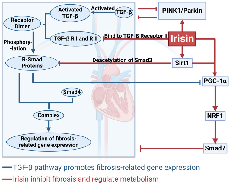
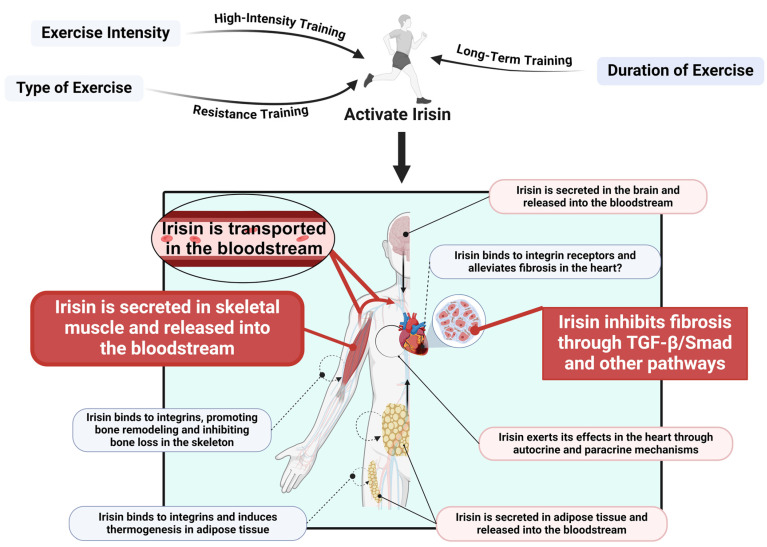
This study systematically elucidates the regulatory mechanisms and potential therapeutic value of the exercise-induced hormone Irisin in the pathological progression of cardiac fibrosis. Through comprehensive analysis and multidimensional data integration, we constructed a complete regulatory network of Irisin within the cardiovascular system, spanning its secretion, signal transduction, and precise regulatory control. Our findings demonstrate that exercise intervention significantly elevates circulating Irisin levels via the skeletal muscle-peroxisome proliferator-activated receptor gamma coactivator 1-alpha (PGC-1α)-fibronectin type III domain-containing protein 5 (FNDC5) signaling axis. Irisin establishes a multidimensional molecular barrier against cardiac fibrosis by targeting Sirtuin 1 (Sirt1) activation, inhibiting the transforming growth factor-beta (TGF-β)/Smad3 signaling pathway, and modulating the transcriptional activity of the mitochondrial biogenesis core factors PGC-1α and nuclear respiratory factor 1 (NRF-1). Moreover, the dual regulatory mechanism of the exercise-skeletal muscle-heart axis not only effectively suppresses the aberrant activation of cardiac fibroblasts but also significantly reduces collagen deposition, oxidative stress, and inflammatory infiltration by restoring mitochondrial dynamics balance. Taken together, this study reveals a novel exercise-mediated cardioprotective mechanism at the molecular interaction network level, thereby providing a theoretical basis for the development of non-pharmacological bio-intervention strategies targeting the Irisin signaling pathway and laying a translational foundation for precise exercise prescriptions in cardiovascular diseases.

Keywords: Irisin; cardiac fibrosis; cardioprotection; exercise.


Conflict of interest statement

The authors declare no conflicts of interest.

Figures

Figure 1
Figure 1
Mechanisms and Current Treatments of Cardiac Fibrosis. This figure outlines the mechanisms, detection methods, and treatment strategies for cardiac fibrosis. miR-21 promotes fibrosis progression by regulating the transforming growth factor-beta (TGF-β) signaling pathway, while miR-29 regulates the metabolic balance of the extracellular matrix (ECM). Oxidative stress and inflammatory factors further exacerbate pathological changes. Magnetic Resonance Imaging (MRI) and serum biomarkers can be used to assess fibrosis. Biomarkers include procollagen type I N-terminal propeptide (PINP), procollagen type II N-terminal propeptide (PIINP), and matrix metalloproteinases (MMPs). Drugs such as Losartan, N-acetylcysteine (NAC), and regenerative medicine techniques provide potential therapeutic options. This figure was created in BioRender. Wang, Z. (2025). https://BioRender.com/n80e582, accessed on 7 April 2025.
Figure 2
Figure 2
Mechanisms Underlying Irisin-Mediated Cardioprotection. This figure outlines how Irisin exerts cardioprotective effects by inhibiting inflammation, alleviating oxidative stress, maintaining mitochondrial dynamics, suppressing apoptosis, and mitigating microvascular damage. Moreover, Irisin protects cardiac function by reducing fibrosis through the inhibition of the TGF-β signaling pathway. This figure was created in BioRender. Wang, Z. (2025) https://BioRender.com/x59p404, accessed on 7 April 2025.
Figure 3
Figure 3
Comprehensive mechanism by which Irisin influences fibrosis Via Sirtuin 1 (Sirt1), peroxisome proliferator-activated receptor gamma coactivator 1-alpha (PGC-1α)/nuclear respiratory factor 1 (NRF-1), and related pathways. This figure summarizes how Irisin significantly reduces the expression of fibrosis-related genes by directly inhibiting TGF-β activation and blocking Smad signaling through receptor binding. Additionally, Irisin activates Sirt1, which mediates the deacetylation of Smad3, further suppressing Smad complex formation. By regulating the PGC-1α/NRF-1 pathway, Irisin helps maintain mitochondrial homeostasis, while enhancing mitophagy through the PTEN-induced kinase 1 (PINK1)/Parkin pathway to clear damaged mitochondria and improve intracellular equilibrium. Collectively, these actions inhibit fibrotic progression, mitigate tissue damage, and offer new insights for the treatment of cardiac fibrosis. This figure was created in BioRender. Wang, Z. (2025) https://BioRender.com/s24j371, accessed on 7 April 2025.
Figure 4
Figure 4
Exercise-Induced Irisin Release and Its Role in Cardiac Fibrosis. Exercise (high-intensity, resistance, and long-term training) activates Irisin secretion from skeletal muscle. Irisin enters the bloodstream and exerts systemic effects. In the heart, it binds to integrin receptors, modulating the TGF-β/Smad signaling pathway to inhibit fibrosis. Additionally, Irisin functions through autocrine and paracrine mechanisms. It also influences thermogenesis in adipose tissue and bone remodeling, highlighting its multifunctional role. This figure was created in BioRender. Wang, Z. (2025) https://BioRender.com/o81x119, accessed on 7 April 2025.

Similar articles

References

    1. Naghavi M., Abajobir A.A., Abbafati C., Abbas K.M., Abd-Allah F., Abera S.F., Aboyans V., Adetokunboh O., Afshin A., Agrawal A., et al. Global, regional, and national age-sex specific mortality for 264 causes of death, 1980–2016: A systematic analysis for the Global Burden of Disease Study 2016. Lancet. 2017;390:1151–1210. doi: 10.1016/S0140-6736(17)32152-9. - DOI - PMC - PubMed
    1. Talman V., Ruskoaho H. Cardiac fibrosis in myocardial infarction-from repair and remodeling to regeneration. Cell Tissue Res. 2016;365:563–581. doi: 10.1007/s00441-016-2431-9. - DOI - PMC - PubMed
    1. McMurray J.J., Pfeffer M.A. Heart failure. Lancet. 2005;365:1877–1889. doi: 10.1016/S0140-6736(05)66621-4. - DOI - PubMed
    1. Zhu L., Wang W., Ren C., Wang Y., Zhang G., Liu J., Wang W. Cellular Phenotypic Transformation in Heart Failure Caused by Coronary Heart Disease and Dilated Cardiomyopathy: Delineating at Single-Cell Level. Biomedicines. 2022;10:402. doi: 10.3390/biomedicines10020402. - DOI - PMC - PubMed
    1. Khan R., Sheppard R. Fibrosis in heart disease: Understanding the role of transforming growth factor-beta in cardiomyopathy, valvular disease and arrhythmia. Immunology. 2006;118:10–24. doi: 10.1111/j.1365-2567.2006.02336.x. - DOI - PMC - PubMed

Substances

LinkOut - more resources