. 2025 Apr 9;26(8):3534.
doi: 10.3390/ijms26083534.

The Yeast Gsk-3 Kinase Mck1 Is Necessary for Cell Wall Remodeling in Glucose-Starved and Cell Wall-Stressed Cells

Affiliations

The Yeast Gsk-3 Kinase Mck1 Is Necessary for Cell Wall Remodeling in Glucose-Starved and Cell Wall-Stressed Cells

Fan Zhang et al. Int J Mol Sci. .

Abstract

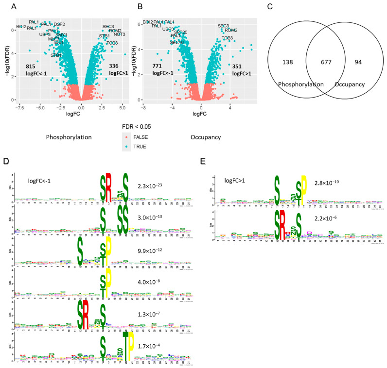
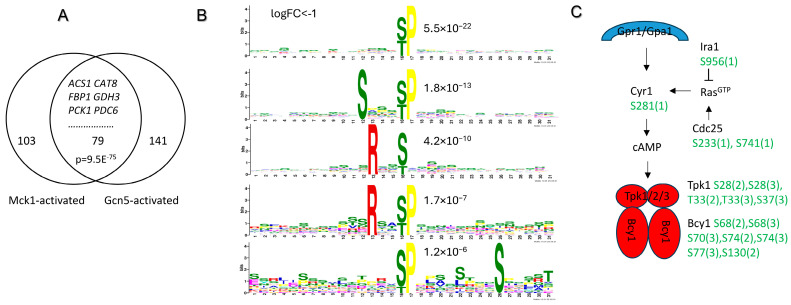
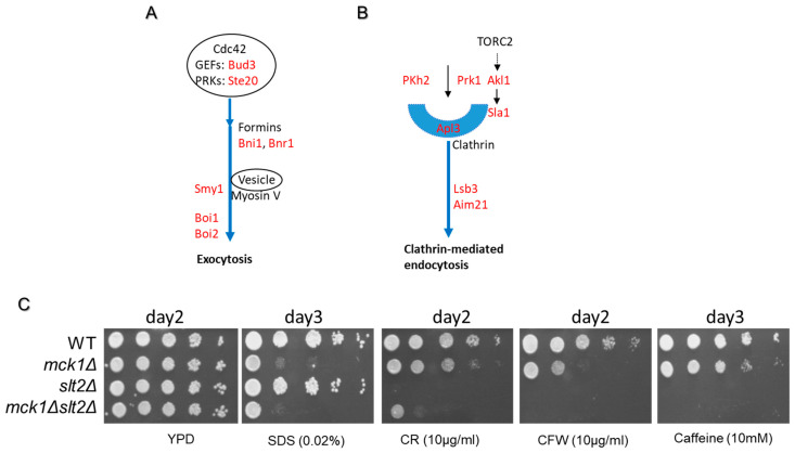
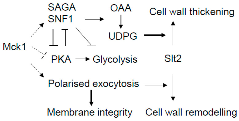
The cell wall integrity (CWI) pathway is responsible for transcriptional regulation of cell wall remodeling in response to cell wall stress. How cell wall remodeling mediated by the CWI pathway is effected by inputs from other signaling pathways is not well understood. Here, we demonstrate that the Mck1 kinase cooperates with Slt2, the MAP kinase of the CWI pathway, to promote cell wall thickening in glucose-starved cells. Integrative analyses of the transcriptome, proteome and metabolic profiling indicate that Mck1 is required for the accumulation of UDP-glucose (UDPG), the substrate for β-glucan synthesis, through the activation of two regulons: the Msn2/4-dependent stress response and the Cat8-/Adr1-mediated metabolic reprogram dependent on the SNF1 complex. Analysis of the phosphoproteome suggests that similar to mammalian Gsk-3 kinases, Mck1 is involved in the regulation of cytoskeleton-dependent cellular processes, metabolism, signaling and transcription. Specifically, Mck1 may be implicated in the Snf1-dependent metabolic reprogram through PKA inhibition and SAGA (Spt-Ada-Gcn5 acetyltransferase)-mediated transcription activation, a hypothesis further underscored by the significant overlap between the Mck1- and Gcn5-activated transcriptomes. Phenotypic analysis also supports the roles of Mck1 in actin cytoskeleton-mediated exocytosis to ensure plasma membrane homeostasis and cell wall remodeling in cell wall-stressed cells. Together, these findings not only reveal the novel functions of Mck1 in metabolic reprogramming and polarized growth but also provide valuable omics resources for future studies to uncover the underlying mechanisms of Mck1 and other Gsk-3 kinases in cell growth and stress response.

Keywords: Mck1; PKA; SAGA; SNF1; Slt2; metabolic reprogramming; polarized growth.


Conflict of interest statement

Author Houjiang Zhou is currently employed by Zhejiang Hisun Pharmaceutical Co., Ltd. The remaining authors declare that the research was conducted in the absence of any commercial or financial relationships that could be construed as a potential conflict of interest.

Figures

Figure 1
Figure 1
Mck1 and Slt2 act cooperatively to promote cell wall thickening in PDS cells. (A): Typical cell growth curve and the sampling timepoints in the study (from the mid-exponential (EXP) to early stationary phase (SP)). (B): Representative TEM images of the cell wall of WT and the mck1Δ mutant cells. (C): Cell wall thickness quantified by TEM imaging analysis. For each strain, cell wall thickness (the electron-transparent layer) at 3 positions of 50 individual cells was determined and averaged. Error bars indicate s.d. among the 50 measurements. Significance of differences between EXP and SP samples was revealed by Student’s t-test. ** 0.001 < p < 0.01, **** p < 0.0001. Bar size: 0.5 µm.
Figure 2
Figure 2
Mck1 is essential to UDPG accumulation in PDS cells. (A) Relative levels of UDPG in WT cells. (BD) Relative levels of UDPG in the mck1Δ/slt2Δ mutants grown at EXP (B), diauxie (C) and mid-PDS (D) phases. (E,F) Relative levels of G1P and G6P (E) and TCA cycle intermediates (F) in mid-PDS cells. Significance of difference between each of the mutants and WT was revealed by Student’s t-test. n.s.: not significant, * 0.01 < p < 0.05, *** 0.001 < p < 0.0001, **** p < 0.0001. OAA: Oxaloacetic acid.
Figure 3
Figure 3
Mck1 regulates a glucose starvation-induced transcription program largely independent of SLT2. (A) Principal component analysis (PCA) of the transcriptome isolated from cells growing at the mid-exponential (EXP) and early-PDS (PDS) phases. (B) Hierarchical clustering and bioinformatics analysis of the differentially expressed (DE) genes regulated by MCK1 in PDS cells (FDR < 0.05, fold change > 1.5). Enriched motifs revealed by RSAT analysis are highlighted in red in the consensus sequences targeted by the indicated TFs. p values indicate the significance of the representation of the GO terms or TFs associated with each gene cluster. (C) Relative Fbp1-GFP and Pck1-GFP levels detected in EXP and PDS cells. Significance of difference between the mutants and WT cells was revealed by Student’s t-test (C). ** 0.001 < p < 0.01, **** p < 0.0001.
Figure 4
Figure 4
Integrative analysis of the proteome and transcriptome regulated by Mck1. (A,B) PCA of the transcriptome (A) and the proteome (B) in all samples. (C) Correlation between the transcript and protein regulation levels. Red dots represent genes significantly up-regulated (top right) or down-regulated (bottom left) at both levels in the mck1Δ mutants. Among those not significantly regulated at the transcript level, blue and yellow dots denote the significantly up-regulated (blue) and down-regulated (yellow) genes at the protein level. Conversely, gray and green dots represent the transcriptionally up-regulated (gray) and down-regulated (orange) genes without corresponding changes at the protein level.
Figure 5
Figure 5
Identifying the Mck1-mediated phosphopeptides. (A,B) Volcano plots displaying differentially regulated phosphopeptides at the phosphorylation (A) and occupancy (B) levels (FDR < 0.05); (C) Overlap between the phosphopeptides exhibiting reduced phosphorylation and occupancy levels in the mck1Δ mutants (logFC < −1). (D,E) Sequence motifs enriched in the peptides with phosphorylation levels significantly reduced (D) or enhanced (E) in the mck1Δ mutants.
Figure 6
Figure 6
Mck1 may act through SAGA and PKA to facilitate transcription activation of the Snf1-mediated metabolic reprogram. (A) Overlap between the Mck1- and Gcn5-activated transcriptome; (B) sequence motifs enriched in the phosphopeptides down-regulated in glucose-depleted mck1Δ mutants; (C) Mck1-mediated phosphorylations in the PKA pathway. The multiplicity of phosphorylation is included in the brackets.
Figure 7
Figure 7
Mck1 is required for polarized growth and cell wall homeostasis in response to plasma membrane and cell wall perturbations. (A,B): Potential Mck1 phosphotargets involved in polarized exocytosis (A) and endocytosis (B); (C) phenotypic assays conducted on plasma membrane or cell wall disturbing agents. Imaging of cell growth was conducted after incubation for 2–3 days, as indicated on top of each image.
Figure 8
Figure 8
The working model demonstrating the potential targets of Mck1 involved in cell wall remodeling. PKA inhibition and SAGA-mediated transcription activation are proposed as the potential mechanisms of Mck1 in facilitating the Snf1-directed metabolic reprogram (including repression of the glycolysis) in glucose-depleted cells. polarized exocytosis may be promoted by Mck1 to ensure plasma membrane homeostasis and to mediate the Slt2-dependent cell wall remodeling program in cell wall-stressed cells. OAA (Oxaloacetate) to UDPG denotes the metabolic reprogram. Arrow: activation; bar: inhibition; dashed line: phosphorylation–function relationship to be established.

Similar articles

References

    1. Gow N.A.R., Latge J.-P., Munro C.A. The Fungal Cell Wall: Structure, Biosynthesis, and Function. Microbiol. Spectr. 2017;5:1–25. doi: 10.1128/microbiolspec.FUNK-0035-2016. - DOI - PMC - PubMed
    1. Orlean P. Architecture and biosynthesis of the Saccharomyces cerevisiae cell wall. Genetics. 2012;192:775–818. doi: 10.1534/genetics.112.144485. - DOI - PMC - PubMed
    1. Levin D.E. Regulation of cell wall biogenesis in Saccharomyces cerevisiae: The cell wall integrity signaling pathway. Genetics. 2011;189:1145–1175. doi: 10.1534/genetics.111.128264. - DOI - PMC - PubMed
    1. Lesage G., Bussey H. Cell wall assembly in Saccharomyces cerevisiae. Microbiol. Mol. Biol. Rev. 2006;70:317–343. doi: 10.1128/MMBR.00038-05. - DOI - PMC - PubMed
    1. Sanz A.B., García R., Pavón-Vergés M., Rodríguez-Peña J.M., Arroyo J. Control of Gene Expression via the Yeast CWI Pathway. Int. J. Mol. Sci. 2022;23:1791. doi: 10.3390/ijms23031791. - DOI - PMC - PubMed

MeSH terms

Substances

LinkOut - more resources