. 2025 Apr 9;26(8):3543.
doi: 10.3390/ijms26083543.

Role of Thalamic CaV3.1 T-Channels in Fear Conditioning

Affiliations

Role of Thalamic CaV3.1 T-Channels in Fear Conditioning

Tamara Timic Stamenic et al. Int J Mol Sci. .

Abstract

The potential contribution of the ion channels that control the excitability of the midline and intralaminar nuclei of the thalamus to the modulation of behaviors has not been well studied. In this study, we used both global genetic deletion (knock-out, KO) and thalamus-specific molecular knock-down (KD) approaches to investigate the role of thalamic CaV3.1 T-type calcium channels (T-channels) in fear learning and fear responses. Previously, we have shown that the dominant subtype of T-channels in the central medial nucleus of the thalamus (CMT) is the CaV3.1 isoform and that CMT neurons from CaV3.1 KO animals have decreased burst firing. By specifically knocking down CaV3.1 T-channels in the CMT using the shRNA approach, we also reduced burst firing without affecting the tonic firing mode of the transfected neurons. We report that global CaV3.1 KO animals showed stronger freezing behaviors during both the conditioning and testing phases of contextual fear conditioning, while CMT-specific CaV3.1 KD mice only had stronger fear responses during testing. In contrast, the cue-mediated fear responses were similar between CaV3.1 KO and CaV3.1 KD mice and the controls. Our findings validate thalamic CaV3.1 T-channels as a potential new target for the development or treatment of different psychiatric diseases, such as post-traumatic stress disorder, schizophrenia, anxiety, and substance abuse disorders.

Keywords: T-type calcium channels; central medial nucleus of thalamus; fear conditioning.


Conflict of interest statement

The authors declare no conflicts of interest.

Figures
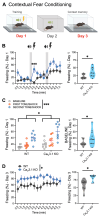
Figure 1
Figure 1
Global deletion of CaV3.1 T-channels increases fear responses during acquisition and expression of contextual fear conditioning. (A) Schematic diagram of contextual fear conditioning: Day 1—training (acquisition); Day 2—different context; Day 3—testing. (B) Percentage of time spent freezing in control wild-type (WT, gray) and CaV3.1 knock-out (KO, blue) mice during training phase of contextual fear conditioning paradigm, analyzed every 30 s (left) and over whole training phase (right). CaV3.1 KO animals showed amplified freezing behaviors after first shock (left; two-way RM ANOVA: interaction F(13,182) = 2.767, p = 0.001; time F(13,182) = 12.32, p < 0.001; genotype F(1,14) = 5.138, p = 0.04; Sidak’s post hoc test) and spent more time freezing overall during 7 min acquisition phase (right; unpaired two-tailed t-test: t(14) = 2.27, p = 0.040; Cohen’s d = 1.13; effect size r = 0.49). (C) Although there was increased freezing after each tone/shock pairing in WT and mutant animals, CaV3.1 KO mice had a higher overall freezing percentage than controls (left, two-way RM ANOVA: interaction F(2,28) = 1.587, p = 0.222; tone F(2,28) = 49.75, p < 0.001; genotype F(1,14) = 4.810, p = 0.046). Baseline freezing behavior was higher in global CaV3.1 KO male mice in comparison to WT animals (right; unpaired two-tailed t-test: t(14) = 1.688, p = 0.026; Cohen’s d = 0.844; effect size r = 0.39). (D) Compared to WT mice, percentage of time freezing of CaV3.1 KO mice during testing significantly increased over time (left; two-way RM ANOVA: interaction F(13,182) = 0.867, p = 0.589; time F(13,182) = 0.875, p = 0.581; genotype F(1,14) = 7.688, p = 0.011) and over whole 7 min testing period (right; unpaired two-tailed t-test: t(14) = 2.94, p = 0.011; Cohen’s d = 1.47; effect size r = 0.59). n = 8 animals per group; * p < 0.05, *** p < 0.001.
Figure 2
Figure 2
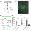
Initial validation of AAV-shRNA injections. (A) Schematic of CMT-specific knock-down (KD) animal generation by injecting Cacna1g shRNA (or scrambled shRNA as control). (B) Image shows GFP-transfected neurons only, counter-stained with DAPI. (C) T-current traces from GFP-positive neurons from animals injected with scrambled shRNA (control, black trace) or Cacna1g shRNA (green trace). (D) Average T-current amplitudes in GFP-positive neurons after control and Cacna1g shRNA injections (unpaired two-tailed t-test: t(16) = 4.023, p < 0.001; Cohen’s d = −2.02; effect size r = -0.71). n = 11 control and 7 Cacna1g shRNA-transfected GFP-positive neurons; *** p < 0.001. (E) q-PCR data pooled from the tissues of 3 Cacna1g shRNA and 4 control animals showing 76.72% decrease in relative CaV3.1 expression in Cacna1g shRNA group in comparison to control (unpaired two-tailed t-test: t(2) = 6.893, p = 0.02; Cohen’s d = −6.89; effect size r = −0.96). n = 2, technical replicates; * p < 0.05.
Figure 3
Figure 3
Different firing properties of GFP-positive thalamic neurons from control (scrambled shRNA) and Cacna1g shRNA-injected animals. (A) Original traces from representative thalamic neurons from GFP-positive control (black) and Cacna1g shRNA-transfected (green) neurons showing active membrane responses to depolarizing (275 pA) and hyperpolarizing (−225 pA) current injections. Note that GFP-positive neurons from Cacna1g shRNA cohort did not show action potentials (APs) with low threshold spikes (LTSs) after membrane hyperpolarization. (B) There was no difference in average tonic AP firing frequency (50–275 pA current injections) between GFP-positive thalamic neurons from control and Cacna1g shRNA mice. (C) Number of APs in rebound burst was statistically significantly smaller in GFP-positive neurons from Cacna1g shRNA animals (two-way RM ANOVA: interaction F(8,88) = 0.963, p = 0.470; injected current F(8,88) = 4.92, p < 0.001; GFP F(1,11) = 5.133, p = 0.045). (D) LTSs were not observed in thalamic GFP-positive Cacna1g shRNA-transfected neurons (two-way RM ANOVA: interaction F(8,88) = 3.50, p = 0.001; injected current F(8,88) = 3.748, p < 0.001; GFP F(1,11) = 15.02, p = 0.003; Sidak’s post hoc test). (E) Threshold for rebound burst firing was higher in thalamic neurons from Cacna1g shRNA-injected animals (unpaired two-tailed t-test: t(7) = 6.314, p < 0.001). Note that this rebound firing had APs without expected T-channel-dependent LTSs. n = 6 control and 7 Cacna1g shRNA-transfected GFP-positive neurons; * p < 0.05, ** p < 0.01, *** p < 0.001.
Figure 4
Figure 4
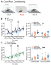
Targeted reduction in thalamic CaV3.1 T-channels increases fear responses during expression but not during acquisition of contextual fear conditioning. (A) Schematic diagram of contextual fear conditioning: Day 1—training (acquisition); Day 2—different context; Day 3—testing. (B) Percentage of time spent freezing in control (scrambled shRNA, gray) and Cacna1g shRNA-injected (blue) mice during training phase of contextual fear conditioning paradigm, analyzed every 30 s (left) and over whole training phase (right). (C) Increased freezing after each tone/shock pairing in control and CaV3.1 KD mice without any differences in freezing behavior between groups (left; two-way RM ANOVA: interaction F(2,36) = 0.182, p = 0.834; tone F(2,36) = 23.56, p < 0.001; shRNA F(1,18) = 0.090, p = 0.768). Control and Cacna1g shRNA-injected animals had similar baseline freezing percentages (right). (D) Compared to control mice, percentage of time freezing in Cacna1g shRNA-injected mice during testing significantly increased over time (left; two-way RM ANOVA: interaction F(13,234) = 0.551, p = 0.891; time F(13,234) = 0.991, p = 0.461; shRNA F(1,18) = 6.642, p = 0.019) and over whole 7 min testing period (right; unpaired two-tailed t-test: t(18) = 2.577, p = 0.019; Cohen’s d = 1.17; effect size r = 0.50). n = 9 control and 11 Cacna1g shRNA-injected mice; * p < 0.05, *** p < 0.001.
Figure 5
Figure 5
Global deletion and targeted reduction in thalamic CaV3.1 T-channels did not change fear responses during cued fear conditioning. (A) Schematic of cued fear conditioning: Day 1—training (acquisition); Day 2—different context; Day 3—testing. (B) Percentage of time spent freezing in control (WT, gray) and CaV3.1 KO (blue) mice during cued fear conditioning paradigm, analyzed every 30 s (left; two-way RM ANOVA: interaction F(13,182) = 1.249, p = 0.248; time F(13,182) = 2.791, p = 0.001; shRNA F(1,14) = 0.156, p = 0.698), and average freezing responses at baseline and after each tone (right; two-way RM ANOVA: interaction F(2,28) = 2.86, p = 0.074; tone F(2,28) = 6.39, p = 0.005; shRNA F(1,14) = 0.039, p = 0.846). (C) Percentage of time freezing in control (gray) and Cacna1g shRNA-injected mice (green) during cued fear conditioning, analyzed over time (left; two-way RM ANOVA: interaction F(13,234) = 0.919, p = 0.534; time F(13,234) = 6.164, p < 0.001; shRNA F(1,18) = 0.124, p = 0.729) and averaged (right; two-way RM ANOVA: interaction F(2,36) = 0.182, p = 0.834; tone F(2,36) = 23.56, p < 0.001; shRNA F(1,18) = 0.090, p = 0.768). n = 8 WT, 8 CaV3.1 KO, 9 control, and 11 Cacna1g shRNA-injected mice; ** p < 0.01, *** p < 0.001.
Figure 6
Figure 6
General activity and anxiety-related behaviors in Cacna1g shRNA-injected mice. (A) Graphic presentation of open-field test. (B) Time spent in the central zone of open-field arena. (C) Number of entries to central zone of open-field arena. (D) Distance traveled in open-field arena in control and Cacna1g shRNA mice. (E) Graphic presentation of zero-maze test. (F) Time spent in open quadrants. (G) Number of entries into open quadrants. (H) Distance traveled during zero-maze test in control and Cacna1g shRNA mice.

References

    1. Wehner J.M., Radcliffe R.A. Cued and Contextual Fear Conditioning in Mice. Curr. Protoc. Neurosci. 2004;27:8.5C.1–8.5C.14. doi: 10.1002/0471142301.ns0805cs27. - DOI - PubMed
    1. Fanselow M.S., Sterlace S.R. The Wiley Blackwell Handbook of Operant and Classical Conditioning. Wiley; Hoboken, NJ, USA: 2014. Pavlovian Fear Conditioning; pp. 117–141. - DOI
    1. Maren S., Phan K.L., Liberzon I. The Contextual Brain: Implications for Fear Conditioning, Extinction and Psychopathology. Nat. Rev. Neurosci. 2013;14:417–428. doi: 10.1038/nrn3492. - DOI - PMC - PubMed
    1. Fanselow M.S., LeDoux J.E. Why We Think Plasticity Underlying Pavlovian Fear Conditioning Occurs in the Basolateral Amygdala. Neuron. 1999;23:229–232. doi: 10.1016/S0896-6273(00)80775-8. - DOI - PubMed
    1. Maren S., Fanselow M.S. The Amygdala and Fear Conditioning: Has the Nut Been Cracked? Neuron. 1996;16:237–240. doi: 10.1016/S0896-6273(00)80041-0. - DOI - PubMed

Substances

LinkOut - more resources