. 2025 Apr 10;26(8):3590.
doi: 10.3390/ijms26083590.

α2-Adrenergic Receptors in Hypothalamic Dopaminergic Neurons: Impact on Food Intake and Energy Expenditure

Affiliations

α2-Adrenergic Receptors in Hypothalamic Dopaminergic Neurons: Impact on Food Intake and Energy Expenditure

Byong Seo Park et al. Int J Mol Sci. .

Abstract

The adrenergic system plays an active role in modulating synaptic transmission in hypothalamic neurocircuitry. While α2-adrenergic receptors are widely distributed in various organs and are involved in various physiological functions, their specific role in the regulation of energy metabolism in the brain remains incompletely understood. Herein, we investigated the functions of α2-adrenergic receptors in the hypothalamus on energy metabolism in mice. Our study confirmed the expression of α2-adrenergic receptors in hypothalamic dopaminergic neurons and assessed metabolic phenotypes, including food intake and energy expenditure, after treatment with guanabenz, an α2-adrenergic receptor agonist. Guanabenz treatment significantly increased food intake (0.25 ± 0.03 g vs. 0.98 ± 0.05 g, p < 0.001) and body weight (-0.1 ± 0.04 g vs. 0.33 ± 0.03 g, p < 0.001) within 6 h post-treatment. Furthermore, guanabenz markedly elevated energy expenditure parameters, including respiratory exchange ratio (RER, 1.017 ± 0.007 vs. 1.113 ± 0.03, p < 0.01) and carbon dioxide production (1.512 ± 0.018 mL/min vs. 1.635 ± 0.036 mL/min, p < 0.05), compared to vehicle-treated controls. Furthermore, using chemogenetic techniques, we demonstrated that the altered metabolic phenotypes induced by guanabenz treatment were effectively reversed by inhibiting the activity of dopaminergic neurons in the hypothalamic arcuate nucleus (ARC) using a chemogenetic technique. Our findings suggest functional connectivity between hypothalamic α2-adrenergic receptor signals and dopaminergic neurons in metabolic controls.

Keywords: alpha-2 adrenergic receptor; energy expenditure; food intake; hypothalamic dopamine neurons.


Conflict of interest statement

The authors declare no conflicts of interest.

Figures

Figure 1
Figure 1
Hypothalamic dopaminergic neurons are activated by norepinephrine. Food intake is measured after intracerebroventricular (i.c.v.) injection of norepinephrine into C57BL/6 mice. (A) The cumulative food intake increases significantly in norepinephrine-treated mice compared to control mice for 6 h after injection (n = 4/group). (B) Norepinephrine treatment increased body weight compared to control mice during the 6-h period (n = 4/group). (C,D) Norepinephrine-induced activation of dopaminergic neurons in ARC is determined by assessing changes in c-Fos immunoreactivity in the hypothalamus of DAT-Cre; GFP mice. (C) Representative photographs (higher magnification image in box) and (D) graphs showing enhanced c-Fos activity in DAT-positive neurons induced by norepinephrine treatment (n = 8 sections/4 mice/group). Results are presented as mean ± SEMs. (A,B,D): * p < 0.05, ** p < 0.01, *** p < 0.001 vs. CTL, as determined using an unpaired two-tailed Student’s t-test. CTL (Control); NE (Norepinephrine). Scale bar = 100 μm.
Figure 2
Figure 2
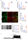
Treatment of α2-adrenergic receptor agonists enhances the activity of dopaminergic neurons in the hypothalamic ARC. (A) qPCR data confirmed the enrichment of DAT or AgRP mRNA expression in RNA purified from hypothalamic DAT- or AgRP-positive neurons using Ribo-tag technique (n = 3/group). (B) α2A, α2B, and α2C subtypes of the α2-adrenergic receptor were expressed in DAT-positive or AgRP neurons (n = 3/group). (C) Representative images show c-Fos immunosignals, a molecular marker of neuronal activity, in the hypothalamic arcuate nucleus (ARC) of mice treated with vehicle or guanabenz, as determined by immunohistochemistry (higher magnification image in box). (D) The number of c-Fos-positive cells increased significantly in DAT-positive neurons within the hypothalamic ARC of guanabenz-treated mice (n = 8 sections/4 mice/group). (E) The bright-field and fluorescent illumination of DAT-GFP neurons are attached to a whole-cell recording pipette (the arrowhead indicates the target cell). (F) Treatment with guanabenz produced high-frequency spikes in DAT-positive neurons. (G) The resting membrane potential and the (H) firing rate increase with guanabenz treatment (n = 10 cells/5 mice/group). Results are presented as means ± SEMs. (D,G,H): ** p < 0.01, *** p < 0.001 vs. CTL, as determined using an unpaired two-tailed Student’s t-test. CTL (Control); GBZ (Guanabenz). Scale bar = 100 μm.
Figure 3
Figure 3
Guanabenz-treated mice show increased food intake and body weight. Food intake and body weight are measured after intracerebroventricular (i.c.v.) injection of guanabenz into C57BL/6 mice (n = 4/group). (A) The cumulative food intake of guanabenz-treated mice is significantly higher than that of vehicle-treated control mice 6 h post-injection, with no alteration observed in 24-h food intake. (B) Guanabenz treatment led to an increase in body weight compared to vehicle-treated control mice at 6 h post-injection, with no significant alteration observed in 24 h changes in body weight. Results are presented as mean ± SEMs. (A,B): *** p < 0.001 vs. CTL, as determined using an unpaired two-tailed Student’s t-test. CTL (Control); GBZ (Guanabenz).
Figure 4
Figure 4
Guanabenz-treated mice exhibit an increase in energy expenditure. Metabolic phenotypes were evaluated after guanabenz injection in C57BL/6 mice using indirect calorimetry (n = 4/group). (A) Oxygen consumption (VO2) did not show significant differences, but (B) carbon dioxide production (VCO2), (C) respiratory exchange ratio (RER), and (D) energy expenditure increased in mice treated with guanabenz compared to vehicle-treated control mice over a 6-h period. Results are presented as mean ± SEMs. A-D: * p < 0.05, ** p < 0.01 vs. CTL, as determined using an unpaired two-tailed Student’s t-test. The arrow indicates the time of the guanabenz injection. CTL (Control); GBZ (Guanabenz).
Figure 5
Figure 5
Guanabenz-induced increase in food intake and energy expenditure was rescued by the inactivation of dopaminergic neurons in ARC. (A) The AAV-hSyn-DIO-hM4D (Gi)-mCherry (hM4D) virus is injected into the hypothalamic arcuate nucleus (ARC) of DAT-cre; GFP mice to inhibit the activity of dopaminergic neurons. (B) Representative images show the hM4D virus (green) in DAT-positive neurons (red) in hypothalamic ARC. The insets show the signals at higher magnifications. (C) The increased food intake induced by guanabenz is decreased in hM4D virus-injected mice (n = 4/group). (D) No significant changes in body weight occurred in guanabenz-treated mice after the inactivation of dopaminergic neurons in the hypothalamic ARC (n = 4/group). (E) In all groups, there were no significant differences in oxygen consumption (VO2). The guanabenz-induced elevation in (F) carbon dioxide production (VCO2), (G) respiratory exchange ratio (RER), and (H) energy expenditure was almost completely rescued by injection of the hM4D virus during the 6-h post-guanabenz treatment period. Results are presented as mean ± SEMs. C-H: * p < 0.05, *** p < 0.001 vs. CTL; # p < 0.05, ### p < 0.001 vs. GBZ, as determined using a one-way ANOVA followed by Sidak’s post hoc multiple comparison test. CTL (Control); GBZ (Guanabenz); hM4D (AAV-hSyn-DIO-hM4D). Scale bar = 100 μm.

Similar articles

References

    1. Qin K., Sethi P.R., Lambert N.A. Abundance and Stability of Complexes Containing Inactive G Protein-coupled Receptors and G Proteins. FASEB J. 2008;22:2920–2927. doi: 10.1096/fj.08-105775. - DOI - PMC - PubMed
    1. Marzo A., Bai J., Otani S. Neuroplasticity Regulation by Noradrenaline in Mammalian Brain. Curr. Neuropharmacol. 2009;7:286–295. doi: 10.2174/157015909790031193. - DOI - PMC - PubMed
    1. Saunders C., Limbird L.E. Localization and Trafficking of A2-Adrenergic Receptor Subtypes in Cells and Tissues. Pharmacol. Ther. 1999;84:193–205. doi: 10.1016/S0163-7258(99)00032-7. - DOI - PubMed
    1. Khan Z.P., Ferguson C.N., Jones R.M. Alpha-2 and Imidazoline Receptor agonistsTheir Pharmacology and Therapeutic Role. Anaesthesia. 1999;54:146–165. doi: 10.1046/j.1365-2044.1999.00659.x. - DOI - PubMed
    1. Berger M., Gray J.A., Roth B.L. The Expanded Biology of Serotonin. Annu. Rev. Med. 2009;60:355–366. doi: 10.1146/annurev.med.60.042307.110802. - DOI - PMC - PubMed

Substances

LinkOut - more resources