. 2025 Apr 10;26(8):3574.
doi: 10.3390/ijms26083574.

Synergistic HDAC4/8 Inhibition Sensitizes Osteosarcoma to Doxorubicin via pAKT/RUNX2 Pathway Modulation

Affiliations

Synergistic HDAC4/8 Inhibition Sensitizes Osteosarcoma to Doxorubicin via pAKT/RUNX2 Pathway Modulation

Anuja Gajanan Magar et al. Int J Mol Sci. .

Abstract

Osteosarcoma is a highly aggressive bone malignancy, particularly challenging in metastatic cases, with a 5-year survival rate remaining under 30%. Although doxorubicin (doxo) is a standard first-line chemotherapeutic agent, its clinical utility is often hindered by the development of drug resistance and associated systemic toxicity. Emerging evidence highlights the role of epigenetic alterations, particularly those involving histone deacetylases (HDACs), in promoting chemoresistance. In this context, the present study aimed to evaluate the therapeutic potential of combining doxo with the selective HDAC inhibitors, tasquinimod (Tas, targeting HDAC4) and PCI-34051 (PCI, targeting HDAC8), in SJSA-1 osteosarcoma cells. Utilizing both 2D and 3D in vitro models, the combination treatment (referred to as the T4 group) significantly reduced cell viability by 57.69% in 2D cultures and decreased spheroid volume by 35.19% in 3D models. The apoptotic response was markedly enhanced, with late apoptosis reaching 64.59% and necrosis at 32.07%, both surpassing the effects observed with doxo alone. Furthermore, wound healing assays demonstrated a 37.74% inhibition of migration, accompanied by a decreased expression of the matrix metalloproteinases MMP9 and MMP13. Mechanistically, the combination therapy led to the downregulation of protein kinase B (pAKT) and RUNX2, along with upregulation of apoptotic markers, including caspase 8, caspase 3, and cleaved caspase 3, indicating a disruption of key survival pathways. These findings suggest that dual HDAC inhibition with Tas and PCI can potentiate doxo efficacy by enhancing apoptosis, inhibiting proliferation, and reducing metastatic potential, thus offering a promising strategy to overcome chemoresistance in osteosarcoma. Further preclinical and clinical studies are required to validate these therapeutic benefits.

Keywords: HDAC inhibitor; anti-cancer; apoptosis; combination therapy; doxorubicin; osteosarcoma.


Conflict of interest statement

The authors declare no conflicts of interest.

Figures

Figure 1
Figure 1
Inhibitory effects of doxorubicin (doxo), tasquinimod (Tas), and PCI-34051 (PCI), individually and in combination, on SJSA-1 osteosarcoma cell proliferation. (A) Representative microscopic images of SJSA-1 cells treated with doxo, Tas, and PCI alone and in combination for 48 h. Blue-colored areas indicate live cells, while white dots represent debris or dead cells. Changes in cell morphology demonstrate treatments’ effects on cell viability and proliferation (images captured at 4X magnification). (B) Cell viability (CCK-8 Assay): CCK-8 assay was performed after 48 h of treatment to evaluate viability of SJSA-1 cells. This assay measures metabolic activity as indicator of cell viability and cytotoxicity. Mean values and % errors are presented to compare cytotoxic effects of each treatment and combination therapy, showing their effectiveness in reducing cell viability. (C) Colony formation assay: long-term proliferative potential of SJSA-1 cells was assessed using colony formation assay. This assay quantifies cells’ ability to form colonies after treatment. Crystal violet staining (blue) was used to visualize colony confluency. A reduced intensity of blue staining indicates a lower number of colonies. Reduced colony numbers or smaller colony sizes indicate inhibitory impact on cell proliferation. (D) Quantification of colony formation assay: number of colonies was counted and analyzed to assess long-term proliferative capacity of cells. Bar graphs represent mean ± SD of colony count from three independent experiments. Statistical significance was determined using one-way ANOVA followed by Tukey’s post hoc test (** p < 0.01 and *** p < 0.001).
Figure 2
Figure 2
Effects of combination therapy of doxo, HDAC4 and HDAC8 inhibitors on apoptosis in osteosarcoma cells. (A) Rate of apoptosis in SJSA-1 osteosarcoma cells following 48 h of treatment with doxo, Tas, PCI, and combination therapies was assessed. This panel shows quantitative data on apoptosis induction, illustrating impact of each treatment alone and in combination on extent of apoptosis in osteosarcoma cells. (Treatment groups: (a-1)—unstained, (b-1)—control, (c-1)—Doxo, (d-1)—Tas, (e-1)—PCI, (f-1)—T1, (g-1)—T2, (h-1)—T3, (i-1)—T4.) (B) Caspase-3/7 activity was measured as indicator of apoptosis. Increased caspase activity points to higher apoptotic signaling, underscoring potential efficacy of combination therapy in promoting cell death in osteosarcoma cells. (Treatment groups: (a-2)—control, (b-2)—Doxo, (c-2)—Tas, (d-2)—PCI, (e-2)—T1, (f-2) –T2, (g-2)—T3, (h-2)—T4).
Figure 3
Figure 3
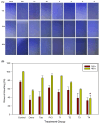
The effects of doxo, Tas, and PCI, individually and in combination, on the metastatic potential of SJSA-1 osteosarcoma cells. (A) Wound healing assay: images from a wound healing assay show the impact of doxo, Tas, PCI, and their combinations on the migratory ability of SJSA-1 cells. The degree of wound closure after 48 h of treatment illustrates the inhibitory effects of each treatment and combination therapy on cell migration, suggesting potential anti-metastatic properties (images captured at 4X magnification). (B) Quantification of wound healing (%) in SJSA-1 cells after 24 and 48 h of treatment. (* p < 0.05).
Figure 4
Figure 4
The evaluation of treatment effects on a 3D tumor spheroid model of SJSA-1 osteosarcoma cells. (A) Spheroid morphology: a representative image of the SJSA-1 3D osteosarcoma spheroid model taken using an Olympus IX73 microscope (magnification: 4X; scale bar: 500 µm) displays the spheroid structure before treatment, providing a baseline for assessing changes in response to treatment. (B) Spheroid volume: the average volume of SJSA-1 spheroids was measured after 48 h of treatment. Reduced spheroid volume indicates the treatment’s efficacy at limiting tumor growth within the 3D model. (C) Cytotoxicity assessment (LIVE/DEAD staining): the cytotoxic effects of doxo, Tas, PCI, and their combinations on the 3D spheroid model were assessed using calcein AM dye (live cells: green fluorescence) and propidium iodide dye (dead cells: red fluorescence) staining (magnification: 4X; scale bar: 500 µm). This staining highlights viable and non-viable cells within the spheroid, visually illustrating the differential cytotoxicity of each treatment approach. (* p < 0.05).
Figure 5
Figure 5
The impact of combination therapy on protein and gene expression in SJSA-1 osteosarcoma cells. (A) A Western blot analysis illustrating the effects of the combination treatment on key proteins involved in metastasis, cell survival, and apoptosis in SJSA-1 osteosarcoma cells. The expression levels of MMP9 and MMP13, which facilitate tumor cell invasion and metastasis through ECM degradation, were significantly reduced, indicating a potential inhibitory effect of the combination therapy on metastatic progression. Additionally, phosphorylated AKT (pAKT), a central regulator of the PI3K/AKT pathway associated with cell survival and chemoresistance, and runt-related transcription factor 2 (RUNX2), a critical modulator of osteosarcoma progression and tumor aggressiveness, were both downregulated, suggesting the suppression of key oncogenic pathways. Conversely, apoptotic markers, including cleaved caspase-8, cleaved caspase-3, and total caspase-3, exhibited increased activation following treatment, indicating enhanced apoptotic signaling. GAPDH was used as a loading control to ensure equal protein loading across samples. (B) A densitometric analysis of the Western blot bands representing the expression levels of target proteins after 48 h of treatment. The band intensities were quantified using Image Lab software (Version 6.1.0 build 7) and normalized to the GAPDH. Relative protein expression levels are expressed as a fold change compared to the untreated control group. The data represent the mean ± the standard deviation from two independent experiments. (C) qRT-PCR analyzed the gene expression of the apoptosis-related markers p16 and p53 following treatment. The results highlight the upregulation of p16 and stable expression of p53 in treated cells. (* p < 0.05 and ** p < 0.01).

Similar articles

References

    1. Chong Z.X., Yeap S.K., Ho W.Y. Unraveling the roles of miRNAs in regulating epithelial-to-mesenchymal transition (EMT) in osteosarcoma. Pharmacol. Res. 2021;172:105818. doi: 10.1016/j.phrs.2021.105818. - DOI - PubMed
    1. Luetke A., Meyers P.A., Lewis I., Juergens H. Osteosarcoma treatment—Where do we stand? A state of the art review. Cancer Treat. Rev. 2014;40:523–532. doi: 10.1016/j.ctrv.2013.11.006. - DOI - PubMed
    1. Chen Y., Liu R., Wang W., Wang C., Zhang N., Shao X., He Q., Ying M. Advances in targeted therapy for osteosarcoma based on molecular classification. Pharmacol. Res. 2021;169:105684. doi: 10.1016/j.phrs.2021.105684. - DOI - PubMed
    1. Jing D., Wu W., Chen X., Xiao H., Zhang Z., Chen F., Zhang Z., Liu J., Shao Z., Pu F. Quercetin encapsulated in folic acid-modified liposomes is therapeutic against osteosarcoma by non-covalent binding to the JH2 domain of JAK2 via the JAK2-STAT3-PDL1. Pharmacol. Res. 2022;182:106287. doi: 10.1016/j.phrs.2022.106287. - DOI - PubMed
    1. Yang C., Choy E., Hornicek F.J., Wood K.B., Schwab J.H., Liu X., Mankin H., Duan Z. Histone deacetylase inhibitor (HDACI) PCI-24781 potentiates cytotoxic effects of doxorubicin in bone sarcoma cells. Cancer Chemother. Pharmacol. 2011;67:439–446. doi: 10.1007/s00280-010-1344-7. - DOI - PubMed

MeSH terms