. 2025 Apr 11;26(8):3612.
doi: 10.3390/ijms26083612.

Claudin-17 Deficiency Drives Vascular Permeability and Inflammation Causing Lung Injury

Affiliations

Claudin-17 Deficiency Drives Vascular Permeability and Inflammation Causing Lung Injury

Mir S Adil et al. Int J Mol Sci. .

Abstract

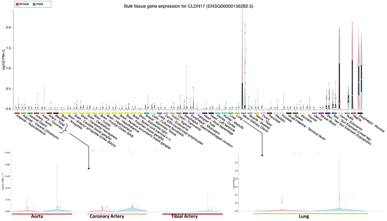
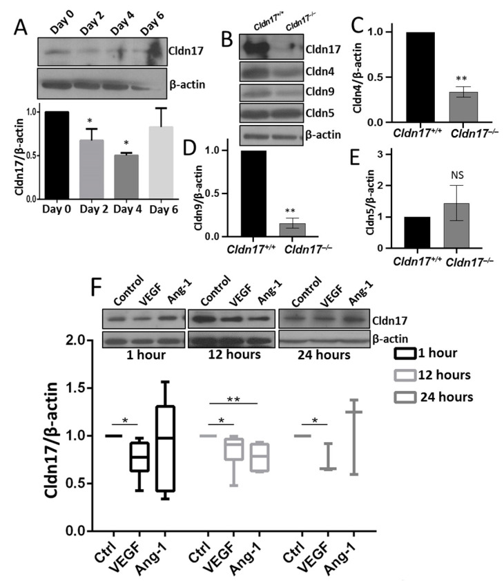
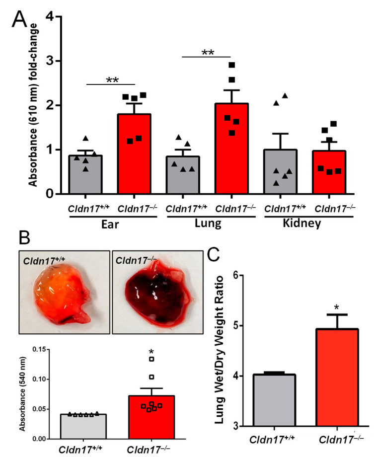
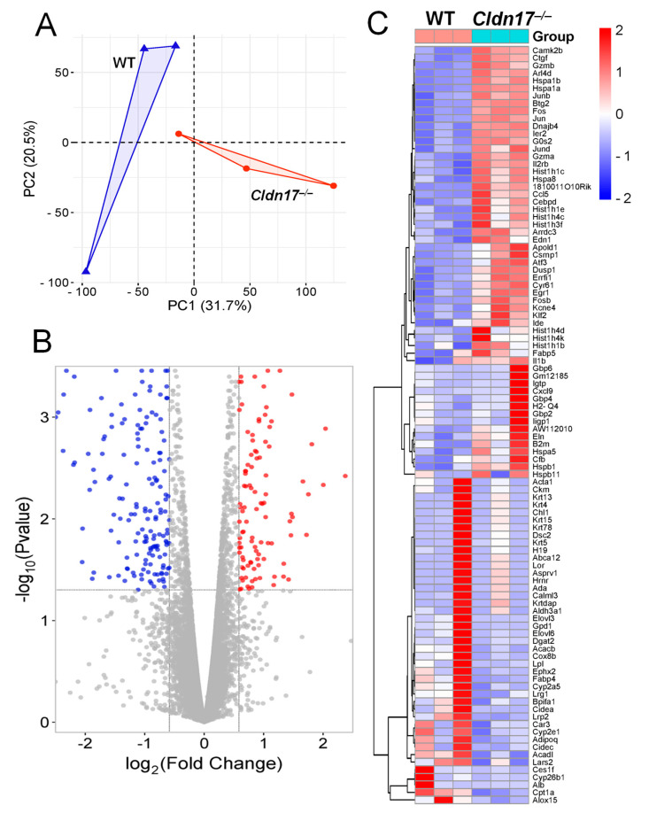
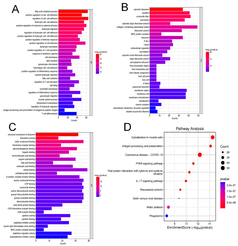
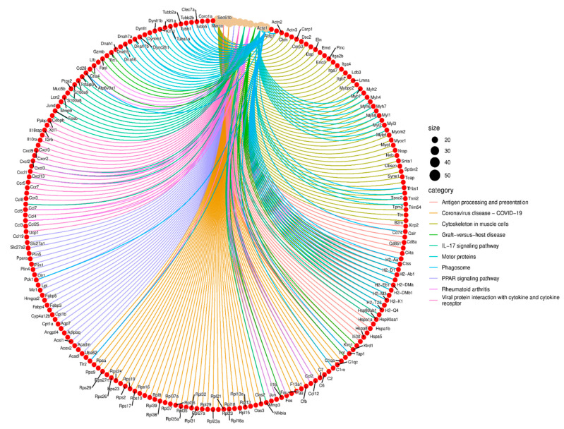
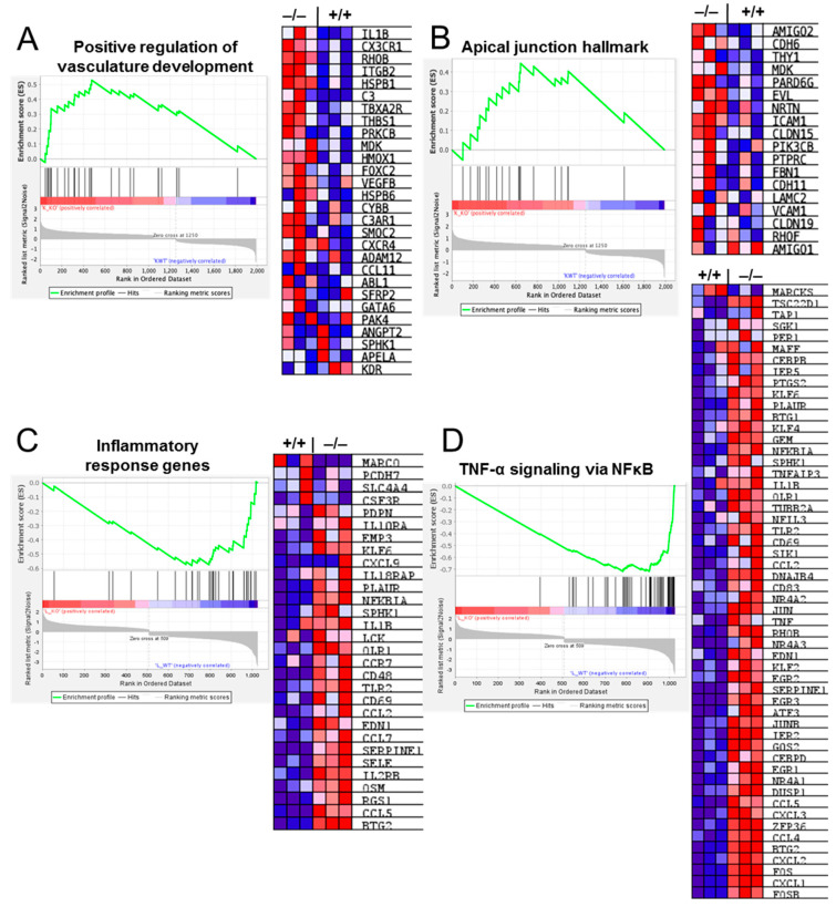
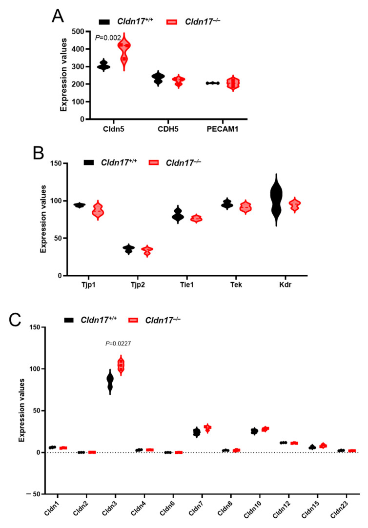
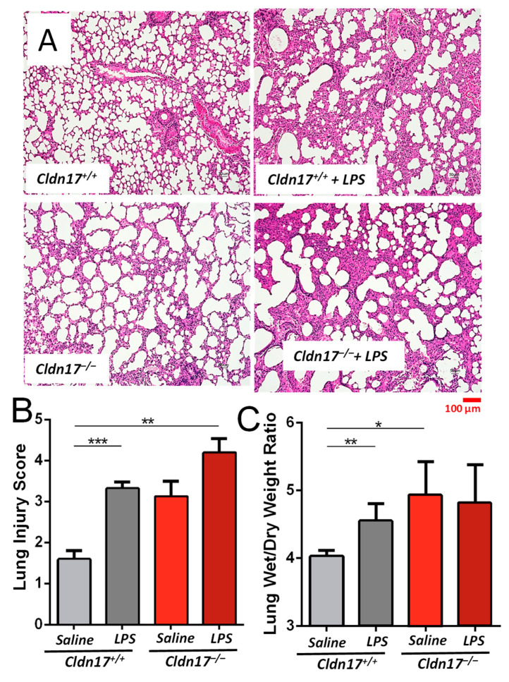
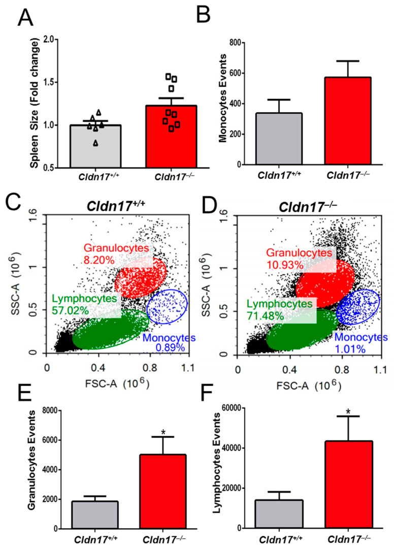
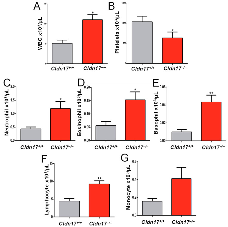
The role of claudin-17 (Cldn17), a tight-junction protein, in vascular permeability remains unclear. We investigated the impact of Cldn17 suppression on vascular permeability. The Miles assay demonstrated significantly increased vascular permeability in the lungs and skin of Cldn17-/- mice, as evidenced by elevated Evan's blue dye extravasation. The Matrigel plug assay demonstrated increased hemoglobin extravasation. Histopathological analysis revealed alveolar flooding, inflammatory cell infiltration, and lung injury in Cldn17-/- lungs. Wet/dry lung weight ratios indicated pulmonary edema, supporting the role of Cldn17 in pulmonary fluid balance, which was exacerbated with lipopolysaccharide administration. Ribosomal nucleic acid sequencing identified distinct transcriptional changes, with the principal component analysis showing clear clustering. Differential gene expression analysis highlighted significant alterations in inflammatory and metabolic pathways. Gene ontology and pathway enrichment analyses revealed the upregulation of immune-related processes, including leukocyte adhesion, interferon-gamma response, and neutrophil degranulation, alongside metabolic dysregulation affecting lipid transport and cytoskeletal organization. Reactome pathway analysis implicated Cldn17 in antigen presentation, interleukin-17 signaling, and inflammatory responses. These findings establish Cldn17 as a critical regulator of vascular permeability and immune homeostasis. Its deficiency drives vascular leakage, exacerbates lung injury, and alters immune signaling pathways, underscoring its potential as a therapeutic target for inflammatory lung diseases.

Keywords: Cldn17; inflammation; lung injury; pulmonary edema; vascular permeability.


Conflict of interest statement

The authors declare no conflicts of interest.

Figures

Figure 1
Figure 1
The Genotype–Tissue Expression (GTEx) portal analysis of Cldn17 in various human tissues shows predominant Cldn17 expression in tissues such as the salivary gland, esophagus, vagina, cervix, testes, and skin and modest expression in organs such as lungs, kidneys, and blood vessels.
Figure 2
Figure 2
LPS-induced sepsis reduces Cldn17 expression in mouse lungs. (A) Representative Western blot images and band densitometry analysis showing reduced expression of Cldn17 in LPS-treated lungs on days 4 and 6 as compared with the vehicle-treated control (n = 3) (BE) Representative Western blot images and quantification graph showing changes in the expression of Cldn4, Cldn5, and Cldn9 in the Cldn17−/− mouse lungs as compared with Cldn17+/+ mouse lungs (n = 3). (F) Representative Western blot images and band densitometry analysis showing reduced expression of Cldn17 in lung endothelial cells treated with 20 ng/mL VEGF and 20 ng/mL angiopoietin-1 (Ang-1) at 1, 12, and 24 h after treatment (n = 3). Data are presented as mean ± SEM; NS, not significant; * p < 0.05; ** p < 0.01.
Figure 3
Figure 3
Matrigel plug and the Miles assay reveals hyperpermeability in Cldn17−/− mice. (A) Extravasation of Evan’s blue dye in various organs determined at a wavelength of 610 nm (n = 5–6). (B) Images of Matrigel plugs isolated from Cldn17+/+ and Cldn17−/− mice and a bar graph showing absorbance of Matrigel plug lysates reflecting hemoglobin concentration (n = 6–7). (C) Pulmonary edema in Cldn17+/+ and Cldn17−/− mice determined by wet/dry weight ratio (n = 6). Data are presented as mean ± SEM; * p ≤ 0.05; ** p < 0.01.
Figure 4
Figure 4
Analysis of DEGs in Cldn17 deficient samples. (A) Principal component analysis (PCA) displaying the fact that the two groups were not overlapped. (B) The volcano plot shows the upregulated and downregulated DEGs in the Cldn17−/− mice compared with the Cldn17+/+ mice. Each dot represents a gene. (C) The heatmap demonstrates the top 100 DEGs among the two groups.
Figure 5
Figure 5
Gene ontology and pathway enrichment analysis revealed key biological processes and pathways affected by Cldn17 deficiency. (AC) Gene Ontology (GO) analysis highlighting biological processes, cellular components, and molecular function significantly enriched in DEGs. (D) Pathway enrichment analysis using KEGG annotations, highlighting significantly affected pathways.
Figure 6
Figure 6
Circular chord diagram visualizing the functional enrichment of DEGs in the Cldn17−/− mouse lungs.
Figure 7
Figure 7
Heat maps and gene set enrichment analysis (GSEA) for altered genes involved in the regulation of (A) vascular development, (B) apical junction, (C) inflammatory response, and (D) TNFα and NFκB signaling. Data represented for genes with foldchange >2; n = 3; +/+; WT; −/−, Cldn17 knockout; p < 0.05.
Figure 8
Figure 8
Cldn17 deficiency had no major impact on the expression of other cell–cell junction proteins in mouse lungs. (A) Violin plots showing changes in the expression of the major endothelial junctional and adhesion proteins Cldn5, Cdh2, and Pecam1 between the Cldn17+/+ and Cldn17−/− mouse lungs. (B) Violin plots showing expression levels of the major endothelial markers Tjp1/Tjp2, Tie1, Tek, and Kdr between the Cldn17+/+ and Cldn17−/− mouse lungs. (C) Violin plots showing no changes in the expression of other claudin isoforms in the Cldn17−/− mouse lungs as compared to the Cldn17+/+ mouse lungs. n = 3; Data are presented as mean ± SEM.
Figure 9
Figure 9
Cldn17−/− mouse lungs exhibit exacerbated LPS-induced ALI. (A) Representative H&E-stained lung sections from the Cldn17+/+ and Cldn17−/− mice under saline and LPS-treated conditions. LPS treatment induced inflammatory cell infiltration and alveolar thickening. The Cldn17−/− lungs showed increased structural disruption and inflammation following LPS exposure. (B) Lung injury scores quantified from histological sections showed that LPS treatment significantly exacerbated lung injury in the Cldn17−/− mice compared with the Cldn17+/+ mice. (C) Bar graph showing lung wet/dry weight ratio of the LPS-treated Cldn17−/− mice, which exhibited significantly higher ratios compared with the Cldn17+/+ mice, indicating increased vascular permeability and fluid accumulation. * p < 0.05; ** p < 0.01; *** p < 0.001. n = 4 for Cldn17+/+; and n = 3 for Cldn17−/−. Data are presented as mean ± SEM.
Figure 10
Figure 10
Cldn17 deficiency is linked to splenomegaly and increased expansion of granulocytes and lymphocytes. (A) Increased spleen size in the Cldn17−/− vs. Cldn17+/+ mice (n = 6–8). (B) Monocytes events. (C,D) Forward vs. side scatter plots for the Cldn17−/− vs. Cldn17+/+ groups. (E,F) Bar graphs showing granulocytes and lymphocyte events. Data are presented as mean ± SEM; WBC, white blood cells; * p < 0.05.
Figure 11
Figure 11
Loss of Cldn17 causes leukocytosis. Bar graphs showing (A) WBCs (n = 7), (B) differential count (n = 7), and (C) platelets (n = 8) in the Cldn17−/− vs. Cldn17+/+ mice. (DG) Bar graphs showing increased eosinophil, basophil, total lymphocytes and monocytes in Cldn17−/− vs. Cldn17+/+ mouse blood, respectively (n=7). Data are presented as mean ± SEM; WBC, white blood cells; * p < 0.05; ** p < 0.01.

Similar articles

References

    1. Park-Windhol C., D’Amore P.A. Disorders of Vascular Permeability. Annu. Rev. Pathol. 2016;11:251–281. doi: 10.1146/annurev-pathol-012615-044506. - DOI - PMC - PubMed
    1. Ferreira A.R., Felgueiras J., Fardilha M. Signaling pathways in anchoring junctions of epithelial cells: Cell-to-cell and cell-to-extracellular matrix interactions. J. Recept. Signal Transduct. Res. 2015;35:67–75. doi: 10.3109/10799893.2014.931426. - DOI - PubMed
    1. Krüger-Genge A., Blocki A., Franke R.P., Jung F. Vascular Endothelial Cell Biology: An Update. Int. J. Mol. Sci. 2019;20:4411. doi: 10.3390/ijms20184411. - DOI - PMC - PubMed
    1. Minami T., Aird W.C. Endothelial cell gene regulation. Trends Cardiovasc. Med. 2005;15:174–184. doi: 10.1016/j.tcm.2005.06.002. - DOI - PubMed
    1. Sahni S.K. Endothelial cell infection and hemostasis. Thromb. Res. 2007;119:531–549. doi: 10.1016/j.thromres.2006.06.006. - DOI - PubMed

MeSH terms

LinkOut - more resources