. 2025 Apr 11;26(8):3630.
doi: 10.3390/ijms26083630.

Segatella copri Outer-Membrane Vesicles Are Internalized by Human Macrophages and Promote a Pro-Inflammatory Profile

Affiliations

Segatella copri Outer-Membrane Vesicles Are Internalized by Human Macrophages and Promote a Pro-Inflammatory Profile

Alison Sepúlveda-Pontigo et al. Int J Mol Sci. .

Abstract

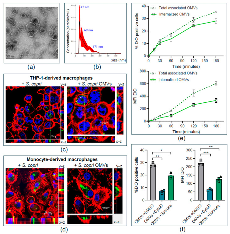
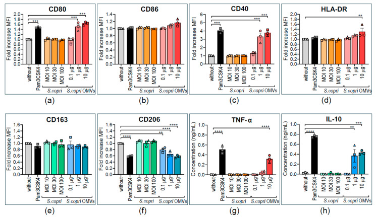
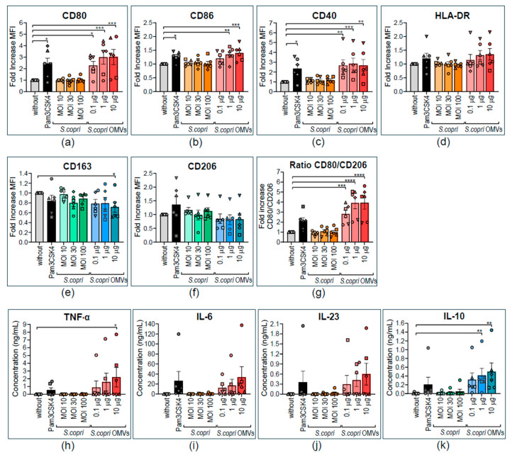
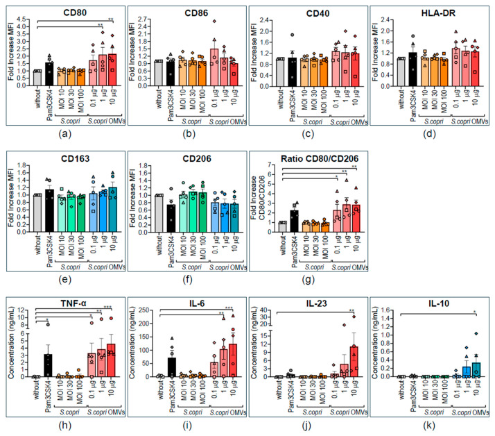
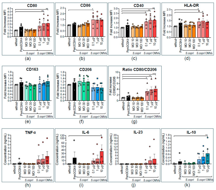
Increased abundance of Segatella copri (S. copri) within the gut microbiota is associated with systemic inflammatory diseases, including rheumatoid arthritis. Although outer-membrane vesicles (OMVs) of Gram-negative bacteria are important players in microbiota-host communication, the effect of S. copri-derived OMVs on immune cells is unknown. Macrophages engulf and eliminate foreign material and are conditioned by environmental signals to promote either homeostasis or inflammation. Thus, we aimed to explore the impact of S. copri-OMVs on human macrophages in vitro, employing THP-1 and monocyte-derived macrophage models. The uptake of DiO-labeled S. copri-OMVs into macrophages was monitored by confocal microscopy and flow cytometry. Furthermore, the effect of S. copri and S. copri-OMVs on the phenotype and cytokine secretion of naïve (M0), pro-inflammatory (M1), and anti-inflammatory (M2) macrophages was analyzed by flow cytometry and ELISA, respectively. We show that S. copri-OMVs enter human macrophages through macropinocytosis and clathrin-dependent mechanisms. S. copri-OMVs, but not the parental bacterium, induced a dose-dependent increase in the expression of M1-related surface markers in M0 and M2 macrophages and activated the secretion of large amounts of pro-inflammatory cytokines in M1 macrophages. These results highlight an important role of S. copri-OMVs in promoting pro-inflammatory macrophage responses, which might contribute to systemic inflammatory diseases.

Keywords: M1/M2 polarization; Segatella copri; endocytosis; macrophages; outer-membrane vesicles.


Conflict of interest statement

The authors declare no conflicts of interest.

Figures

Figure 1
Figure 1
Characterization of Segatella copri-derived OMVs and their internalization by human macrophages. (a) Transmission electron microscopy (TEM) image of S. copri-OMVs, captured at a total magnification of 73,000×. (b) Size distribution of S. copri-OMVs, measured by nanoparticle tracking analysis (NTA), n = 3. (c,d) Orthogonal projection of confocal sections 630× of human THP-1-derived macrophages (c) and peripheral blood monocyte-derived macrophages with 3× digital zoom (d) that were exposed for 3 h to DiO-labeled S. copri at MOI 100 (left) or DiO-labeled OMVs at 50 µg/mL (right). DiO shows green fluorescence; plasma membrane was counterstained with WGA (red) and nuclei with Hoechst 33,342 (blue). (e) Kinetics of S. copri-OMV uptake by THP-1-derived macrophages over a time of 3 h. Percentage of DiO-positive cells (upper panel) and mean fluorescence intensity (MFI) of DiO (lower panel), determined by flow cytometry after exposure to 50 µg/mL DiO-labeled S. copri-OMVs. Fluorescence was measured before (dotted line) and after the addition of trypan blue (continuous line) to quench the DiO signal of S. copri-OMVs adhered to the macrophage membrane. (f) Uptake mechanism of S. copri-OMVs into macrophages was determined by pre-incubation of THP-1-derived macrophages for 1 h with 1 µg/mL Cytochalasin D, 45 mM sucrose or vehicle (DMSO) before exposure to 50 µg/mL DiO-labeled S. copri-OMVs for further 3 h. Data (n = 3) are displayed as mean ± SEM. Each symbol represents an independent experiment. Significant differences, according to one-way ANOVA and the Dunnett multiple comparison test, are indicated (* p ≤ 0.05, ** p ≤ 0.01, *** p ≤ 0.001).
Figure 2
Figure 2
Altered expression of pro- and anti-inflammatory surface markers and cytokines by human THP1-derived macrophages in response to Segatella copri and its OMVs. THP-1 macrophages were stimulated for 24 h with Pam3CSK4 (0.5 μg/mL); S. copri at multiplicity of infection (MOI) 10, 30, or 100; or S. copri-OMVs at 0.1 μg/mL, 1 μg/mL, or 10 μg/mL. (ac) Expression of surface markers related to an M1 phenotype, CD80 (a), CD86 (b), CD40 (c), and HLA-DR (d), as well as the M2 markers CD163 (e) and CD206 (f), were determined by flow cytometry. Alterations of mean fluorescent intensity (MFI) with respect to unstimulated control were displayed as fold increase. The pro-inflammatory cytokine TNF-α (g) and anti-inflammatory IL-10 (h) were quantified in culture supernatants by ELISA. Each symbol represents an independent experiment. All results are expressed as mean ± SEM. Statistical significance was determined by one-way ANOVA and the Dunnett post-test, with ** p < 0.01, *** p < 0.001, and **** p < 0.0001 (n = 3).
Figure 3
Figure 3
Altered expression of pro- and anti-inflammatory surface markers and cytokines by human monocyte-derived M0 macrophages in response to Segatella copri and its OMVs. Peripheral blood monocytes were differentiated into M0 macrophages during 10 days in the presence of 100 U/mL M-CSF and subsequently exposed for a further 24 h to Pam3CSK4 (0.5 μg/mL, positive control); S. copri at MOI 10, 30, or 100; or S. copri-OMVs at concentrations of 0.1 μg/mL, 1 μg/mL, or 10 μg/mL. Expression of CD80 (a), CD86 (b), CD40 (c), and HLA-DR (d) was characteristic of an M1 profile, as well as CD163 (e) and CD206 (f), M2-related markers, and the CD80-to-CD206 ratio (g) was determined by flow cytometry. Alterations of mean fluorescent intensity (MFI) with respect to the control without stimulus were displayed as fold increase. Secretion of the pro-inflammatory cytokines TNF-α (h), IL-6 (i), and IL-23 (j) and anti-inflammatory IL-10 (k) was measured in culture supernatants by ELISA. Each symbol represents a different blood donor. Results are displayed as mean ± SEM. Statistical significance was determined by one-way ANOVA and the Dunnett post-test, with * p ≤ 0.05, ** p ≤ 0.01, *** p ≤ 0.001, and **** p ≤ 0.0001 (n = 6).
Figure 4
Figure 4
Altered expression of pro- and anti-inflammatory surface markers and cytokines by human M1 macrophages in response to Segatella copri and its OMVs. Peripheral blood monocytes were differentiated into macrophages during 8 days in the presence of 1000 U/mL GM-CSF. M1 polarization was induced by adding GM-CSF and IFN-γ (200 U/mL) for the last 2 days of culture. M1 macrophages were exposed to Pam3CSK4 (0.5 μg/mL, positive control); S. copri at MOI 10, 30, or 100; or S. copri-OMVs at concentrations of 0.1 μg/mL, 1 μg/mL, or 10 μg/mL for a further 24 h, and expression of CD80 (a), CD86 (b), CD40 (c), and HLA-DR (d) was characteristic of a M1 profile, as well as CD163 (e) and CD206 (f), M2-related markers, and the CD80-to-CD206 ratio (g) was determined by flow cytometry. Alterations of mean fluorescent intensity (MFI) with respect to the control without stimulus were displayed as fold increase. Secretion of the pro-inflammatory cytokines TNF-α (h), IL-6 (i), and IL-23 (j) and anti-inflammatory IL-10 (k) was measured in culture supernatants by ELISA. Each symbol represents a different blood donor. Results are expressed as mean ± SEM. Statistical significance was determined by one-way ANOVA and the Dunnett post-test, with * p ≤ 0.05, ** p ≤ 0.01, and *** p ≤ 0.001 (n = 5).
Figure 5
Figure 5
Altered expression of pro- and anti-inflammatory surface markers and cytokines by human M2 macrophages in response to Segatella copri and its OMVs. Peripheral blood monocytes were differentiated into macrophages in the presence of 100 U/mL M-CSF, and on day 8, M2 profile was induced by adding M-CSF and IL-4 (20 ng/mL) for a further 48 h. After stimulation with Pam3CSK4 (0.5 μg/mL, positive control); S. copri at MOI 10, 30, or 100; or S. copri-OMVs at concentrations of 0.1 μg/mL, 1 μg/mL, or 10 μg/mL for 24 h, the surface expression of the M1-related markers CD80 (a), CD86 (b), CD40 (c), and HLA-DR (d), and the M2 markers CD163 (e) and CD206 (f), as well as the CD80/CD206 ratio (g), was determined by flow cytometry. Alterations of mean fluorescent intensity (MFI) with respect to the control without stimulus were displayed as fold increase. Secretion of the pro-inflammatory cytokines TNF-α (h), IL-6 (i), and IL-23 (j) and anti-inflammatory IL-10 (k) was measured in culture supernatants by ELISA. Each symbol represents a different blood donor. All results are expressed as mean ± SEM. Statistical significance was determined by one-way ANOVA and the Dunnett post-test, with * p ≤ 0.05, ** p ≤ 0.01, and *** p ≤ 0.001 (n = 5).

Similar articles

References

    1. Ruff W.E., Greiling T.M., Kriegel M.A. Host–microbiota interactions in immune-mediated diseases. Nat. Rev. Microbiol. 2020;18:521–538. doi: 10.1038/s41579-020-0367-2. - DOI - PubMed
    1. Jiao Y., Wu L., Huntington N.D., Zhang X. Crosstalk Between Gut Microbiota and Innate Immunity and Its Implication in Autoimmune Diseases. Front. Immunol. 2020;11:282. doi: 10.3389/fimmu.2020.00282. - DOI - PMC - PubMed
    1. Tiku V., Tan M.-W. Host immunity and cellular responses to bacterial outer membrane vesicles. Trends Immunol. 2021;42:1024–1036. doi: 10.1016/j.it.2021.09.006. - DOI - PubMed
    1. Kaparakis-Liaskos M., Ferrero R.L. Immune modulation by bacterial outer membrane vesicles. Nat. Rev. Immunol. 2015;15:375–387. doi: 10.1038/nri3837. - DOI - PubMed
    1. Mozaheb N., Mingeot-Leclercq M.-P. Membrane Vesicle Production as a Bacterial Defense Against Stress. Front. Microbiol. 2020;11:600221. doi: 10.3389/fmicb.2020.600221. - DOI - PMC - PubMed

LinkOut - more resources