. 2025 Apr 11;26(8):3632.
doi: 10.3390/ijms26083632.

Enzymes Drive Glutathione Shunt to Explain Oxidative State Using an In-Parallel Multi-Omic Method

Affiliations

Enzymes Drive Glutathione Shunt to Explain Oxidative State Using an In-Parallel Multi-Omic Method

Valerie C Wasinger et al. Int J Mol Sci. .

Abstract

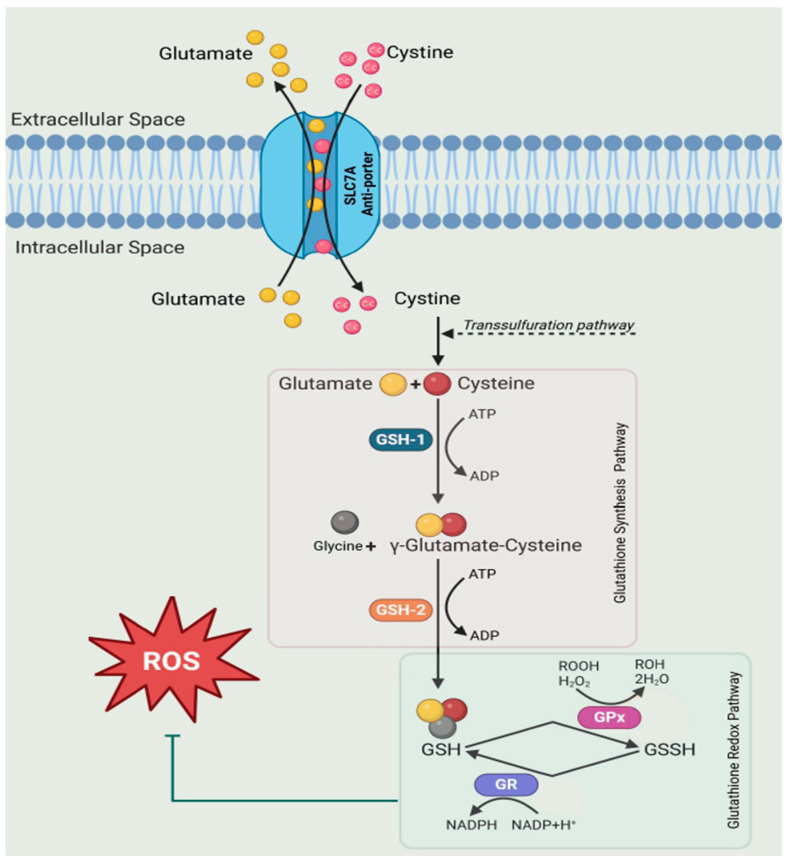
The glutathione shunt is one of the most important contributors to the cellular redox state, with implications across cancer, chronic diseases, diseases of ageing, and autoimmune diseases, including inflammatory bowel disease (IBD). Traditionally, the redox state is gauged by the ratio of the surrogate metabolites GSH and GSSG. However, this presents methodological challenges and offers a constrained illustration of metabolites without a systems-level understanding of redox dynamics, failing to elucidate variations across an entire biochemical network. Targeted proteomics can fill this void. Here, we describe an in-parallel metabolomic and proteomic targeted method to encompass measurements directly related to the shunt. Samples are simultaneously prepared to extract the substrate building blocks, cysteine, cystine, methionine, glutamic acid, and kynurenine; and the proteins, SLC7A11 (xCT), Glutamate Cysteine Ligase (GSH1), Glutathione Synthetase (GSH2), Glutathione Peroxidase (GPx), and Glutathione Reductase (GSHR) for targeted mass spectrometry. We demonstrate the method by targeted analysis of proteins in plasma, serum, nasal swab, and saliva and apply the multi-omic method to assess changes in the glutathione shunt in the serum of patients diagnosed with IBD. This allows for a broader narrative to establish context at which the glutathione shunt is operating.

Keywords: GPx; GSH; GSSG; IBD; SLCA7A1; biomarker; glutathione shunt; liquid biopsy.


Conflict of interest statement

The authors declare no conflicts of interest.

Figures

Figure A1
Figure A1
Relative GPx3 levels in patients categorised by confocal laser endoscopy scoring, showing an inflexion point at a CLS score of ~10. Definitions for this form of defining IBD by gut leakiness has previously been described [42].
Figure A2
Figure A2
Multiple sequence alignment of the GPx. Highlighted sequences indicate unique peptides. Indications of identity are represented by *, similarity are represented by dots, gaps in the identity are represented by dash.
Figure A3
Figure A3
Mass chromatogram of serum sample (8-fold dilution) precipitated with TCA using a 20 min gradient. (A) shows Glutamic acid (m/z 148) and labelled glutamate (m/z 154). The metabolites cysteine (m/z 122) and cystine (m/z 241) were quantitated using labelled glutamate, which elutes at similar retention time. (B) shows methionine (m/z 150) and labelled methionine (m/z 153); (C) shows kynurenine (m/z 209) and labelled kynurenine (m/z 213).
Figure 1
Figure 1
GSH exists as a tri-amino acid combination of glycine, cysteine, and glutamine, with a readily oxidisable thiol group. Increased synthesis and turnover of glutathione as well as an increase in NADPH consumption from the re-generation of the oxidised form (GSSG) supports the antioxidant defence system. In Eukaryotes, GSH1 catalyses the formation of γ-GC, while GSH2 conjugates glycine to form GSH [9]. The antiporter SLC7A transports cystine, cysteine, glutamate, and kynurenine to restore redox homeostasis.
Figure 2
Figure 2
Evidence for the detection of the substrate anti-porter xCT (SLC7A) across the biological matrices of (A) saliva, (B) serum, (C) plasma, and (D) nasal swabs. Area under the peak (AUC) was assessed for the peptide KPVVSTISK with an MH2+ m/z of 479.8008.
Figure 3
Figure 3
Evidence for the detection of the enzymes GSH1 and GSH2 across the biological matrices of (A) saliva, (B) serum, (C) plasma, and (D) nasal swabs. AUC was assessed for the GSH1 peptide DPLTLFEEK (Grey box) with an MH2+ m/z of 546.2846. Serum GSH1 abundances had a median AUC of 10,000. AUC was assessed for GSH2 peptide (Green box) AIENELLAR with an MH2+ m/z of 514.7909 in saliva, serum and plasma while ALAEGVLLR with an MH2+ m/z of 471.2926 was detected in nasal swabs.
Figure 4
Figure 4
Evidence for the detection of the GSH peroxidase enzyme GPx3 and the reductase GSHR across the biological matrices of (A) saliva, (B) serum, (C) plasma, and (D) nasal swabs. AUC was assessed for the GPx3 peptide QEPGENSEILPTLK (Grey box) with an MH2+ m/z of 777.9041, GSHR peptides LNAIYQNNLTK (Green box) with an MH2+ m/z of 646.3539, and ALLTPVAIAAGR (white box) with an MH2+ m/z of 576.8586.
Figure 5
Figure 5
PRM targeted confirmation of protein markers of redox potential applied to IBD as a demonstrative pathological utility. (A) xCT (SLC7A); (B) GSH1 and GSH2; and (C) GPx3 and GSHR. Patient matched (n = 4) flare and remission data demonstrate a similar trend for (D) xCT, (E) GSH1 and GSH2, and (F) GSHR. Significance p < 0.05 represented by *; p < 0.005 represented by **.
Figure 6
Figure 6
Metabolomic quantitation of substrate building blocks of the GSH shunt. (A) The amino acids methionine and glutamic acid, (B) the amino acid cysteine, cystine, and an important SLC7A anti-ported tryptophan metabolism product Kynurenine in an NRF2-dependent manner with links to ferroptosis. All measurements are in μM.

References

    1. Bustamante S., Yau Y., Boys V., Chang J., Paramsothy S., Pudipeddi A., Leong R.W., Wasinger V.C. Tryptophan Metabolism ‘Hub’ Gene Expression Associates with Increased Inflammation and Severe Disease Outcomes in COVID-19 Infection and Inflammatory Bowel Disease. Int. J. Mol. Sci. 2022;23:14776. doi: 10.3390/ijms232314776. - DOI - PMC - PubMed
    1. Ballatori N., Krance S.M., Notenboom S., Shi S., Tieu K., Hammond C.L. Glutathione dysregulation and the etiology and progression of human diseases. Biol. Chem. 2009;390:191–214. doi: 10.1515/BC.2009.033. - DOI - PMC - PubMed
    1. Ferguson G.D., Bridge W.J. The glutathione system and the related thiol network in Caenorhabditis elegans. Redox Biol. 2019;24:101171. doi: 10.1016/j.redox.2019.101171. - DOI - PMC - PubMed
    1. Rahman I., Kode A., Biswas S.K. Assay for quantitative determination of glutathione and glutathione disulfide levels using enzymatic recycling method. Nat. Protoc. 2006;1:3159–3165. doi: 10.1038/nprot.2006.378. - DOI - PubMed
    1. Aniya Y., Imaizumi N. Mitochondrial glutathione transferases involving a new function for membrane permeability transition pore regulation. Drug Metab. Rev. 2011;43:292–299. doi: 10.3109/03602532.2011.552913. - DOI - PubMed

MeSH terms

LinkOut - more resources