. 2025 Apr 11;26(8):3626.
doi: 10.3390/ijms26083626.

Piceatannol Inhibits the Immunostimulatory Functions of Dendritic Cells and Alleviates Experimental Arthritis

Affiliations

Piceatannol Inhibits the Immunostimulatory Functions of Dendritic Cells and Alleviates Experimental Arthritis

Luyang Han et al. Int J Mol Sci. .

Abstract

Rheumatoid arthritis (RA) is a highly prevalent systemic autoimmune disease. Recently, natural small molecules have been explored as alternative therapeutic agents. Iris halophila Pall is the traditional herbal medicine, and it is rich in active ingredients with anti-inflammatory and immunomodulatory effects. In our previous study, LC-MS analysis revealed that piceatannol (PIC) is one of the primary active ingredients in the root of Iris tectorum. The purpose of this study was to explore the immunomodulatory effects of PIC on the maturation and function of dendritic cells, as well as on experimental arthritis induced by complete Freund's adjuvant (CFA) and incomplete Freund's adjuvant (IFA). Additionally, we aimed to probe into the potential mechanisms underlying the effects of PIC. We first verified the immunosuppressive effect of PIC using flow cytometry and an ELISA. The immunosuppressive mechanism of PIC on dendritic cells (DCs) was investigated through a joint analysis of network pharmacology and Western blotting. Our findings revealed that under Lipopolysaccharide (LPS)-induced inflammatory conditions, PIC could restrain the maturation and function of DCs (p < 0.001) and decrease the secretion of inflammatory cytokines (p < 0.001) compared to the LPS group. Furthermore, PIC suppressed the activation and polarization of CD4+ T cells, resulting in a decreased proportion of Th1 and Th17 cells (p < 0.001), ultimately improving the symptoms of CFA-induced arthritis in comparison to the model group. The PIC-induced shift in the T helper cell differentiation correlated with the secretion of polarizing cytokines from DCs in the AIA model. Mechanistically, PIC exerted its immunosuppressive function mainly by down-regulating the Mitogen-Activated Protein Kinase (MAPK) and Nuclear Factor kappa-B (NF-κB) signaling pathways. Collectively, these data unveil the anti-inflammatory mechanisms of a traditional medicine via the inhibition of the immune activation function of DCs in vivo and open up a therapeutic approach for autoinflammatory diseases.

Keywords: dendritic cells; piceatannol; rheumatoid arthritis.


Conflict of interest statement

The authors declare that they have no known competing financial interests or personal relationships that could have appeared to influence the work reported in this paper.

Figures
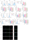
Figure 1
Figure 1
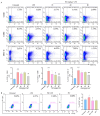
The effects of PIC on LPS-induced DC maturation and function in vitro. DCs were treated with varying doses of PIC in the presence or absence of LPSs and detected by flow cytometry and ELISA: (A) the expression levels of CD40, CD86, and MHC II on the surfaces of DCs; (B) the secretion levels of cytokines IL-12p40 and IL-6; (C) cytokine TGF-β and IL-10 expression; (D) dextran co-cultured with DCs to detect the phagocytosis of antigens; (E) the expression of CCR7 on DC surface; (F) the morphology of DCs under the laser confocal microscope (Scale bar is 100 μm). Statistical significance was noted as ## p < 0.01 and ### p < 0.001 compared to the untreated group, and as * p < 0.05, ** p < 0.01 and *** p < 0.001 compared to the LPS group.
Figure 2
Figure 2
The effects of PIC on the maturation and migration of DCs in vivo. PIC (20, 40, and 80 mg/kg) was mixed with or without LPS and injected into the paws of mice for 24 h: (A) the proportions of CD11c+CD40+, CD11c+CD86+, and CD11c+MHCII+ cells in the lymph nodes of the right legs; (B) the proportions of DCs stained with FITC-CFSE in lymph nodes. # p < 0.05, ### p < 0.001 compared to the untreated group. * p < 0.05 and ** p < 0.0 compared to the LPS group.
Figure 3
Figure 3
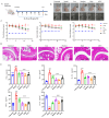
Therapeutic effects of PIC on arthritis in mice. After the CFA-induced arthritic mouse model was successfully constructed, PIC or MTX was injected subcutaneously every other day. (A) Flowchart of arthritic mouse modeling and treatment; (B) swelling degree of mouse foot and ankle joints; (C) from the first day of treatment, the foot and ankle joints of mice were measured every other day; (D) joint pathology histological section stained with H&E; (E) the expression levels of cytokines TNF-α, IL-6, IL-17 A, IFN-γ, and IL-10 in sera of mice were detected by ELISA. ### p < 0.001 compared to the untreated group. ** p < 0.01 and *** p < 0.001 compared to the LPS group.
Figure 4
Figure 4
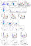
The effects of PIC on immune cells in lymph nodes and spleens of arthritic mouse model. (A) The expression levels of DC co-stimulatory molecules CD40 and CD86 in mouse leg lymph nodes were detected; (B) the levels of the cytokines TNF, IL-23, and IL-6 secreted by DCs in the spleens of mice were detected; (C) different cytokines secreted by Th1, Th2, and Th17 in the spleens of mice and the percentages of Th2/Th1, nTreg/Th17, and iTreg/Th17 were analyzed; (D) the correlations between IL-23-secreting- and IL-6-secreting DCs with Th17 cells and TNF-α-secreting DCs with Th1 cells in arthritic mice in vivo. ### p < 0.001 compared to the untreated group. * p < 0.05 ** p < 0.01 and *** p < 0.001 compared to the LPS group.
Figure 5
Figure 5
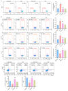
PIC inhibited the DC-stimulated proliferation of OVA-specific CD4+ T cells and the differentiation of T cells from the spleens of arthritic mice in vitro. (A) DCs were treated with PIC in the presence of absence of LPSs for 12 h, mixed with CFSE-labeled OT II mouse spleen cells at a ratio of 1:5 before 0.1 mg/mL OVA antigen was added, and co-cultured in 24-well plates. The effect of PIC on the proliferation of specific CD4+ T cells was detected after 72 h. (B) The treated DCs were mixed with spleen cells from arthritic mice at a ratio of 1:5. After 72 h of co-culturing in 24-well plates, the effect of the PIC on the polarization of CD4+ T cells into the Th1, Th2, Th17, and Treg cell subtypes was detected by flow cytometry. ## p < 0.01, ### p < 0.001 compared to the untreated group. ** p < 0.01 and *** p < 0.001 compared to the LPS group.
Figure 6
Figure 6
Network pharmacology prediction and molecular docking technology. (A) Venn diagram showing the intersection target between the active ingredient and the disease; (B,C) the protein interaction network diagram greater than the average degree value in the common targets of PIC and RA; (D) GO functional enrichment analysis; (E) analysis of KEGG pathway enrichment; (F) a network showcasing the primary pathways and targets involved in the treatment of RA with PIC.
Figure 7
Figure 7
(A) Cytochalasin D inhibits the phagocytosis of DCs by interfering with the polymerization of microfilaments and destroying the dynamic structure of the cytoskeleton. We verified the endocytosis of PIC by DCs with or without cytochalasin D; (B) molecular docking of drugs and targets: (ae) molecular docking of PIC with MAPK14, PTGS2, KIT, EGFR, and RELA; (fj) molecular docking between PIC and the corresponding inhibitors for each target; (C) effects of drugs on the MAPK and NF-κB signaling pathways in DCs. Statistical significance is indicated as follows: # p < 0.05, ## p < 0.01, ### p < 0.001 compared to the untreated group; * p < 0.05 and *** p < 0.001 compared to the LPS group, ns compared to LPS and Cy-D group.

Similar articles

References

    1. Petrelli F., Mariani F.M., Alunno A., Puxeddu I. Pathogenesis of rheumatoid arthritis: One year in review 2022. Clin. Exp. Rheumatol. 2022;40:475–482. doi: 10.55563/clinexprheumatol/l9lyen. - DOI - PubMed
    1. Scherer H.U., Häupl T., Burmester G.R. The etiology of rheumatoid arthritis. J. Autoimmun. 2020;110:102400. doi: 10.1016/j.jaut.2019.102400. - DOI - PubMed
    1. Jang S., Kwon E.J., Lee J.J. Rheumatoid Arthritis: Pathogenic Roles of Diverse Immune Cells. Int. J. Mol. Sci. 2022;23:905. doi: 10.3390/ijms23020905. - DOI - PMC - PubMed
    1. Firestein G.S. The disease formerly known as rheumatoid arthritis. Arthritis Res. Ther. 2014;16:114. doi: 10.1186/ar4593. - DOI - PMC - PubMed
    1. Yang Y.H., Morand E., Leech M. Annexin A1: Potential for glucocorticoid sparing in RA. Nat. Rev. Rheumatol. 2013;9:595–603. doi: 10.1038/nrrheum.2013.126. - DOI - PubMed

LinkOut - more resources