. 2025 Apr 14;26(8):3687.
doi: 10.3390/ijms26083687.

JEG-3 Trophoblast Cells Influence ILC-like Transformation of NK Cells In Vitro

Affiliations

JEG-3 Trophoblast Cells Influence ILC-like Transformation of NK Cells In Vitro

Valentina Mikhailova et al. Int J Mol Sci. .

Abstract

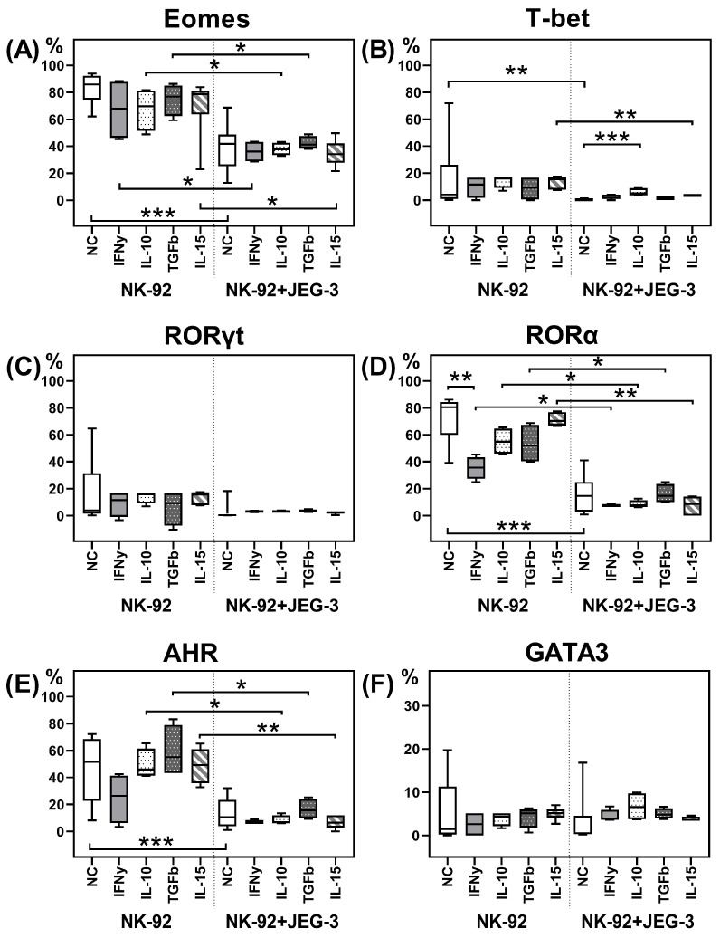
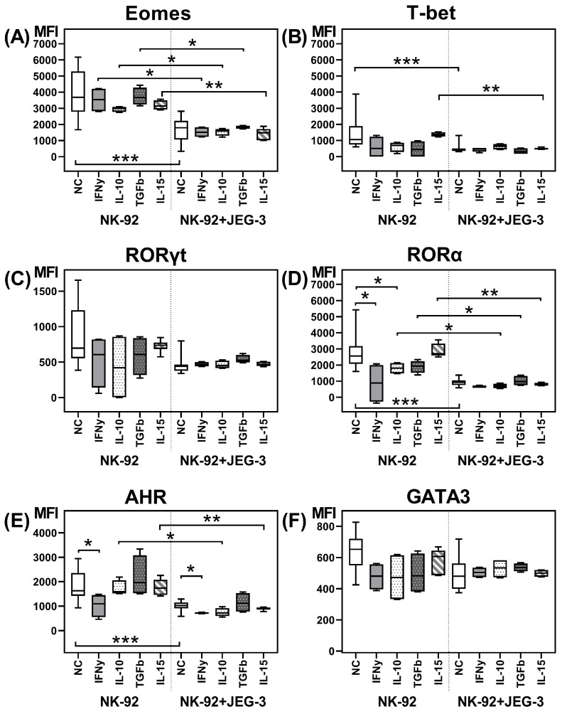
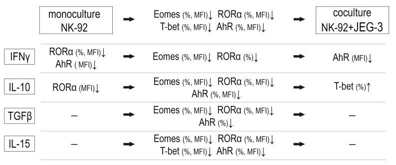
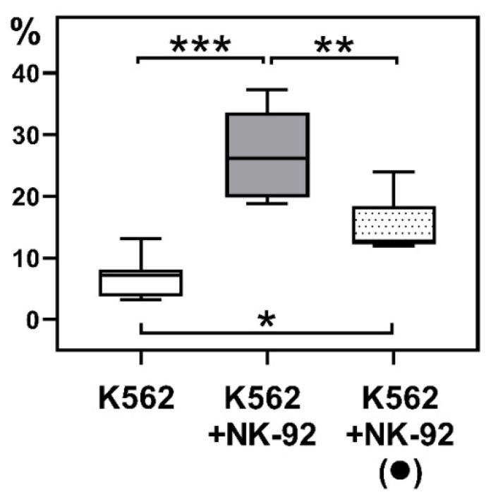
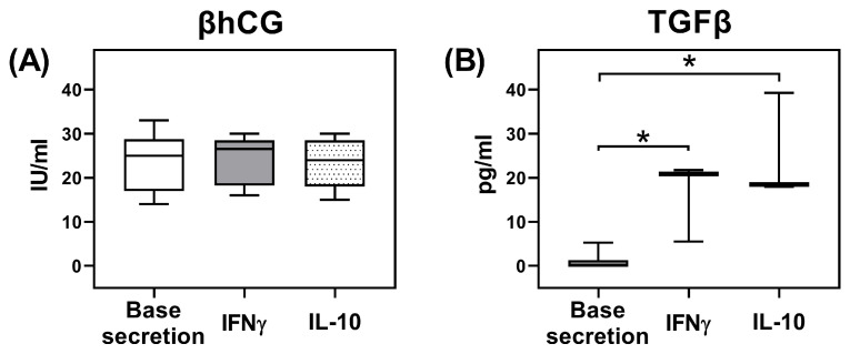
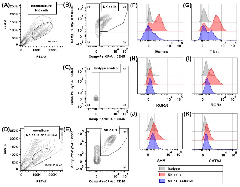
The uterine decidua contains NK cells differing in their characteristics from classical NK cells, as well as other populations of innate lymphoid cells (ILCs). ILC differentiation depends on the active transcription factors: ILC1 is characterized by T-bet expression, ILC2 is defined by RORα and GATA3, ILC3 expresses RORγt and AhR. We analyzed in vitro the expression of transcription factors by NK cells in the presence of trophoblast cells and cytokines and changes in NK cell cytotoxic activity. We used NK-92 and JEG-3 cell lines, which we cocultured in the presence of IFNγ, IL-10, IL-15, and TGFβ. Then, cells were treated with antibodies to AhR, Eomes, GATA-3, RORα, RORγt, and T-bet and were analyzed. We determined NK cell cytotoxicity towards K562 cells. To characterize the functional state of trophoblast cells, we estimated their secretion of TGFβ and βhCG. We showed that in the presence of trophoblasts, the expression of the classical NK cell transcription factors-Eomes, T-bet, as well as RORα, regulating ILC2 differentiation, and AhR, participating in NCR+ ILC3 formation-decreased in NK cells. RORγt expression typical for NCR- ILC3 remained unchanged. IFNγ inhibited AhR expression. IL-10 stimulated an increase in the number of T-bet+ ILC1-like cells. Both IL-10 and IFNγ suppressed RORα expression by NK cells and stimulated TGFβ secretion by trophoblasts. After coculture with trophoblast cells, NK cells reduced their cytotoxicity. These results indicated trophoblast cell influence on the acquisition of ILC1 and ILC3 characteristics by NK cells.

Keywords: AhR; Eomes; ILC; JEG-3; NK cells; NK-92; RoRα; T-bet; transcription factors; trophoblast.


Conflict of interest statement

The authors declare no conflicts of interest.

Figures

Figure 1
Figure 1
Relative number of NK-92 cells containing the transcription factors Eomes (A), T-bet (B), RORγt (C), RORα (D), AhR (E), and GATA3 (F), after cultivation without and in the presence of JEG-3 cells, without and in the presence of cytokines. NC—cultivation without cytokines. Statistical significance of differences: *—p < 0.05, **—p < 0.01, ***—p < 0.001.
Figure 2
Figure 2
Expression intensity of the transcription factors Eomes (A), T-bet (B), RORγt (C), RORα (D), AhR (E), and GATA3 (F) by NK-92 cells after cultivation without and in the presence of JEG-3 cells, without and in the presence of cytokines. NC—cultivation without cytokines. Statistical significance of differences: *—p < 0.05, **—p < 0.01, ***—p < 0.001.
Figure 3
Figure 3
The resultant scheme of NK-92 cell expression of transcription factors in the presence of trophoblast cells and cytokines. The decrease of the transcription factor—↓, the increase of the transcription factor—↑, the coculture comparison to the monoculture—horizontal arrow.
Figure 4
Figure 4
K-562 cell death after incubation with intact NK-92 cells (K562+NK-92) and NK-92 cells pre-cocultured with JEG-3 trophoblast cells (K562+NK-92 (●)). K562—baseline K562 cell death. Statistical significance of differences: *—p < 0.05, **—p < 0.01, ***—p < 0.001.
Figure 5
Figure 5
Concentration of βhCG (A) and TGFβ (B) secreted by JEG-3 trophoblast cells in the presence of IL-10 and IFNγ. Statistical significance of difference from the baseline secretion level: *—p < 0.05.
Figure 6
Figure 6
Transcription factor content in NK cells: gating strategy for NK-92 cells after cultivation without and in the presence of JEG-3 trophoblast cells. NK-92 cells in monoculture treated with antibodies to CD45 and CD56 in FSC/SSC coordinates (A), in CD45 PerCP/CD56 PE-Cy7 coordinates (B); NK-92 cells treated with isotype antibodies in PerCP/PE-Cy7 coordinates (C); NK-92 cells in coculture with JEG-3 trophoblast cells treated with antibodies to CD45 and CD56 in FSC/SSC coordinates (D), in CD45 PerCP/CD56 PE-Cy7 coordinates (E). Expression of transcription factors Eomes (F), T-bet (G), RORγt (H), RORα (I), AhR (J), and GATA3 (K) by NK-92 cells in monoculture (red) and coculture with trophoblast cells (blue).

Similar articles

References

    1. Stokic-Trtica V., Diefenbach A., Klose C.S.N. NK Cell Development in Times of Innate Lymphoid Cell Diversity. Front. Immunol. 2020;11:813. doi: 10.3389/fimmu.2020.00813. - DOI - PMC - PubMed
    1. Vacca P., Chiossone L., Mingari M.C., Moretta L. Heterogeneity of NK Cells and Other Innate Lymphoid Cells in Human and Murine Decidua. Front. Immunol. 2019;10:170. doi: 10.3389/fimmu.2019.00170. - DOI - PMC - PubMed
    1. Kiekens L., Van Loocke W., Taveirne S., Wahlen S., Persyn E., Van Ammel E., De Vos Z., Matthys P., Van Nieuwerburgh F., Taghon T., et al. T-BET and EOMES Accelerate and Enhance Functional Differentiation of Human Natural Killer Cells. Front. Immunol. 2021;12:732511. doi: 10.3389/fimmu.2021.732511. - DOI - PMC - PubMed
    1. Zhang J., Le Gras S., Pouxvielh K., Faure F., Fallone L., Kern N., Moreews M., Mathieu A.L., Schneider R., Marliac Q., et al. Sequential actions of EOMES and T-BET promote stepwise maturation of natural killer cells. Nat. Commun. 2021;12:5446. doi: 10.1038/s41467-021-25758-2. - DOI - PMC - PubMed
    1. Cortez V.S., Colonna M. Diversity and function of group 1 innate lymphoid cells. Immunol. Lett. 2016;179:19–24. doi: 10.1016/j.imlet.2016.07.005. - DOI - PMC - PubMed

Substances

LinkOut - more resources