Case Reports
. 2025 Apr 12;26(8):3670.
doi: 10.3390/ijms26083670.

Uncovering a Novel Pathogenic Mechanism of BCS1L in Mitochondrial Disorders: Insights from Functional Studies on the c.38A>G Variant

Affiliations
Case Reports

Uncovering a Novel Pathogenic Mechanism of BCS1L in Mitochondrial Disorders: Insights from Functional Studies on the c.38A>G Variant

Valeria Capaci et al. Int J Mol Sci. .

Abstract

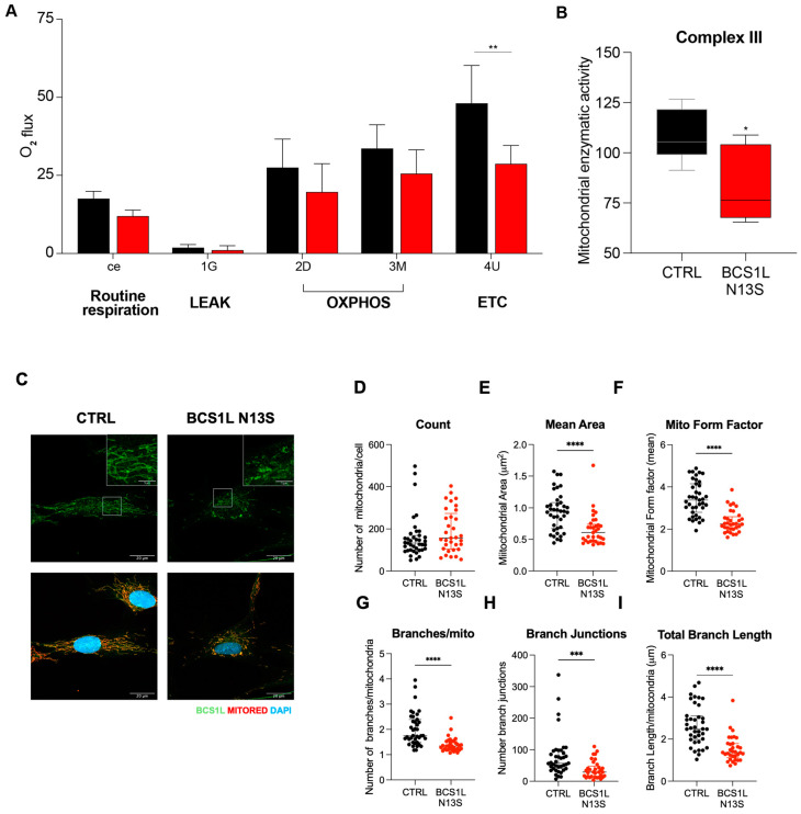
The BCS1L gene encodes a mitochondrial chaperone which inserts the Fe2S2 iron-sulfur Rieske protein into the nascent electron transfer complex III. Variants in the BCS1L gene are associated with a spectrum of mitochondrial disorders, ranging from mild to severe phenotypes. Björnstad syndrome, a milder condition, is characterized by sensorineural hearing loss (SNHL) and pili torti. More severe disorders include Complex III Deficiency, which leads to neuromuscular and metabolic dysfunctions with multi-systemic issues and Growth Retardation, Aminoaciduria, Cholestasis, Iron Overload, and Lactic Acidosis syndrome (GRACILE). The severity of these conditions varies depending on the specific BCS1L mutation and its impact on mitochondrial function. This study describes a 27-month-old child with SNHL, proximal renal tubular acidosis, woolly hypopigmented hair, developmental delay, and metabolic alterations. Genetic analysis revealed a homozygous BCS1L variant (c.38A>G, p.Asn13Ser), previously reported in a patient with a more severe phenotype that, however, was not functionally characterized. In this work, functional studies in a yeast model and patient-derived fibroblasts demonstrated that the variant impairs mitochondrial respiration, complex III activity (CIII), and also alters mitochondrial morphology in affected fibroblasts. Interestingly, we unveil a new possible mechanism of pathogenicity for BCS1L mutant protein. Since the interaction between BCS1L and CIII is increased, this suggests the formation of a BCS1L-containing nonfunctional preCIII unable to load RISP protein and complete CIII assembly. These findings support the pathogenicity of the BCS1L c.38A>G variant, suggesting altered interaction between the mutant BCS1L and CIII.

Keywords: BCS1L; assembly chaperone; complex III; electron transfer chain; mitochondrial disorder.


Conflict of interest statement

The authors declare no conflicts of interest.

Figures

Figure 1
Figure 1
Case report. (A) Pedigree of the family. The black symbol indicates the subject carrying the homozygous variant c.38A>G in BCS1L (NM_004328.5) gene. The parents are carriers of the same variant. (B) Schematic structure of BCS1L protein NP_001073335.1 indicating the aminoacidic substitution.
Figure 2
Figure 2
Heterologous complementation study in Saccharomyces cerevisiae using the human BCS1L cDNA. (A) Growth test: a bcs1Δ yeast strain harboring either the wild-type human BCS1L, the mutant allele (bcs1lN13S), or the empty vector were serially diluted and spotted on SC agar plates supplemented with the fermentable carbon source glucose (2%) or the non-fermentable carbon source glycerol (2%) and incubated at 36° C. (B) Respiratory activity: yeast strains were grown at 36 °C in SC medium supplemented with 0.6% glucose. Data are the mean of at least three values ± SD. The black bar indicates the wild-type strain; the grey bar indicates the strain carrying the alleged pathological mutation; the white bar indicates the null mutant strain. Statistical analysis was performed using the Mann–Whitney test: * p < 0.05; ** p < 0.01. (C) NADH-cytochrome c oxidoreductase (NCCR) activity: recorded on a mitochondrial-enriched fraction from yeast strains grown at 36° C in SC medium supplemented with 0.6% glucose. Data were normalized to the wild-type and represented as the mean of at least four values ± SD. Statistical analysis was performed using the Mann–Whitney test: * p < 0.05. (D) Iron accumulation: quantified in yeast strains grown up to the early stationary phase in SC medium supplemented with 0.6% glucose; 2 mM of ferrous sulfate was added. Statistical analysis was performed using the Mann–Whitney test: * p < 0.05.
Figure 3
Figure 3
Protein analyses in Saccharomyces cerevisiae. (A) Protein quantification: Representative Western blot on total protein extract using antibodies recognizing Bcs1l or Por1 as loading control; signals were first normalized to Por1 and then to the wild-type signal to which the value 1.0 was assigned. Densitometric analysis is reported in the histogram below the blot and was performed on at least three independent blots using Image Lab Software v6.1 (Bio-Rad, Hercules, CA, USA). Statistical analysis was performed using the Mann–Whitney test: * p < 0.05. (B) Proteins localization: Western blot on denaturing SDS–PAGE of mitochondrial (M) and cytosolic (C) proteins from bcs1Δ strains expressing wild-type BCS1L or bcs1lN13S. An antibody against Bcs1l was used to detect Bcs1l; antibodies against Por1 and Pgk1 were used as markers of the mitochondrial and cytosolic fractions, respectively.
Figure 4
Figure 4
BCS1L N13S affects mitochondrial metabolic activity and morphology in human dermal fibroblasts (HDFs). (A) Specific O2 flux and (B) enzymatic activity of CIII normalized to the activity of citrate synthase in HDF primary cells, in either the control or mutated BCS1L N13S. Graph represents the mean ± SEM of N ≥ 3 independent experiments. (C) Representative images of immunofluorescence analysis of BCS1L (green) in in HDF primary cells in either the control or mutated BCS1L N13S. Mitochondria were counterstained with mitochondrial marker MITORED (red) and nuclei DAPI staining (blue) Scale bar 20 mm (inset scale bar 5 mm). (DI) Graphs show mitochondrial count, area, forma factor and branches/mitochondria, branches junction, and length calculated by Mitochondrial Analyzer image J Plugin. (n = 35 cells for each condition). p value (* p < 0.05, ** p < 0.01, *** p < 0.001, **** p < 0.0001) was calculated by paired two-tailed Mann–Whitney test.
Figure 5
Figure 5
BCS1L N13S affects CIII assembly. (A) Representative images of Western blot analysis of the BCS1L in HDF primary cells in the either control or mutated BCS1L N13S. Graph shows the means ± SEM of three independent experiments, where the relative expression level of the proteins was obtained by densitometry measures and normalized to HSP90. (B) Representative images of PLA between BCS1L and CIII in HDF cells. Right: Graph showing the number of dots/cell. (n ≥ 100 cells/condition). (C) Representative images of BN-PAGE of digitized HDF primary cells in either the control or mutated BCS1L N13S followed by Western blot analysis of RISP, BCS1L, and Cyt-b proteins. Right: Graph shows the means ± SEM of three independent experiments, where the relative expression level of the proteins was obtained by densitometry measures and normalized to Cyt-B. p value (* p < 0.05, ** p < 0.01, **** p <0.0001) was calculated by an unpaired two-tailed Mann–Whitney test.

References

    1. Fernandez-Vizarra E., Zeviani M. Mitochondrial Disorders of the OXPHOS System. FEBS Lett. 2021;595:1062–1106. doi: 10.1002/1873-3468.13995. - DOI - PubMed
    1. Gusic M., Prokisch H. Genetic Basis of Mitochondrial Diseases. FEBS Lett. 2021;595:1132–1158. doi: 10.1002/1873-3468.14068. - DOI - PubMed
    1. De Vivo D.C., DiMauro S. Mitochondrial Diseases. 6th ed. Elsevier Inc.; Amsterdam, The Netherlands: 2017.
    1. Morán M., Marín-Buera L., Gil-Borlado M.C., Rivera H., Blázquez A., Seneca S., Vázquez-López M., Arenas J., Martín M.A., Ugalde C. Cellular Pathophysiological Consequences of BCS1L Mutations in Mitochondrial Complex III Enzyme Deficiency. Hum. Mutat. 2010;31:930–941. doi: 10.1002/humu.21294. - DOI - PubMed
    1. Zhan J., Zeher A., Huang R., Tang W.K., Jenkins L.M., Xia D. Conformations of Bcs1L Undergoing ATP Hydrolysis Suggest a Concerted Translocation Mechanism for Folded Iron-Sulfur Protein Substrate. Nat. Commun. 2024;15:4655. doi: 10.1038/s41467-024-49029-y. - DOI - PMC - PubMed

Publication types

MeSH terms

Substances

LinkOut - more resources