. 2025 Apr 14;26(8):3709.
doi: 10.3390/ijms26083709.

Conditional Overexpression of Neuritin in Supporting Cell Protects Cochlear Hair Cell and Delays Age-Related Hearing Loss by Enhancing Autophagy

Affiliations

Conditional Overexpression of Neuritin in Supporting Cell Protects Cochlear Hair Cell and Delays Age-Related Hearing Loss by Enhancing Autophagy

Shanshan Wang et al. Int J Mol Sci. .

Abstract

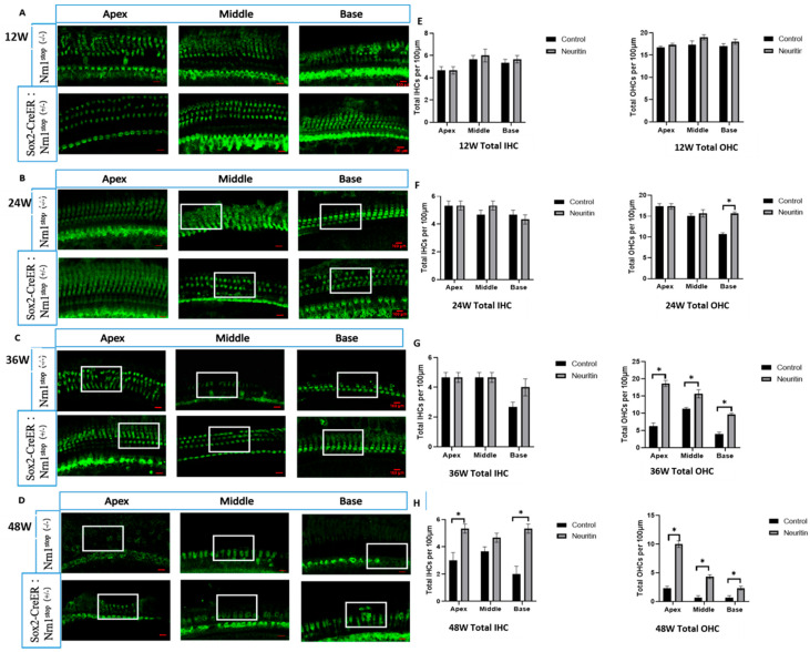
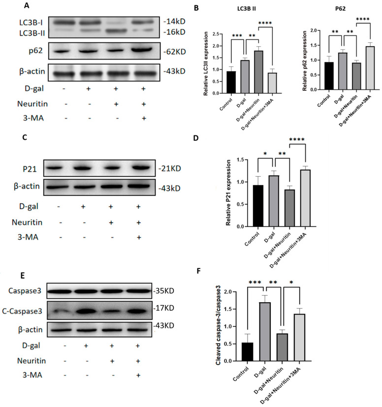
Age-related hearing loss (ARHL) is a highly prevalent, burdensome sensorineural hearing loss closely associated with impaired autophagic influx. Our previous studies revealed that neuritin, a neurotrophic factor primarily expressed in the central nervous system, could alleviate drug-induced damages in hair cells (HCs) and spiral ganglion neurons. However, its effects on ARHL and whether these effects are closely related to autophagy remain unclear. Using the Nrn1 knock-in mice and cultured cochlear basilar membrane (CBM) of the neonatal mouse, we show that neuritin could restore aging-associated hearing loss and alleviate senescence-associated damage in the cochlea. Overexpression of neuritin in support cells (SCs) alleviates the loss of cochlear HCs and nerve fibers, reducing the damage to spiral ganglion neurons and the shifts in ABR's high-frequency threshold. Furthermore, conditional overexpression of neuritin in SCs improves autophagic influx by upregulating the expression of microtubule-associated protein 1 light chain 3 type B (LCB3) protein and downregulating the expression of p21 protein. In cultured neonatal mouse CBM, neuritin administration significantly inhibits D-galactose-induced HC loss, cellular apoptosis, and ROS production and promotes autophagic influx. These effects were weakened when the autophagy inhibitor 3-MA was added. In summary, our results confirm the therapeutic potential of neuritin treatment for ARHL.

Keywords: LCB3; P21; age-related hearing loss; autophagy; hair cells; neuritin.


Conflict of interest statement

The authors declare no conflicts of interest.

Figures

Figure 1
Figure 1
The cellular localization and conditional overexpression of neuritin in the cochlea of mice. (A,B) The expression levels and cellular location of neuritin protein in the wild-type mouse cochlea (5-week-old). Green represents anti-EGFP labeling neuritin; red represents anti-Sox2 labeling supporting cells (SCs) (scale bar = 50 µm). (C) Schematic timeline of conditional overexpression and detection time of neuritin protein in Sox2CreER/+Neuritinstop/stop mouse (neuritin). (D,E) Western blots and quantitative analysis of neuritin expression levels in the cochlear of Nrn1stop mouse (control) and Sox2CreER/+Neuritinstop/stop mouse (neuritin) at different time points. Statistical analyses were performed by Student’s t-test; *, p < 0.05.
Figure 2
Figure 2
Conditional overexpression of neuritin in supporting cells (SCs) ameliorates aging-induced hair cell (HC) damage in the cochlear. (AD) The HCs in the apex, middle, and base loops of the cochlear basilar membrane in Sox2CreER/+ Nrn1stop (+/-) mice (neuritin) and Nrn1stop (-/-) mice (control) at the time points of 12, 24, 36, and 48 weeks; the white rectangles show the typical zones of HCs in the basal, middle, and apical regions of the cochlear basilar membrane. Green represents anti-Myo7A labeling HCs (scale bar = 100 μm). (EH) Quantity analysis of HCs in the apex, middle, and base loops of the cochlear basement membrane in mice with different ages. IHC, inner hair cell; OHC, outer hair cell. Statistical analyses were performed by Student’s t-test; *, p < 0.05.
Figure 3
Figure 3
Conditional overexpression of neuritin in supporting cells (SCs) reduces aging-induced nerve fiber damages in the cochlear. (AD) The nerve fibers in the apex, middle, and base loops of the cochlear basilar membrane in Sox2CreER/+ Nrn1stop (+/-) mice (neuritin) and Nrn1stop (-/-) mice (control) at the time points of 12, 24, 36, and 48 weeks. Red represents NF200-positive nerve fibers; scale bar = 100 μm. Yellow rectangles show the typical zone of nerve fibers in the apex, middle, and base loops of the cochlear basilar membrane. (EH) Quantification of nerve fibers in the apex, middle, and base loops of cochlear basement membrane in mice at different time points (n = 6 for each group). Statistical analyses were performed by Student’s t-test; *, p < 0.05.
Figure 4
Figure 4
Conditional overexpression of neuritin in supporting cells (SCs) reduces aging-induced spiral ganglion neuron (SGN) damage in the cochlear. (AD) HE staining results of spiral ganglion neurons in the apex, middle, and base of the cochlear basilar membrane in Sox2CreER/+ Nrn1stop (+/-) mice (neuritin) and Nrn1stop (-/-) mice (control) at the time points of 12, 24, 36, and 48 weeks observed by H&E staining; scale bar = 100 μm. Yellow rectangles show the typical zone of SGNs in the cochlear basilar membrane’s apex, middle, and base loops. (EH) Quantification of SGN density in the apex, middle, and base loops of the cochlear basement membrane in mice with different ages (n = 6 for each group). Statistical analyses were performed by Student’s t-test; *, p < 0.05.
Figure 5
Figure 5
Neuritin overexpression rescues high-frequency hearing threshold shifts in mice with age-related hearing loss. (AC) The hearing thresholds elicited by click or high-frequency (8000 Hz, 16,000 Hz, and 32,000 Hz) stimuli in Sox2CreER/+ Nrn1stop (+/-) mice (neuritin) and Nrn1stop (-/-) mice (control) at the time points of 12, 24, 36, and 48 weeks. (D) Line chart of ABR hearing threshold shifts at different frequencies in Sox2 CreER/+ Nrn1stop (+/-) mice (neuritin) and Nrn1stop (-/-) mice (control) at the time points of 12, 24, 36, and 48 weeks. Statistical analyses were performed by Student’s t-test; *, p < 0.05.
Figure 6
Figure 6
Conditional overexpression of neuritin in supporting cells (SCs) improves autophagic influx in the cochlea in mice with age-related hearing loss. (A,B) The changes in LC3B and p62/SQSTM1 protein expression levels in the cochlear of Sox2CreER/+ Nrn1stop (+/-) mice (neuritin) and Nrn1stop (-/-) mice (control) at different time points (12, 24, 36, and 48 weeks) detected by Western blot analysis. (C) Quantitative analysis of LC3B and p62/SQSTM1 protein expression levels in the cochlear of Sox2CreER/+ Nrn1stop (+/-) mice (neuritin) and Nrn1stop (-/-) mice (control) (n = 6 per group). Statistical analyses were performed using Student’s t-test. *, p < 0.05; **, p < 0.01; ***, p < 0.001. (D) Fluorescence immunohistopathologic evaluation of the localization and expression levels of LC3B protein in cochlea of Sox2CreER/+ Nrn1stop (+/-) mice (neuritin) and Nrn1stop (-/-) mice (control). Green represents fluorescently labeled LC3B protein; red represents phalloidin-stained hair cells (HCs); blue is DAPI staining of cell nuclei and white rectangles represent phalloidin staining hair cells. DAPI, 4′,6-diamidino-2-phenylindole. Scale bar = 50 μm. Statistical analyses were performed by Student’s t-test; *, p < 0.05; **, p < 0.01; ***, p < 0.001.
Figure 7
Figure 7
Establishment and characterization of senescence-associated damages in the cultured neonatal mouse cochlear basilar membranes (CBMs). (A,B) Senescence-associated D-galactosidase (SA-D-gal) staining and the relative levels of D-gal-positive staining cells in cultured CBMs of neonatal mice treated with different dosages of D-gal (20 mg/mL, 40 mg/mL, and 60 mg/mL) (n = 6 for each group). Scale bar = 50 μm. Statistical analyses were performed by one-way ANOVA followed by the Bonferroni post hoc test. *, p < 0.05; **, p< 0.01; ***, p < 0.001; ****, p < 0.0001. (C) The relative reactive oxidative species (ROS) levels detected by DCFH-DA in cultured CBMs of neonatal mice treated with different dosages of D-gal (20 mg/mL, 40 mg/mL, and 60 mg/mL) (n = 6 for each group). The red border implies the dose of D-gal (40 mg/mL) used in the subsequent experiments. (D,E) Fluorescence immunostaining and quantitative analysis of p21 protein expression levels in the apex, middle, and base of cultured CBMs in D-gal-treated (40 mg/mL) and control groups (n = 6 per group). Scale bar = 20 μm. Green represents fluorescently labeled p21 protein; red represents phalloidin-stained hair cells (HCs); purple represents DAPI-stained nucleus. The dotted rectangle represents the enlarged area of the cross-section for each condition. (F,G) Fluorescence immunohistopathologic evaluation and quantitative analysis of the expression level of TUNEL-positive cells (green) in D-gal-treated (40 mg/mL) and control groups (n = 6 for each group); scale bar = 50 μm. DAPI, 4′,6-diamidino-2-phenylindole.
Figure 8
Figure 8
Neuritin ameliorates d-galactose-induced senescence damage in the cultured neonatal mouse cochlear basilar membranes (CBMs). (A) Representative images of SA-β-Gal staining (blue) in the cultured neonatal mouse cochlear basilar membranes (CBMs) from different groups (control, 40 mg/mL D-gal, and 40 mg/mL D-gal + 16 µg/mL neuritin). Scale bar = 50 μm. (B) The relative levels of SA-β-gal-positive staining cells in the different groups. Data are shown as mean ± SD (n = 6 in each group). (C,D) Fluorescence immunohistopathologic evaluation and quantitative analysis of the expression levels of P21 protein in different groups (n = 6 in each group); scale bar = 20 μm. Green represents fluorescently labeled p21 protein; red represents phalloidin-stained hair cells (HCs); purple represents DAPI-stained nucleus. (E) ROS levels detected by DCFH-DA in different groups (n = 6 per group). The dotted rectangle represents the enlarged area of the cross-section for each condition. Statistical analyses were performed by one-way ANOVA followed by LSD post hoc test. *, p < 0.05; **, p< 0.01; ***, p < 0.001; ****, p < 0.0001; ns, no statistical significance. DAPI, 4′,6-diamidino-2-phenylindole.
Figure 9
Figure 9
Neuritin ameliorates d-gal-induced damages in cultured neonatal mouse cochlear basilar membranes (CBMs) by enhancing autophagic influx. (A,B) Western blot and quantitative analysis of the expression levels of LC3BI/II, p62/SQSTM1 proteins in the CBMs of different groups (control, d-gal, neuritin, and neuritin +d-gal) (n = 6 per group). (C,D) Fluorescence immunohistopathologic evaluation and quantitative analysis of the expression level of LC3B protein in the CBMs of different groups; scale bar = 50 μm. Green represents fluorescently labeled LC3B protein, and red represents phalloidin-stained hair cells (HCs). (E,F) Fluorescence immunohistopathologic evaluation and quantitative analysis of the expression levels of TUNEL-positive cells in the CBMs in different groups; scale bar = 50 μm. Green represents fluorescently labeled TUNEL-positive cells. (G,H) Fluorescence immunohistopathologic evaluation and quantitative analysis of mitochondrial reactive oxidative species (ROS) levels evaluated by MitoSOX red staining in different groups. Red represents MitoSOX-stained mitochondria in hair cells (HCs); purple represents DAPI-stained nucleus. Scale bar = 20 μm. The dotted rectangle represents the enlarged area of the cross-section for each condition. (I,J) Western blot and quantitative analysis of the expression level of caspase3 and C-caspase3 proteins in the CBMs of different groups (n = 6 in each group). Statistical analyses were performed by one-way ANOVA followed by LSD post hoc test. *, p < 0.05; **, p< 0.01; ***, p < 0.001; ****, p < 0.0001; ns, no statistical significance.
Figure 10
Figure 10
The protective effects of neuritin against d-gal-induced damages in cultured neonatal mouse cochlear basilar membranes (CBMs) were weakened by the administration of autophagy inhibitor 3-MA. Western blot and quantitative analysis of the expression level of LC3BI/II, p62/SQSTM1 (A,B), p21 (C,D), and caspase3 and C-caspase3 proteins (E,F) in the cultured neonatal mouse cochlear basilar membranes (CBMs) receiving different treatments (control, 40 mg/mL D-gal, 40 mg/mL D-gal + 16 μg/mL neuritin, and 40 mg/mL D-gal + 16 μg/mL neuritin + 2 nmol/mL 3-MA; n = 6 per group). Statistical analyses were performed by one-way ANOVA followed by LSD post hoc test. *, p < 0.05; **, p< 0.01; ***, p < 0.001; ****, p < 0.0001.

Similar articles

References

    1. Lin F.R. Age-Related Hearing Loss. N. Engl. J. Med. 2024;390:1505–1512. doi: 10.1056/NEJMcp2306778. - DOI - PubMed
    1. Cunningham L.L., Tucci D.L. Hearing Loss in Adults. N. Engl. J. Med. 2017;377:2465–2473. doi: 10.1056/NEJMra1616601. - DOI - PMC - PubMed
    1. Liu H., Giffen K.P., Chen L., Henderson H.J., Cao T.A., Kozeny G.A., Beisel K.W., Li Y., He D.Z. Molecular and Cytological Profiling of Biological Aging of Mouse Cochlear Inner and Outer Hair Cells. Cell Rep. 2022;39:110665. doi: 10.1016/j.celrep.2022.110665. - DOI - PMC - PubMed
    1. Sun Z., Cheng Z., Gong N., Xu Z., Jin C., Wu H., Tao Y. Neural Presbycusis at Ultra-High Frequency in Aged Common Marmosets and Rhesus Monkeys. Aging. 2021;13:12587–12606. doi: 10.18632/aging.202936. - DOI - PMC - PubMed
    1. Man J., Chen H., Zhang T., Yin X., Yang X., Lu M. Global, Regional, and National Burden of Age-Related Hearing Loss from 1990 to 2019. Aging. 2021;13:25944–25959. doi: 10.18632/aging.203782. - DOI - PMC - PubMed

Substances

LinkOut - more resources