. 2025 Apr 15;26(8):3734.
doi: 10.3390/ijms26083734.

Single-Cell Transcriptomic Analysis of the Potential Mechanisms of Follicular Development in Stra8-Deficient Mice

Affiliations

Single-Cell Transcriptomic Analysis of the Potential Mechanisms of Follicular Development in Stra8-Deficient Mice

Han Wang et al. Int J Mol Sci. .

Abstract

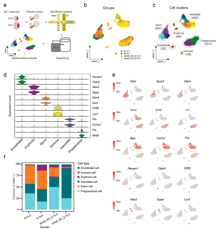
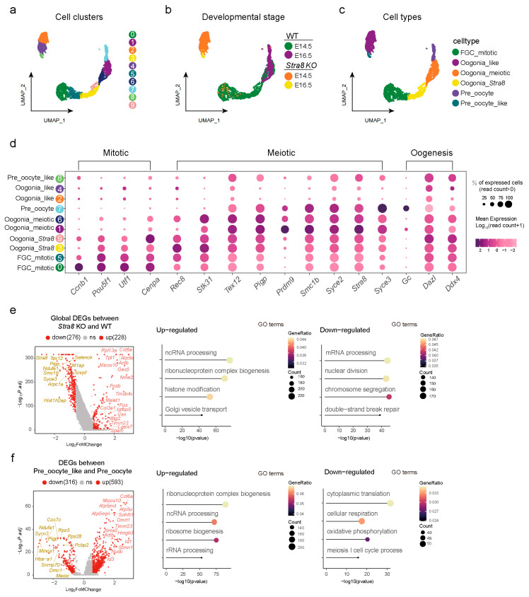
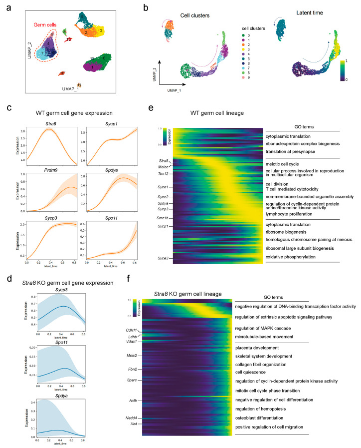
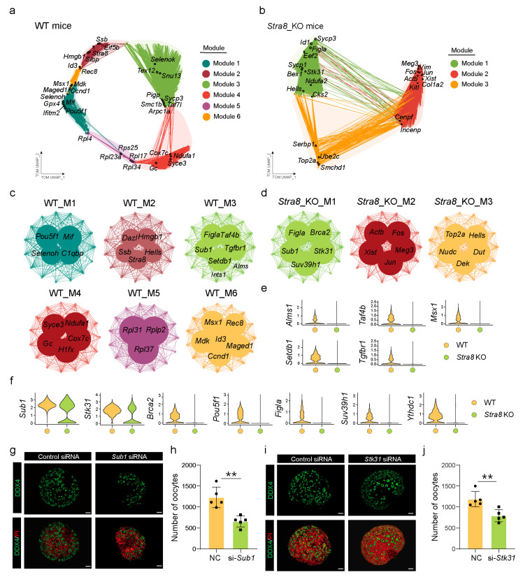
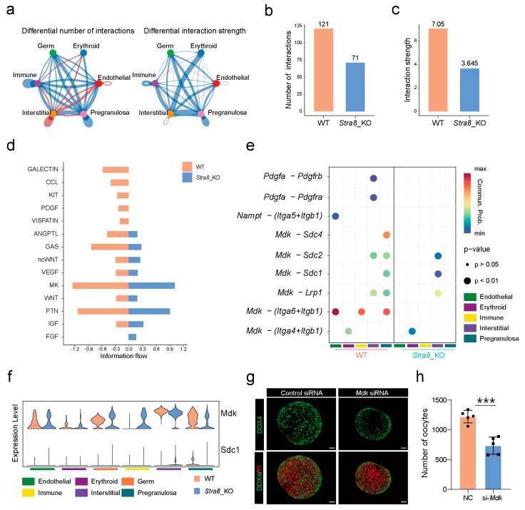
Follicle development is a critical process in mammalian reproduction, with significant implications for ovarian reserve and fertility. Stra8 is a known key factor regulating the initiation of meiosis; however, oocyte-like cells still appear in Stra8-deficient mice. Nevertheless, the underlying mechanism remains unclear and requires further investigation. Therefore, we used single-cell RNA sequencing to construct a comprehensive transcriptional atlas of ovarian cells from both wild-type and Stra8-deficient mice at embryonic stages E14.5 and E16.5. With stringent quality control, we obtained a total of 14,755 single cells of six major cell types. A further fine-scale analysis of the germ cell clusters revealed notable heterogeneity between wild-type and Stra8-deficient mice. Compared to the wild-type mice, the deficiency in Stra8 led to the downregulation of meiosis-related genes (e.g., Pigp, Tex12, and Sycp3), and the upregulation of apoptosis-related genes (e.g., Fos, Jun, and Actb), thereby hindering the meiotic process. Notably, we observed that, following Stra8 deficiency, the expression levels of Sub1 and Stk31 remained elevated at this stage. Furthermore, an RNA interference analysis confirmed the potential role of these genes as regulatory factors in the formation of primordial follicle-like cells. Additionally, Stra8 deficiency disrupted the signaling between germ cells and pregranulosa cells that is mediated by Mdk-Sdc1, leading to the abnormal expression of the PI3K/AKT signaling pathway. Together, these results shed light on the molecular processes governing germ cell differentiation and folliculogenesis, emphasizing the complex role of Stra8 in ovarian function.

Keywords: Stra8; oogenesis; single-cell RNA sequencing.


Conflict of interest statement

The authors declare no conflicts of interest.

Figures

Figure 1
Figure 1
Construction of single-cell transcriptional atlases of ovaries in WT and Stra8-deficient mice. (a) Schematic diagram of genital ridge collection in WT and Stra8-deficient mice at different time points and scRNA-seq analysis. (b) Distribution of ovarian cells in E14.5, E16.5, Stra8-deficient E14.5 and Stra8-deficient E16.5 groups. (c) Distribution of ovarian cells in the UMAP plot. The numbers and colors in the figure represent different cell clusters identified by Seurat. (d) Violin diagram of signature genes in different cell types. (e) Distribution of signature genes across different cell types in the UMAP plot. (f) The proportion of different cell types in the four groups.
Figure 2
Figure 2
Analysis of differentially expressed genes (DEGs) among germ cell subsets. (a) Distribution of germ cell subpopulations in the UMAP plot. The numbers in the figure represent different cell subclusters identified by Seurat. (b) UMAP diagram at different developmental stages (E14.5, E16.5, Stra8-deficient E14.5, and Stra8-deficient E16.5). (c) Annotations on germ cell subpopulations (FGC_mitotic, Oogonia_Stra8, Oogonia_meiotic, Pre_oocyte, Oogonia_like, and Pre_oocyte_like). (d) Dot plot showing germ cell cluster-specifically expressed genes. (e) Left panel: volcano plot illustrating global DEGs between Stra8-deficient and WT groups (left); right panel: corresponding GO enrichment results of up-regulated and down-regulated genes (right). (f) Left panel: volcano plot illustrating DEGs between Pre_oocyte_like and Pre_oocyte cell clusters (left); right panel: corresponding GO enrichment results of up-regulated and down-regulated genes (right).
Figure 3
Figure 3
Differential analysis of germ cell fate trajectories between WT and Stra8-deficient mice. (a) Distribution of three clusters of germ cells in the UMAP plot. The numbers in the figure represent different cell subclusters identified by Seurat. (b) Pseudo-temporal trajectory of germ cell development (left) and distribution of germ cells in latent time (right). (c) Pseudo-temporal developmental trajectories of representative meiotic genes in the WT group. (d) Pseudo-temporal developmental trajectories of representative meiotic genes in Stra8-deficient group. (e) Expression trend of the top 50 genes in latent time and GO term enrichment in the WT group. (f) Expression trend of the top 50 genes in latent time and GO term enrichment in the Stra8-deficient group.
Figure 4
Figure 4
Construction of gene co-expression networks to identify key genes involved in follicle formation. (a) UMAP plot of the co-expression network of germ cells from WT mice. (b) UMAP plot of the gene co-expression network in germ cells from Stra8-deficient mice. (c) Gene co-expression network diagram of different modules in the WT group. (d) Gene co-expression network diagram of different modules in the Stra8-deficient group. (e) Violin plots of transcripts that disappear after Stra8 elimination. (f) Violin plots showing genes with decreased expression following Stra8 deficiency. (g) Representative images of ovaries in the control and Sub1 siRNA groups cultured for six days. Germ cells are stained with DDX4 (green) and nuclei are stained with PI (red). (h) The number of oocytes in the control and Sub1 siRNA groups. (i) Representative images of ovaries in the control and Stk31 siRNA groups cultured for six days. Germ cells are stained with DDX4 (green) and nuclei are stained with PI (red). (j) The number of oocytes in the control and Stk31 siRNA groups. The percentage of each group is presented as the mean ± SD. All experiments were repeated at least three times (** p < 0.01).
Figure 5
Figure 5
Decreased Mdk–Sdc1 ligand–receptor signaling affects primordial follicle formation in Stra8-deficient mice. (a) Ovarian cell signaling network. (b) The number of interactions between different cell types. (c) The interaction strength between different cell types. (d) The information flow of WT and Stra8-deficient groups. (e) The ligand–receptor pairs between germ cells and other somatic cells. (f) Expression of ligands and receptors between WT and Stra8-deficient groups. (g) Representative images of ovaries in the control and Mdk–siRNA groups cultured for six days. Germ cells are stained with DDX4 (green) and nuclei are stained with PI (red). (h) The number of oocytes in the control group and Mdk–siRNA group. The percentage of each group is presented as the mean ± SD. All experiments were repeated at least three times (*** p < 0.001).

Similar articles

References

    1. Cai H., Liu B., Wang H., Sun G., Feng L., Chen Z., Zhou J., Zhang J., Zhang T., He M., et al. SP1 governs primordial folliculogenesis by regulating pregranulosa cell development in mice. J. Mol. Cell Biol. 2020;12:230–244. doi: 10.1093/jmcb/mjz059. - DOI - PMC - PubMed
    1. Li R., Albertini D.F. The road to maturation: Somatic cell interaction and self-organization of the mammalian oocyte. Nat. Rev. Mol. Cell Biol. 2013;14:141–152. doi: 10.1038/nrm3531. - DOI - PubMed
    1. Fan X., Bialecka M., Moustakas I., Lam E., Torrens-Juaneda V., Borggreven N.V., Trouw L., Louwe L.A., Pilgram G.S.K., Mei H., et al. Single-cell reconstruction of follicular remodeling in the human adult ovary. Nat. Commun. 2019;10:3164. doi: 10.1038/s41467-019-11036-9. - DOI - PMC - PubMed
    1. Peters H., Byskov A.G., Himelstein-Braw R., Faber M. Follicular growth: The basic event in the mouse and human ovary. J. Reprod. Fertil. 1975;45:559–566. doi: 10.1530/jrf.0.0450559. - DOI - PubMed
    1. Tingen C., Kim A., Woodruff T.K. The primordial pool of follicles and nest breakdown in mammalian ovaries. Mol. Hum. Reprod. 2009;15:795–803. doi: 10.1093/molehr/gap073. - DOI - PMC - PubMed

MeSH terms

Substances

LinkOut - more resources