. 2025 May 1;5(5):881-895.
doi: 10.1158/2767-9764.CRC-24-0025.

P4HA1 Mediates Hypoxia-Induced Invasion in Human Pancreatic Cancer Organoids

Affiliations

P4HA1 Mediates Hypoxia-Induced Invasion in Human Pancreatic Cancer Organoids

Bernat Navarro-Serer et al. Cancer Res Commun. .

Abstract

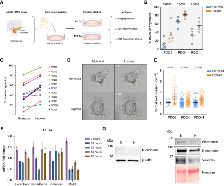
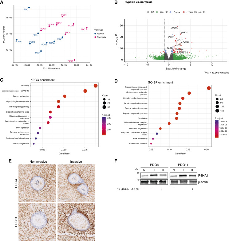
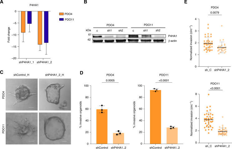
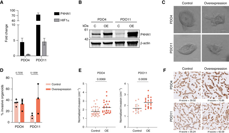
Pancreatic ductal adenocarcinoma (PDAC) is a highly aggressive malignancy with dismal prognosis. PDAC develops in a hypoxic environment in which cells adapt and activate processes to allow survival under low-oxygen conditions, some of which may enhance the ability of cancer cells to invade locally or metastasize distantly. Using human PDAC organoids, we determined that hypoxia consistently enhanced invasion across 11 patient-derived models. Using RNA sequencing of hypoxic invasive organoids (compared with matched invasive normoxic organoids from the same patients), we identified prolyl 4-hydroxylase subunit alpha 1 (P4HA1) as a potential regulator of PDAC invasion in hypoxia. Leveraging publicly available datasets from human tissue, we determined that P4HA1 is more highly expressed in PDAC compared with normal pancreatic tissue and that high P4HA1 expression correlates with poor patient prognosis. To further interrogate the role of P4HA1 in invasion of hypoxic patient-derived organoids, we quantified invasion in organoids modified to knockdown or overexpress P4HA1, demonstrating that P4HA1 is necessary for hypoxia-enhanced invasion and sufficient to increase invasion in normoxia in PDAC organoids. Our results identify P4HA1 as a driver of PDAC organoid invasion in hypoxia.

Significance: This study demonstrates that hypoxia increases invasion across a cohort of human pancreatic cancer organoids and identifies the collagen-modifying enzyme P4HA1 as a driver of hypoxia-enhanced invasion. These results characterize a molecular mechanism by which the microenvironment alters tumor cell behavior and underscore new strategies to inhibit invasion.


Conflict of interest statement

M.F. Wissler reports grants from NIH/NCI during the conduct of the study. V. Wang reports grants from NIH T32CA153952 during the conduct of the study. H. Knutsdottir reports other support from deCode genetics outside the submitted work. J.S. Bader reports grants from NIH and Jayne Koskinas Ted Giovanis Foundation during the conduct of the study, as well as other support from Opentrons Labworks and Dextera Biosciences outside the submitted work. E.J. Fertig reports grants from NIH/NCI, Lustgarten Foundation, and Break Through Cancer during the conduct of the study, as well as personal fees from ResistanceBio/Viosera Therapeutics and Mestag Therapeutics and grants from AbbVie and Roche/Genentech outside the submitted work. D.M. Gilkes reports grants from Johns Hopkins University during the conduct of the study. L.D. Wood reports grants from NIH, Sol Goldman Pancreatic Cancer Research Center, Buffone Family Gastrointestinal Cancer Research Fund, Allegheny Health Network-Johns Hopkins Cancer Research Fund, American Cancer Society, American Association for Cancer Research, Emerson Collective Cancer Research Fund, Rolfe Pancreatic Cancer Foundation, Joseph C Monastra Foundation, The Gerald O Mann Charitable Foundation, and Dennis Troper and Susan Wojcicki during the conduct of the study. No disclosures were reported by the other authors.

Figures

Figure 1
Figure 1
Hypoxia increases invasion in patient-derived PDAC organoids. A, Schematic of PDAC organoid culture generation, collection, study, and analysis timeline. B, Effect of hypoxia on invasion in PDO1, PDO4, and PDO11 organoids. Percent invasion represents a mean of minimum three biological replicates with SD. Each biological replicate n > 30 organoids (P = 0.015, P = 0.0032, and P = 0.1636, respectively, t test). C, Fold change of PDAC PDO sample invasion in normoxia and hypoxia. D, Representative images of PDO4 invasive organoids in normoxia and hypoxia. Scale bars, 100 μm. E, Inverse circularity scores of normoxic and hypoxic PDO1 (n = 15 and n = 29, respectively; P = 0.0122, t test), PDO4 (both n = 71; P = 0.0025, t test), and PDO11 (n = 44 and n = 54, respectively; P = 0.0024, t test) invasive organoids. F, Expression level of EMT markers (E-cadherin, N-cadherin, vimentin, and SNAIL) in PDO4 measured by qRT-PCR at multiple time points. Fold changes, expressed relative to normoxic control samples, represent a mean of three technical replicates with SD. G, Western blot analysis of EMT markers (fibronectin, E-cadherin, N-cadherin, and vimentin) in PDO4 in normoxia and hypoxia. H, hypoxia; N, normoxia.
Figure 2
Figure 2
Comparison of transcriptomes and molecular pathways of invasive normoxic and hypoxic organoids. A, Principal component analysis plot of gene expression in hypoxic invasive organoid samples (n = 8). PC, principal component. B, Volcano plot of differentially expressed genes between normoxic and hypoxic invasive organoids. FC, fold change. C, Pathway analysis of normoxic and hypoxic invasive organoids using KEGG processes. D, Pathway analysis of normoxic and hypoxic invasive organoids using GO Biological Processes (GO-BP). E, IHC for hydroxyproline surrounding invasive and noninvasive organoids in PDO4 and PDO11. F, Western blot analysis assessing P4HA1 levels in 2D cultures prepared from PDO4 and PDO11 organoids 24 hours after treatment with a HIF1a inhibitor (10 μmol/L PX-478). H, hypoxia; N, normoxia.
Figure 3
Figure 3
P4HA1 expression is increased in PDAC and is clinically relevant. A, Gene set enrichment analysis showing a correlation between a high P4HA1 expression and the expression of genes in a hypoxia gene signature (P = 3 × 10⁻²⁰). B, scRNA-seq analysis shows increased expression levels of P4HA1 in human pancreatic tumors compared with normal tissue (P = 9 × 10⁻¹⁶). C, Protein expression of P4HA1 in PDAC using the Clinical Proteomic Tumor Analysis Consortium database, accessed through the University of Alabama at Birmingham Cancer data analysis Portal. D, Kaplan–Meier analysis of patients with PDAC shows increased risk of death associated with a high expression of P4HA1. Plot generated using KMPlot. The median survival time for low expression and high expression cohorts is 72.73 and 18.17 months, respectively (HR = 2.4; 95% confidence interval, 1.42–4.05; P = 0.00077).
Figure 4
Figure 4
Knockdown of P4HA1 decreases invasion in PDAC PDOs in hypoxia. A, Expression level of P4HA1 measured by qRT-PCR. Fold changes, expressed relative to the modified control samples, represent a mean of three technical replicates with SDs (PDO4 P = 0.0547, t test; PDO6 P = 0.0816, t test). B, Western blot analysis to confirm effectiveness of P4HA1 knockdown in PDO4 and PDO11 compared with the empty vector. C, Representative images of invasive shControl and shP4HA1_2-modified organoids in hypoxia. Scale bars, 100 μm. D, Effect of P4HA1 knockdown on invasion in modified PDO4 and PDO11 organoids. Percent invasion represents a mean of three biological replicates with SDs. Each biological replicate n > 30 organoids. (P = 0.0005 and P < 0.0001, t test). E, Inverse circularity scores of empty vector–modified and P4HA1 knockdown–modified PDO4 (top; n = 41 and n = 27, respectively; P = 0.0079, t test) and PDO11 (down; both n = 40; P < 0.0001, t test) organoids in hypoxia.
Figure 5
Figure 5
Overexpression of P4HA1 increases invasiveness in PDAC PDOs in normoxia. A, Expression level of P4HA1 measured by qRT-PCR. Fold changes, expressed relative to the modified control samples, represent a mean of three technical replicates with SDs. B, Western blot analysis to confirm the effectiveness of P4HA1 overexpression in PDO4 and PDO11 compared with the empty vector. C, Representative images of invasive empty vector–modified and overexpressed P4HA1–modified organoids in normoxia. Scale bars, 100 μm. D, Effect of P4HA1 overexpression on invasion in modified PDO4 and PDO11 organoids. Percent invasion represents a mean of three biological replicates with SDs. Each biological replicate n > 30 organoids (P = 0.7235 and P = 0.1209, t test). E, Inverse circularity scores of empty vector–modified and P4HA1 overexpression–modified PDO4 (n = 31 and n = 25, respectively; P = 0.0069, t test) and PDO11 (n = 15 and n = 21, respectively; P = 0.0009, t test) organoids in normoxia. F, IHC for hydroxyproline in HistoGel-embedded cell pellets prepared from 2D cultures of PDO4 and PDO11 P4HA1 overexpression and control lines. C, control; OE, overexpression.

References

    1. Siegel RL, Miller KD, Fuchs HE, Jemal A. Cancer statistics, 2022. CA Cancer J Clin 2022;72:7–33. - PubMed
    1. Koong AC, Mehta VK, Le QT, Fisher GA, Terris DJ, Brown JM, et al. . Pancreatic tumors show high levels of hypoxia. Int J Radiat Oncol Biol Phys 2000;48:919–22. - PubMed
    1. Yokoi K, Fidler IJ. Hypoxia increases resistance of human pancreatic cancer cells to apoptosis induced by gemcitabine. Clin Cancer Res 2004;10:2299–306. - PubMed
    1. Bao B, Ali S, Ahmad A, Azmi AS, Li Y, Banerjee S, et al. . Hypoxia induced aggressiveness of pancreatic cancer cells is due to increased expression of VEGF, IL-6 and miR-21, which can be attenuated by CDF treatment. PLoS One 2012;7:e50165. - PMC - PubMed
    1. Büchler P, Reber HA, Büchler M, Shrinkante S, Büchler MW, Friess H, et al. . Hypoxia-inducible factor 1 regulates vascular endothelial growth factor expression in human pancreatic cancer. Pancreas 2003;26:56–64. - PubMed

MeSH terms

Substances