. 2025 Apr 16;26(8):3750.
doi: 10.3390/ijms26083750.

TMEM16A Maintains Acrosomal Integrity Through ERK1/2, RhoA, and Actin Cytoskeleton During Capacitation

Affiliations

TMEM16A Maintains Acrosomal Integrity Through ERK1/2, RhoA, and Actin Cytoskeleton During Capacitation

Ana L Roa-Espitia et al. Int J Mol Sci. .

Abstract

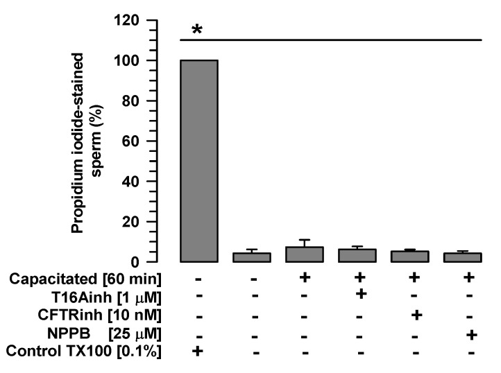
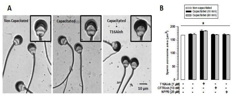
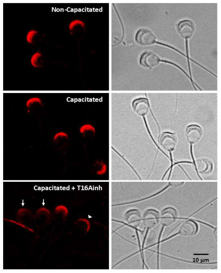
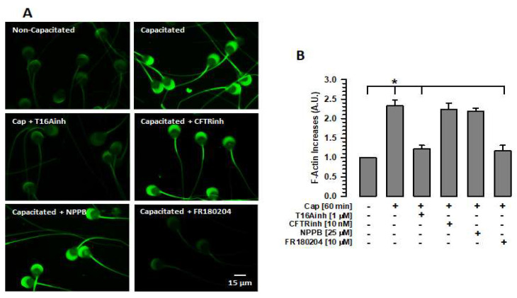
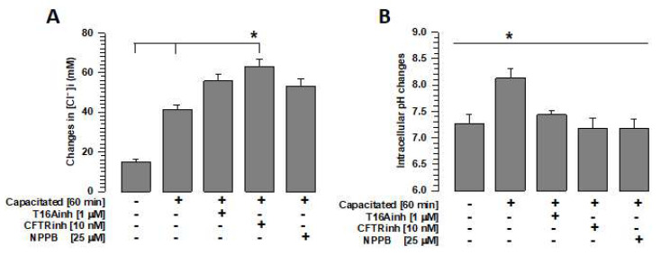
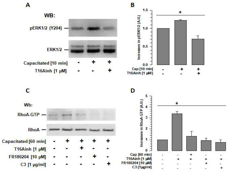
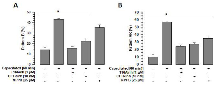
Mammalian spermatozoa undergo a series of physiological and biochemical changes in the oviduct that lead them to acquire the ability to fertilize eggs. During their transit in the oviduct, spermatozoa face a series of environmental changes that can affect sperm viability. A series of ion channels and transporters, as well as the sperm cytoskeleton, allow spermatozoa to remain viable and functional. Cl- channels such as TMEM16A (calcium-activated chloride channel), CFTR (cystic fibrosis transmembrane conductance regulator), and ClC3 (chloride voltage-gated channel 3) are some of the ion transporters involved in maintaining cellular homeostasis. They are expressed in mammalian spermatozoa and are associated with capacitation, acrosomal reaction, and motility. However, little is known about their role in maintaining sperm volume. Therefore, this study aimed to determine the mechanism through which TMEM16A maintains sperm volume during capacitation. The effects of TMEM16A were compared to those of CFTR and ClC3. Spermatozoa were capacitated in the presence of specific TMEM16A, CFTR, and ClC3 inhibitors, and the results showed that only TMEM16A inhibition increased acrosomal volume, leading to changes within the acrosome. Similarly, only TMEM16A inhibition prevented actin polymerization during capacitation. Further analysis showed that TMEM16A inhibition also prevented ERK1/2 and RhoA activation. On the other hand, TMEM16A and CFTR inhibition affected both capacitation and spontaneous acrosomal reaction, whereas ClC3 inhibition only affected the spontaneous acrosomal reaction. In conclusion, during capacitation, TMEM16A activity regulates acrosomal structure through actin polymerization and by regulating ERK1/2 and RhoA activities.

Keywords: acrosome structure; bicarbonate influx; chloride channels; fertilization; sperm.


Conflict of interest statement

The authors declare no conflicts of interest.

Figures

Figure 1
Figure 1
Sperm viability is not affected by TMEM16A, CFTR, or ClC3 inhibitors. Guinea pig spermatozoa were capacitated for 60 min in the presence of one of the following Cl channel inhibitors: T16Ainh (1 µM), CFTRinh (10 nM), or NPPB (25 µM). Viability was assessed using propidium iodide, and the number of stained and unstained spermatozoa was counted (500 cells per sample). Images were recorded and analyzed using Niko Element 3.1 software. Results are expressed as mean ± S.E. (n = 3), * p < 0.05.
Figure 2
Figure 2
TMEM16A inhibition affects the volume of the acrosomal region. Guinea pig spermatozoa were capacitated for 60 min in the presence of T16Ainh, CFTRinh, or NPPB inhibitors. Spermatozoa were then fixed and stained with Coomassie blue G250. The acrosomal area of 100 spermatozoa was carefully assessed using Nis Element 3.1 software. (A) Images of non-capacitated and capacitated spermatozoa in the absence and presence of T16Ainh. The inset shows the acrosomal changes experienced by spermatozoa capacitated in the presence of T16Ainh (right panel) compared to non-capacitated (left panel) and spermatozoa capacitated in the absence of T16Ainh (middle panel). (B) Quantification of changes in the acrosome area of capacitated spermatozoa in the absence and presence of the different inhibitors. Results are expressed as means ± S.E. (n = 3), * p < 0.05.
Figure 3
Figure 3
TMEM16A inhibition disrupts acrosomal structure. A specific antibody for the acrosomal protein calreticulin was used to define the changes in the acrosome produced when spermatozoa were capacitated for 60 min in the absence or presence of the inhibitor T16Ainh (1 µM). The localization of calreticulin is shown in the left panels. The top panel shows non-capacitated spermatozoa, the middle panel shows capacitated spermatozoa, and the bottom panel shows spermatozoa capacitated in the presence of T16Ainh. The right panels show bright field images of spermatozoa. Images are representative of three different experiments. Arrow: pattern with dim fluorescence dispersed throughout the acrosome. Arrowhead: pattern with fluorescence concentrated in the apical region of the acrosome.
Figure 4
Figure 4
Inhibition of TMEM16A or ERK1/2 prevents actin polymerization during capacitation. To define the changes in F-actin levels produced by inhibition of Cl channels or ERK1/2, spermatozoa were capacitated in the absence or presence of one of the following inhibitors: T16Ainh, CFTRinh, NPPB, or FR180204 (ERK1/2). After 60 min of incubation, spermatozoa were fixed, and F-actin was visualized by staining with phalloidin-FITC. (A) Localization of F-actin using phalloidin-FITC. Images represent three independent experiments. (B) Quantification of fluorescence emitted by phalloidin-FITC-stained spermatozoa. Cap: capacitated. Fluorescence levels were assessed using Nis Element 3.1 software. The fluorescence values were normalized with respect to values of non-capacitated spermatozoa. Results are expressed as mean ± S.E. (n = 3), * p < 0.05.
Figure 5
Figure 5
Inhibition of chloride ion channels alters intracellular Cl and pH homeostasis. To define the effects of TMEM16A, CFTR, and ClC3 channel inhibition on intracellular Cl and pH homeostasis, spermatozoa were capacitated for 60 min in the absence or presence of the inhibitors T16Ainh, CFTRinh, or NPPB, and intracellular Cl and intracellular pH were assessed using specific probes. (A) Quantification of changes in intracellular Cl concentrations using MQAE. Results are expressed as mean ± S.E. (n = 5), * p < 0.05. (B) Quantification of intracellular pH changes using BCECF-AM. Results are expressed as mean ± S.E. (n = 5), * p < 0.05.
Figure 6
Figure 6
TMEM16A regulates RhoA activity through ERK1/2 during capacitation. (A) Total extracts of spermatozoa capacitated for 10 min in the absence or presence of the TMEM16A inhibitor (T16Ainh) were used to determine its effect on ERK1/2 activity. Ten minutes of capacitation is the time when ERK1/2 reaches their maximum activity. Specific antibodies were used to detect ERK1/2 (pERK) phosphorylated at Y204 (upper panel) and total ERK1/2 in each sample (lower panel). WBs represent three independent experiments. (B) WBs of ERK1/2 and pERK1/2 were analyzed using densitometry. Results were first normalized using the ratio N/N0, where N is the amount of pERK1/2 and N0 is the total amount of ERK1/2. The results obtained were then normalized with respect to the results of non-capacitated sperm. Results are expressed as mean ± S.E. (n = 3), * p < 0.05. (C) Total extracts of spermatozoa capacitated for 60 min in the absence or presence of the ERK1/2-specific inhibitor FR180204 were used to determine its effect on RhoA activity. RhoA-GTP was isolated via pull-down using a Rhotekin column and analyzed using WB and a RhoA-specific antibody (upper panel). The lower panel shows the total amount of RhoA in each sample. WBs represent three independent experiments. (D) WBs for RhoA-GTP and RhoA were analyzed using densitometry. Cap: capacitated. Results were first normalized as the ratio N/N0, where N is the amount of RhoA-GTP and N0 is the total amount of RhoA, and then normalized to the results of non-capacitated spermatozoa. Results are expressed as mean ± S.E. (n = 3), * p < 0.05. Cap: capacitated.
Figure 7
Figure 7
Inhibition of Cl channels, TMEM16A, CFTR, and ClC3 affects capacitation and the acrosome reaction. To define the effects of inhibition of TMEM16A, CFTR, and ClC3 channels on capacitation and the spontaneous acrosome reaction, spermatozoa were capacitated for 60 min in the absence or presence of the inhibitors T16Ainh, CFTRinh, or NPPB, and the B and AR patterns were assessed by CTC staining. (A) Effects of the inhibitors T16Ainh, CFTRinh, and NPPB on the B pattern. Results are expressed as mean ± S.E. (n = 3), * p < 0.05. (B) Effects of the inhibitors T16Ainh, CFTRinh, and NPPB on the AR pattern. Results are expressed as mean ± S.E. (n = 3), * p < 0.05.

Similar articles

References

    1. Nicolli A.R., Cesari A. The Other Side of Capacitation: Role of Mouse Male Molecules in the Regulation of Time and Place of Capacitation. Reproduction. 2023;166:R73–R85. doi: 10.1530/REP-23-0188. - DOI - PubMed
    1. Rogers B.J., Bastias C., Coulson R.L., Russell L.D. Cytochalasin D Inhibits Penetration of Hamster Eggs by Guinea Pig and Human Spermatozoa. J. Androl. 1989;10:275–282. doi: 10.1002/j.1939-4640.1989.tb00102.x. - DOI - PubMed
    1. Castellani-Ceresa L., Mattioli M., Radaelli G., Barboni B., Brivio M.F. Actin Polymerization in Boar Spermatozoa: Fertilization Is Reduced with Use of Cytochalasin D. Mol. Reprod. Dev. 1993;36:203–211. doi: 10.1002/mrd.1080360211. - DOI - PubMed
    1. Sanchez-Gutierrez M., Contreras R.G., Mujica A. Cytochalasin-D Retards Sperm Incorporation Deep into the Egg Cytoplasm but Not Membrane Fusion with the Egg Plasma Membrane. Mol. Reprod. Dev. 2002;63:518–528. doi: 10.1002/mrd.10203. - DOI - PubMed
    1. Brener E., Rubinstein S., Cohen G., Shternall K., Rivlin J., Breitbart H. Remodeling of the Actin Cytoskeleton During Mammalian Sperm Capacitation and Acrosome Reaction. Biol. Reprod. 2003;68:837–845. doi: 10.1095/biolreprod.102.009233. - DOI - PubMed

Substances

LinkOut - more resources