

Proteome Alterations in Cardiac Fibroblasts: Insights from Experimental Myocardial Infarction and Clinical Ischaemic Cardiomyopathy
- PMID: 40332511
- PMCID: PMC12028142
- DOI: 10.3390/ijms26083846
Proteome Alterations in Cardiac Fibroblasts: Insights from Experimental Myocardial Infarction and Clinical Ischaemic Cardiomyopathy
Abstract
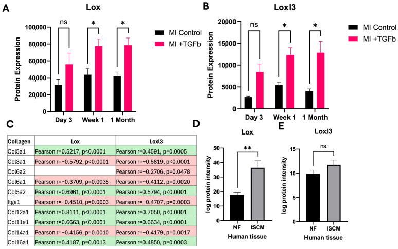
Ischaemic heart disease (IHD) is a chronic condition that can cause pathological cardiac remodelling and heart failure (HF). In this study, we sought to determine how cardiac fibroblasts were altered post-experimental myocardial infarction (MI). Female C57BL6 mice underwent experimental MI by permanent left coronary artery ligation. Cardiac fibroblasts were isolated from extracted heart tissue of experimental MI mice and subsequently treated with the pro-fibrotic cytokine, TGF-β, for 24 h and analysed using high throughput LC-MS/MS analysis. Findings were validated using mass spectrometry data generated from human left ventricular tissue analysis, which were collected from patients with ischaemic cardiomyopathy (ISCM) and age/sex-matched patients without clinical HF (NF). Proteomic analysis revealed significant protein expression changes in mouse cardiac fibroblasts after MI. These changes were most pronounced at 1 month post-MI, compared to earlier time points (3 days and 1 week). TGF-β treatment profoundly affected fibroblast cells extracted from MI mice, indicating a heightened sensitivity to pro-fibrotic factors after myocardial injury. Extracellular matrix (ECM) proteins significantly altered in MI fibroblasts following TGF-β treatment were significantly associated with cardiac remodelling. Notably, Lox was significantly changed in both isolated fibroblasts treated with TGF-β from experiment MI mice and human ISCM. Isolated cardiac fibroblasts from MI mice are more susceptible to developing pathogenic traits following TGF-β treatment than isolated fibroblasts from normal heart tissue. ECM proteins associated with these enhanced fibroblast activities and functions are evident. These altered proteins may play a functional role in MI-associated cardiac dysfunction.
Keywords: TGF-β; cardiac remodelling; extracellular matrix; ischaemic heart disease; myocardial infarction; proteomics.
Conflict of interest statement
The authors declare no conflicts of interest.
Figures


Similar articles
-
ITIH5-mediated fibroblast/macrophage crosstalk exacerbates cardiac remodelling after myocardial infarction.J Transl Med. 2025 Feb 24;23(1):224. doi: 10.1186/s12967-025-06244-5. J Transl Med. 2025. PMID: 39994656 Free PMC article.
-
Cortical bone stem cell-derived exosomes' therapeutic effect on myocardial ischemia-reperfusion and cardiac remodeling.Am J Physiol Heart Circ Physiol. 2021 Dec 1;321(6):H1014-H1029. doi: 10.1152/ajpheart.00197.2021. Epub 2021 Oct 8. Am J Physiol Heart Circ Physiol. 2021. PMID: 34623184 Free PMC article.
-
Notch3 Ameliorates Cardiac Fibrosis After Myocardial Infarction by Inhibiting the TGF-β1/Smad3 Pathway.Cardiovasc Toxicol. 2016 Oct;16(4):316-24. doi: 10.1007/s12012-015-9341-z. Cardiovasc Toxicol. 2016. PMID: 26487518
-
Fibroblasts and the extracellular matrix in right ventricular disease.Cardiovasc Res. 2017 Oct 1;113(12):1453-1464. doi: 10.1093/cvr/cvx146. Cardiovasc Res. 2017. PMID: 28957531 Free PMC article. Review.
-
Transforming growth factor (TGF)-β signaling in cardiac remodeling.J Mol Cell Cardiol. 2011 Oct;51(4):600-6. doi: 10.1016/j.yjmcc.2010.10.033. Epub 2010 Nov 6. J Mol Cell Cardiol. 2011. PMID: 21059352 Free PMC article. Review.
Cited by
-
In depth characterisation of the proteome of MIS-C and post COVID-19 infection in children reveals inflammatory pathway activation and evidence of tissue damage.J Transl Med. 2025 Aug 18;23(1):929. doi: 10.1186/s12967-025-06826-3. J Transl Med. 2025. PMID: 40826070 Free PMC article.
References
-
- Sun Z., Yun Z., Lin J., Sun X., Wang Q., Duan J., Li C., Zhang X., Xu S., Wang Z., et al. Comprehensive mendelian randomization analysis of plasma proteomics to identify new therapeutic targets for the treatment of coronary heart disease and myocardial infarction. J. Transl. Med. 2024;22:404. doi: 10.1186/s12967-024-05178-8. - DOI - PMC - PubMed
MeSH terms
Substances
Grants and funding
LinkOut - more resources
Full Text Sources
Medical
Research Materials
Miscellaneous
