. 2025 Apr 19;26(8):3871.
doi: 10.3390/ijms26083871.

Establishment of a Human iPSC Line from Mucolipidosis Type II That Expresses the Key Markers of the Disease

Affiliations

Establishment of a Human iPSC Line from Mucolipidosis Type II That Expresses the Key Markers of the Disease

Maria Eduarda Moutinho et al. Int J Mol Sci. .

Abstract

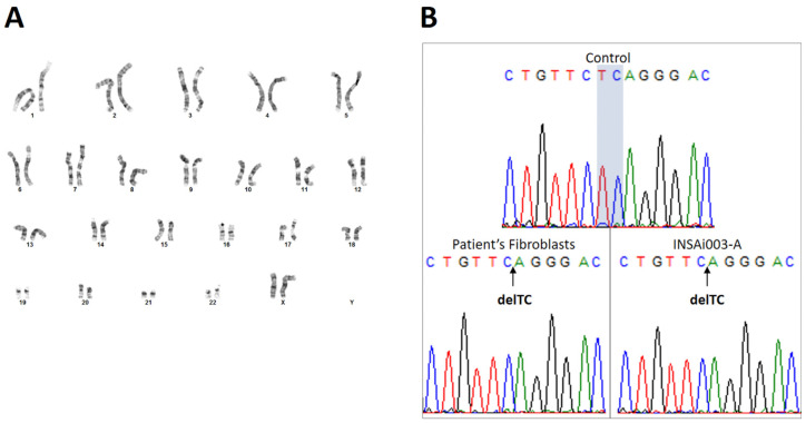
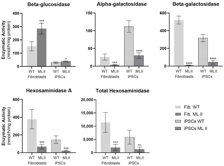
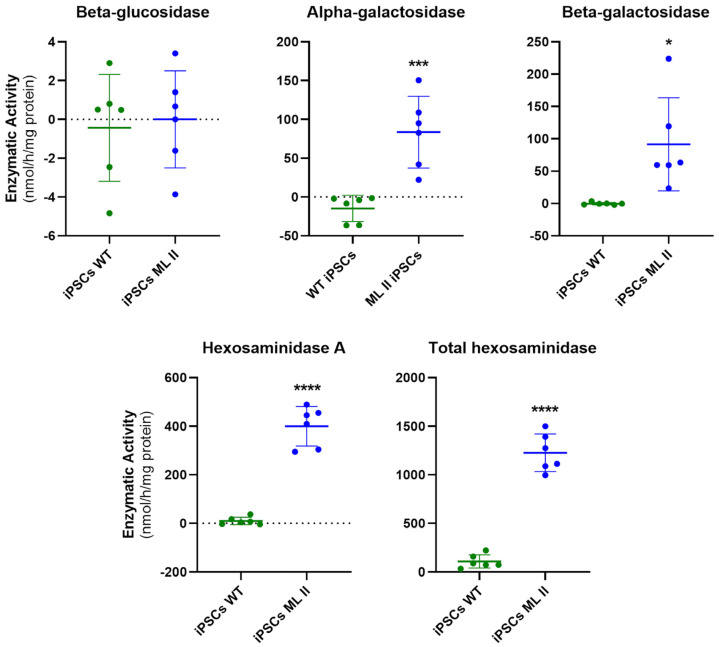
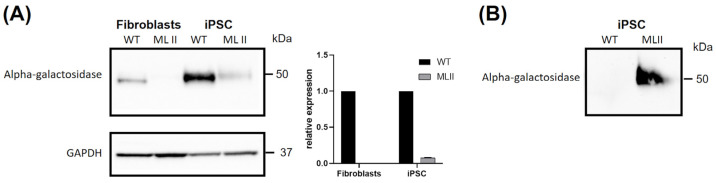
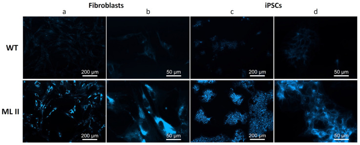
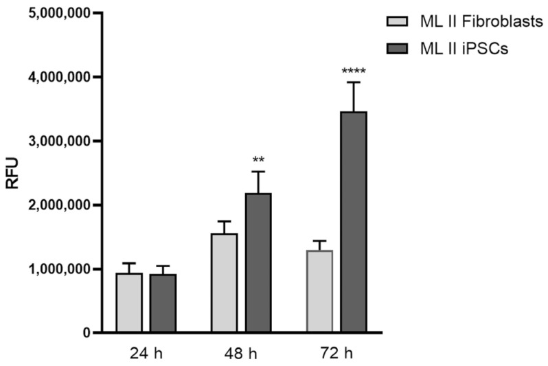
Mucolipidosis type II (ML II) is a rare and fatal disease of acid hydrolase trafficking. It is caused by pathogenic variants in the GNPTAB gene, leading to the absence of GlcNAc-1-phosphotransferase activity, an enzyme that catalyzes the first step in the formation of the mannose 6-phosphate (M6P) tag, essential for the trafficking of most lysosomal hydrolases. Without M6P, these do not reach the lysosome, which accumulates undegraded substrates. The lack of samples and adequate disease models limits the investigation into the pathophysiological mechanisms of the disease and potential therapies. Here, we report the generation and characterization of an ML II induced pluripotent stem cell (iPSC) line carrying the most frequent ML II pathogenic variant [NM_024312.5(GNPTAB):c.3503_3504del (p.Leu1168fs)]. Skin fibroblasts were successfully reprogrammed into iPSCs that express pluripotency markers, maintain a normal karyotype, and can differentiate into the three germ layers. Furthermore, ML II iPSCs showed a phenotype comparable to that of the somatic cells that originated them in terms of key ML II hallmarks: lower enzymatic activity of M6P-dependent hydrolases inside the cells but higher in conditioned media, and no differences in an M6P-independent hydrolase and accumulation of free cholesterol. Thus, ML II iPSCs constitute a novel model for ML II disease, with the inherent iPSC potential to become a valuable model for future studies on the pathogenic mechanisms and testing potential therapeutic approaches.

Keywords: LSD; ML II; cellular model; iPSCs; lysosomal storage disorder; stem cells.


Conflict of interest statement

The authors declare no conflicts of interest.

Figures

Figure 1
Figure 1
ML II fibroblast culture after reprogramming, showing a colony with clear edges and a typical pluripotent stem cell morphology. Cells were observed using a Leica DMIL inverted contrast microscope (Leica Microsystems, Wetzlar, Germany) with 100× magnification.
Figure 2
Figure 2
PCR amplification of oriP and EBNA-1 transgenes for detection of Epi5™ reprogramming vectors in ML II iPSCs in passages (P) 2, 8, and 16. As a negative control, ML II fibroblasts (Fib., lane 3) were included. Positive controls (+Ctrl) are the reprogramming vectors containing oriP and EBNA-1 genes. The 100 bp DNA ladder, stained with 6x TriTrack Loading Dye, was used as a molecular weight DNA ladder (lane 1). +Ctrl—positive control; Fib—ML II fibroblasts; iPSCs—ML II iPSCs; B—blank (no DNA template).
Figure 3
Figure 3
Endogenous expression of pluripotency markers in ML II iPSCs. (A) Expression levels of the pluripotency genes NANOG, OCT4, and SOX2 in ML II iPSCs relative to the parental ML II fibroblasts, measured by RT-qPCR. The results were normalized to the housekeeping gene GAPDH, and expression levels were calculated relative to the expression in the fibroblasts by the 2−∆∆Ct method. As a positive control, an FD iPSC line previously published [INSAi002-A (FD)] [16] was used. Graphs were constructed in GraphPad Prism 8.0.2 and represent the mean and standard deviation of three independent experiments. (B) Immunofluorescence in ML II iPSCs showed positive staining for the nuclear pluripotency markers NANOG, OCT4, and SOX2, as well as for the surface pluripotency marker TRA-1-60. Cells were analyzed on a DM400 M fluorescence microscope (Leica), and images were captured and processed in Leica Application Suite v.3.7.0 software.
Figure 4
Figure 4
Immunofluorescence showing positive staining for the three germ layer markers OTX2, SOX17, and Brachyury after ML II iPSCs differentiation into ectoderm, endoderm, and mesoderm, respectively. Cells were analyzed on a DM400 M fluorescence microscope (Leica), and images were captured and processed in the Leica Application Suite v.3.7.0 software.
Figure 5
Figure 5
Genetic characterization of the ML II iPSCs. (A) G-banded standard karyotype showing a normal female karyotype (46, XX). (B) Sanger sequencing chromatograms showing a TC deletion (delTC) both in ML II patient’s fibroblasts and in ML II iPSCs INSAi003-A, which corresponds to the pathogenic variant NM_024312.5(GNPTAB):c.3503_3504del (p.Leu1168fs). The TC nucleotides are present (highlighted) in the WT control sample. The arrows indicate the position in the exon 19 sequence where the TC deletion occurs.
Figure 6
Figure 6
Enzymatic activities of specific hydrolases in ML II and WT fibroblasts and iPSCs. Graphics were constructed in GraphPad Prism 8.0.2. and represent the mean and standard deviation of five independent experiments. Data were analyzed by the Student’s t-test between the respective cell types (ML II fibroblasts vs. WT fibroblasts and ML II iPSCs vs. WT iPSCs). * p ≤ 0.05, ** p ≤ 0.01, *** p ≤ 0.001 and **** p ≤ 0.0001 vs. WT.
Figure 7
Figure 7
Enzymatic activities of specific hydrolases in conditioned culture media of ML II and WT iPSCs. Graphics were constructed in GraphPad Prism 8.0.2. and represent individual values, as well as the mean and standard deviation of six independent samples. Data were analyzed by the Student’s t-test (ML II iPSCs vs. WT iPSCs). * p ≤ 0.05, *** p ≤ 0.001 and **** p ≤ 0.0001 vs. WT.
Figure 8
Figure 8
Western blotting for alpha-galactosidase. (A) Cellular extracts from WT and ML II fibroblasts and iPSCs. GAPDH was used as a loading control, and densitometric analyses of alpha-galactosidase levels were normalized for GAPDH. Two independent experiments were performed. (B) Conditioned media from WT and ML II iPSCs analyzed for alpha-galactosidase.
Figure 9
Figure 9
Filipin staining in WT and ML II fibroblasts (a,b) and WT and ML II iPSCs (c,d). Cells were analyzed on a DM400 M fluorescence microscope (Leica), and images were captured and processed in Leica Application Suite v.3.7.0 software.
Figure 10
Figure 10
Cell proliferation was assessed by Alamar Blue assay on ML II fibroblasts and iPSCs after 24 h, 48 h, and 72 h of culture. Data were analyzed by the Student’s t-test (ML II iPSCs vs. ML II fibroblasts). ** p ≤ 0.01 and **** p ≤ 0.0001 vs. ML II fibroblasts.

Similar articles

References

    1. Dogterom E.J., Wagenmakers M.A.E.M., Wilke M., Demirdas S., Muschol N.M., Pohl S., Meijden J.C.v.d., Rizopoulos D., Ploeg A.T.v.d., Oussoren E. Mucolipidosis Type II and Type III: A Systematic Review of 843 Published Cases. Genet. Med. 2021;23:2047–2056. doi: 10.1038/s41436-021-01244-4. - DOI - PubMed
    1. Velho R.V., Harms F.L., Danyukova T., Ludwig N.F., Friez M.J., Cathey S.S., Filocamo M., Tappino B., Güneş N., Tüysüz B., et al. The Lysosomal Storage Disorders Mucolipidosis Type II, Type III Alpha/Beta, and Type III Gamma: Update on GNPTAB and GNPTG Mutations. Hum. Mutat. 2019;40:842–864. doi: 10.1002/HUMU.23748. - DOI - PubMed
    1. Matos L., Vilela R., Rocha M., Santos J.I., Coutinho M.F., Gaspar P., Prata M.J., Alves S. Development of an Antisense Oligonucleotide-Mediated Exon Skipping Therapeutic Strategy for Mucolipidosis II: Validation at RNA Level. Hum. Gene Ther. 2020;31:775–783. doi: 10.1089/hum.2020.034. - DOI - PubMed
    1. Coutinho M., Encarnação M., Gomes R., da Silva Santos L., Martins S., Sirois-Gagnon D., Bargal R., Filocamo M., Raas-Rothschild A., Tappino B., et al. Origin and Spread of a Common Deletion Causing Mucolipidosis Type II: Insights from Patterns of Haplotypic Diversity. Clin. Genet. 2011;80:273–280. doi: 10.1111/j.1399-0004.2010.01539.x. - DOI - PubMed
    1. Takahashi K., Tanabe K., Ohnuki M., Narita M., Ichisaka T., Tomoda K., Yamanaka S. Induction of Pluripotent Stem Cells from Adult Human Fibroblasts by Defined Factors. Cell. 2007;131:861–872. doi: 10.1016/j.cell.2007.11.019. - DOI - PubMed

MeSH terms

Substances

LinkOut - more resources