. 2025 Jun;120(3):489-507.
doi: 10.1007/s00395-025-01109-w. Epub 2025 May 7.

Altered protein homeostasis in cardiovascular diseases contributes to Alzheimer's-like neuropathology

Affiliations

Altered protein homeostasis in cardiovascular diseases contributes to Alzheimer's-like neuropathology

Nirjal Mainali et al. Basic Res Cardiol. 2025 Jun.

Abstract

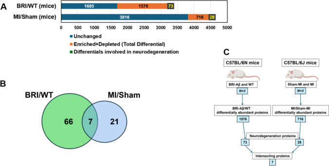
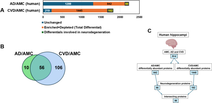
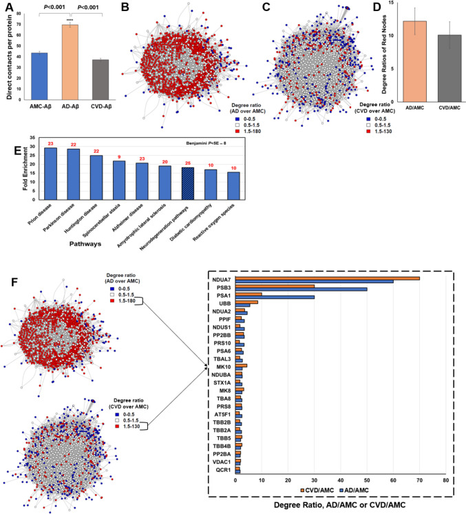
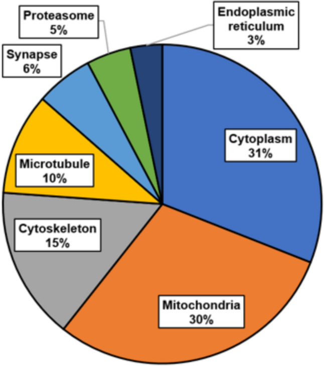
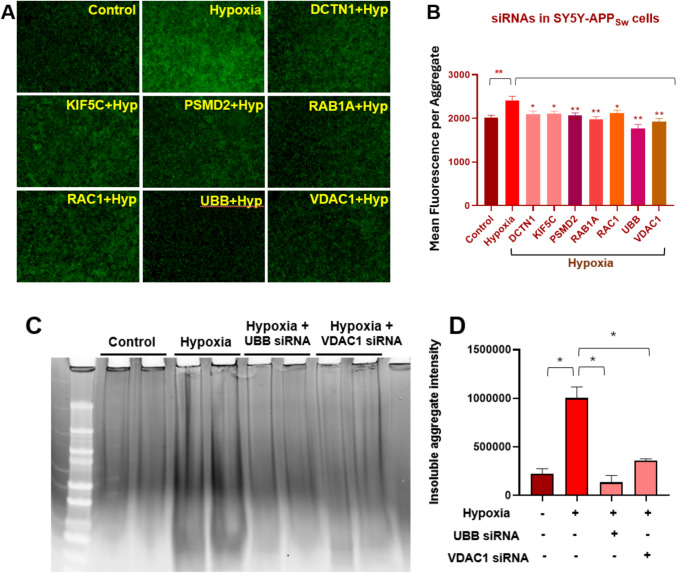
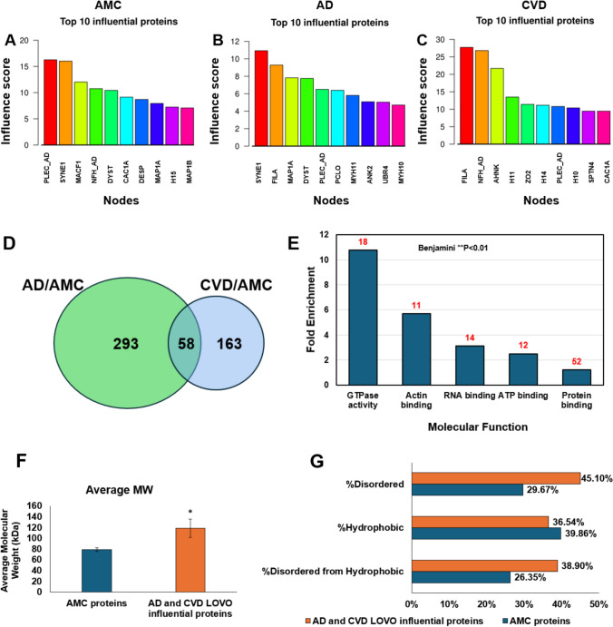
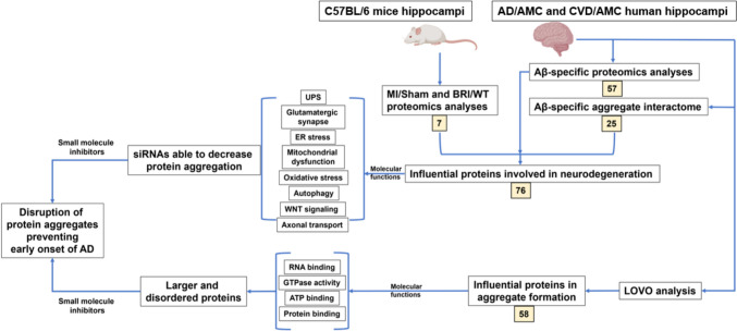
Cardiovascular diseases (CVDs) are the leading cause of death worldwide. CVD is known to increase the risk of subsequent neurodegeneration but the mechanism(s) and proteins involved have yet to be elucidated. We previously showed that myocardial infarction (MI), induced in mice and compared to sham-MI mice, leads to increases in protein aggregation, endoplasmic reticulum (ER) stress in both heart and brain, and changes in proteostatic pathways. In this study, we further investigate the molecular mechanisms altered by induced MI in mice, which were also implicated by proteomics of postmortem human hippocampal aggregates from Alzheimer's disease (AD) and cardiovascular disease (CVD) patients, vs. age-matched controls (AMC). We utilized intra-aggregate crosslinking to identify protein-protein contacts or proximities, and thus to reconstruct aggregate "contactomes" (nonfunctional interactomes). We used leave-one-out analysis (LOOA) to determine the contribution of each protein to overall aggregate cohesion, and gene ontology meta-analyses of constituent proteins to define critical organelles, processes, and pathways that distinguish AD and/or CVD from AMC aggregates. We identified influential proteins in both AD and CVD aggregates, many of which are associated with pathways or processes previously implicated in neurodegeneration such as mitochondrial, oxidative, and endoplasmic-reticulum stress; protein aggregation and proteostasis; the ubiquitin proteasome system and autophagy; axonal transport; and synapses.

Keywords: Alzheimer’s disease; Cardiovascular disease; Crosslinking studies; Leave-one-out analysis; Protein aggregates.


Conflict of interest statement

Declarations. Conflict of interest: The authors declare that they have no conflict of interest. Ethical approval: The research presented here was reviewed by the Institutional Review Board of the Central Arkansas Veterans Healthcare System (CAVHS IRB), which deemed it non-human-subject research due to our use of de-identified brain tissue from an approved brain bank, plus mechanistic studies of cultured human cell lines model of AD-like aggregation.

Figures

Fig. 1
Fig. 1
Proteomic and GO analyses for proteins differentially abundant in MI vs. sham-MI aggregates, and in BRI-Aβ42 vs. wildtype C57BL/6N mice. Aggregates were isolated from hippocampi of MI, sham-MI, BRI-Aβ42, and wildtype (WT) mice and their proteomes analyzed. Based on these comparisons (MI vs. sham-MI, and BRI-Aβ42 vs. WT), we identified differentially abundant proteins (enriched + depleted) in BR-Aβ42 and MI aggregates compared to their respective controls. A 1685 proteins were unchanged, 1576 proteins were differential, and 73 of these differential proteins were annotated as involved in neurodegeneration in BRI-Aβ42 mice relative to WT. Similarly, 3816 proteins were unchanged, 716 proteins were differential, and 28 differential proteins were involved in neurodegeneration in MI mice relative to sham-MI mice. For BRI-Aβ42 vs. WT, χ2 (2) = [494], P < 2.2E – 16; whereas for MI vs. sham-MI, χ2 (2) = [5149], P < 2.2E – 16 employing Chi-squared tests. B Venn diagram showing 7 differentially abundant shared by both comparisons. C Flowchart of mouse animal-model comparisons and results
Fig. 2
Fig. 2
Proteomic and GO analyses for human hippocampal-aggregate proteins differentially abundant in AD and CVD, each relative to age-matched controls (AMC). Aggregates were isolated from hippocampal tissues of AD, CVD, and AMC individuals, and subjected to proteomic analyses. Comparisons of AD vs. AMC and CVD vs. AMC aggregate proteins identified those enriched or depleted in AD and CVD, relative to AMC. A Comparing AD to AMC aggregates, 1299 proteins were unchanged, 842 proteins were differentially abundant, and 66 differential proteins (7.8%) were annotated as involved in neurodegeneration. Comparing CVD to AMC aggregates, 219 proteins were unchanged, 1445 proteins were differential, and 162 differential proteins (11.2%) were annotated as involved in neurodegeneration. For AD vs. AMC, χ2 (2) = [794], P < 2.2E – 16; for CVD vs. AMC, χ2 (2) = [1657], P < 2.2E – 16 by Chi-squared tests. B Venn diagram showing 56 common proteins from differentially abundant AD/AMC and CVD/AMC comparisons. C Flow chart for AMC, AD, and CVD human hippocampal-aggregate comparisons and results
Fig. 3
Fig. 3
Aβ-IP aggregate interactome analysis from AMC, AD, and CVD hippocampi. Protein foci were recovered after immuno-pulldown (IP) from AD, CVD, and AMC hippocampal tissue using Aβ antibody and crosslinking with chemical crosslinkers; sarcosyl-insoluble aggregates were then isolated from them. After LC–MS/MS analysis, we used an R program to visualize the contactomes and determine the degree (number of direct interactions) of each protein. Degree ratios were determined for AD vs. AMC and CVD vs. AMC. A Mean degree (direct contacts) of all interactome proteins: 42 for AMC, 70 for AD, and 38 for CVD. AD interactions per protein (mean ± SEM) differed from AMC or CVD at P < 0.001 by 2-tailed heteroscedastic t tests. B Visualization of aggregate interactome shared by AD and AMC; red nodes have AD/AMC degree ratios ≥ 1.5, and blue nodes have ratios < 0.5 (less abundant in AD). C Aggregate interactome shared by CVD and AMC is displayed as in B, with node colors here indicating CVD/AMC degree ratios. D Mean ± SEM degree ratios were calculated for proteins with more interacting partners (degree) than AMC, in CVD (grey) or AD (orange). E KEGG pathway analysis, using 182 common proteins from those most differential in AD/AMC and CVD/AMC comparisons, implicate 25 proteins involved in neurodegeneration (Alzheimer’s, Parkinson’s, Huntington’s, Prion disease, etc.) with Benjamini-adjusted P value < 0.01 for annotation enrichment in DAVID (https://david.ncifcrf.gov/home.jsp). Numbers over bars indicate the number of differential aggregate proteins in each category. F Degree ratios are plotted for comparisons of shared differential proteins contrasting AD/AMC (blue bars) or CVD/AMC (orange bars)
Fig. 4
Fig. 4
Cellular localization of differential proteins common to CVD and AD aggregates in human hippocampi and mouse models of AD or CVD, based on KEGG pathway analysis. Cellular localizations of 76 differential proteins implicate chiefly cytoplasm and mitochondria, followed by cytoskeleton, microtubule, synapse, proteasome, and sarcoplasmic reticulum
Fig. 5
Fig. 5
Hypoxia-induced protein aggregation in neuroblastoma cells is reduced by RNAi knockdown of genes encoding highly differential aggregate proteins. After 7 h of exposure to hypoxia, SY5Y-APPSw cells were transfected with siRNA or shRNA constructs (via RNAiMax lipofection) targeting DCTN1, KIF5C, PSMD2, RAB1A, RAC1, UBB, and VDAC1. Cells were returned to a normoxic incubator (reperfusion for 48 h), after which they underwent Thioflavin T staining or were harvested for aggregate isolation. A, B Thioflavin-T fluorescence is shown, for SY5Y-APPSw cells after transfection with siRNAs targeting 7 differential proteins. All knockdowns reduced aggregation significantly. *P < 0.05; **P < 0.01 by 2-tailed heteroscedastic t tests. C, D Detergent-insoluble aggregates from control and anoxia-treated SY5Y-APPSw cells are reduced tenfold by prior RNAi knockdown with UBB siRNA, and 3.5-fold after KD with VDAC1 siRNA. Reduction in aggregate protein after each siRNA was significant at *P < 0.01 by 2-tailed heteroscedastic t tests
Fig. 6
Fig. 6
Leave-one-vertex-out analysis (LOVO) in AMC, AD, and CVD interactomes ranks proteins by predicted influence on aggregate stability and growth. Crosslink-based interactomes, constructed for AMC, AD and CVD aggregates, were used to conduct leave-one-vertex-out (LOVO) analysis using R programming. Top influential proteins after LOVO analysis were plotted, and molecular functions and structural properties of influential AD and CVD proteins (each relative to AMC) were listed. A–C LOVO analysis of AMC, AD, and CVD interactomes, showing top influential proteins and their influence scores in each interactome. D Influential proteins based on higher influence ratio of AD/AMC and CVD/AMC shows 58 common proteins between them. E GO term analysis of these common influential proteins implicates functions related to GTPase binding, RNA binding, actin binding, ATP binding, and protein binding with Benjamini-adjusted P value < 0.01 for annotation enrichment in DAVID (https://david.ncifcrf.gov/home.jsp). F Average molecular weight is increased > 60% in AD and CVD aggregates, relative to AMC (*P < 0.05, by 2-tailed heteroscedastic t tests). G Percent disordered proteins is elevated > 50% among influential proteins involved in aggregates from AD and CVD consensus interactomes relative to AMC
Fig. 7
Fig. 7
Summary of research implicating differential aggregate proteins in disease models

Similar articles

References

    1. Adesuyan M, Jani YH, Alsugeir D, Howard R, Wong ICK, Wei L, Brauer R (2023) Trends in the incidence of dementia in people with hypertension in the UK 2000 to 2021. Alzheimers Dement (Amst) 15:e12466. 10.1002/dad2.12466 - PMC - PubMed
    1. Ayyadevara S, Balasubramaniam M, Parcon PA, Barger SW, Griffin WS, Alla R, Tackett AJ, Mackintosh SG, Petricoin E, Zhou W, Shmookler Reis RJ (2016) Proteins that mediate protein aggregation and cytotoxicity distinguish Alzheimer’s hippocampus from normal controls. Aging Cell 15:924–939. 10.1111/acel.12501 - PMC - PubMed
    1. Ayyadevara S, Ganne A, Balasubramaniam M, Shmookler Reis RJ (2022) Intrinsically disordered proteins identified in the aggregate proteome serve as biomarkers of neurodegeneration. Metab Brain Dis 37:147–152. 10.1007/s11011-021-00791-8 - PMC - PubMed
    1. Ayyadevara S, Mercanti F, Wang X, Mackintosh SG, Tackett AJ, Prayaga SV, Romeo F, Shmookler Reis RJ, Mehta JL (2016) Age- and hypertension-associated protein aggregates in mouse heart have similar proteomic profiles. Hypertension 67:1006–1013. 10.1161/hypertensionaha.115.06849 - PMC - PubMed
    1. Balasubramaniam M, Ayyadevara S, Ganne A, Kakraba S, Penthala NR, Du X, Crooks PA, Griffin ST, Shmookler Reis RJ (2019) Aggregate interactome based on protein cross-linking interfaces predicts drug targets to limit aggregation in neurodegenerative diseases. iScience 20:248–264. 10.1016/j.isci.2019.09.026 - PMC - PubMed

LinkOut - more resources