. 2025 Aug 1;64(2):236-244.
doi: 10.1097/SHK.0000000000002611. Epub 2025 Apr 28.

N-ACETYLCYSTEINE REDUCES VON WILLEBRAND FACTOR MULTIMER SIZE AND IMPROVES RENAL MICROVASCULAR BLOOD FLOW IN RATS AFTER SEVERE TRAUMA

Affiliations

N-ACETYLCYSTEINE REDUCES VON WILLEBRAND FACTOR MULTIMER SIZE AND IMPROVES RENAL MICROVASCULAR BLOOD FLOW IN RATS AFTER SEVERE TRAUMA

Alexander St John et al. Shock. .

Abstract

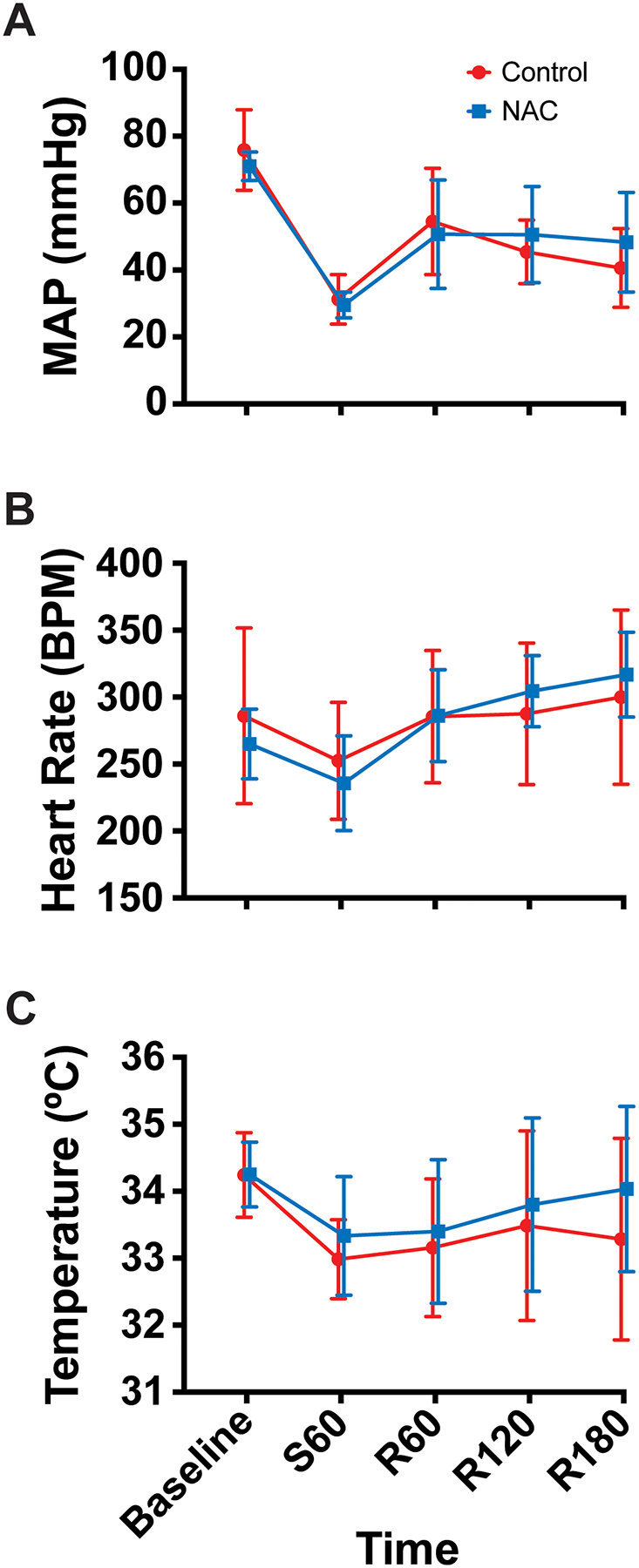
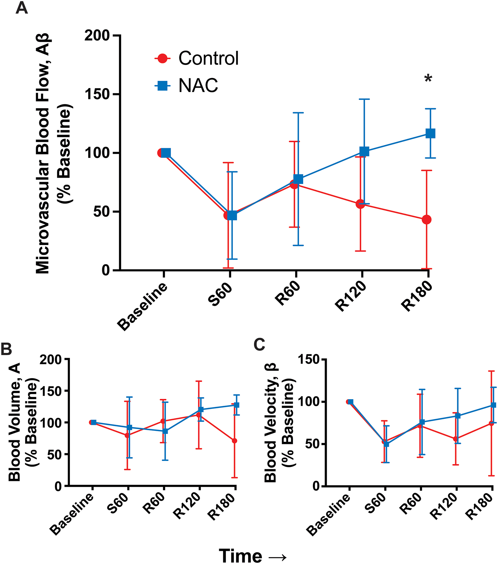
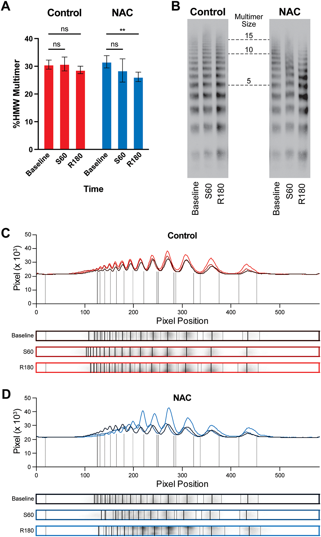
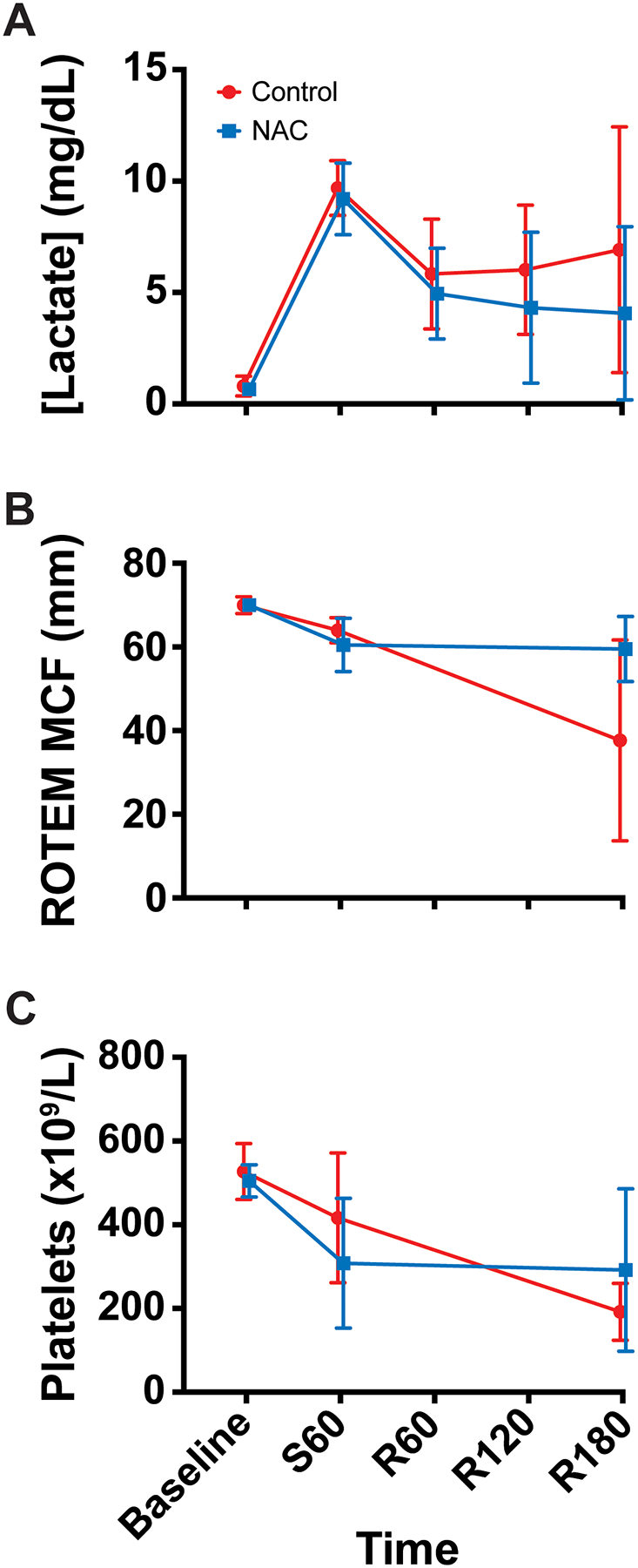
Background: Severe injury induces systemic microvascular impairment that reduces microvascular blood flow (MBF), even after resuscitation to normal blood pressure. These changes are associated with organ dysfunction and death, but the underlying causes and potential therapeutic approaches to address them remain unclear. Two possible contributors are hyperadhesive von Willebrand factor (VWF) secretion from an activated endothelium and oxidative modification of hemostatic proteins. N-acetylcysteine has been shown to address both of these processes and increase MBF in other disease states with similar features. Methods: Anesthetized, male Sprague-Dawley rats were subjected to a standardized polytrauma and pressure-targeted catheter hemorrhage. They then received either no treatment (control) or a single bolus of N-acetylcysteince (NAC), followed by autologous whole blood transfusion. Renal MBF was measured using contrast-enhanced ultrasound at prespecified time points. VWF multimer gels and other laboratory studies were performed. Histologic analysis of vascular thrombi was also performed on uninjured tissue from rats undergoing either this trauma protocol or a sham procedure. Results: NAC increased MBF at 3 h after resuscitation. This was accompanied by a decrease in VWF multimer size that was not seen in the control group. Histologic data showed an overall increase in systemic thrombus burden associated with trauma. Conclusions: NAC improves renal MBF, possibly by reducing VWF multimer size and reducing microthrombus burden. This is significant both mechanistically and therapeutically. It sheds light on the possible pathways involved in causing microvascular obstruction after trauma and identifies possible treatment approaches that could be developed further. Ultimately, targeting these pathways could move us closer to resuscitation strategies that optimize vital organ MBF.

Keywords: Microvascular blood flow; antioxidant; rats; trauma; von Willebrand factor.


Conflict of interest statement

The authors report no conflicts of interest.

Figures

Figure 1.
Figure 1.
Vital sign data (mean, standard deviation) (Control n=7, NAC n=6). (A) Mean arterial pressure (MAP), (B) Heart rate (HR), and (C) Temperature over time. Time points are denoted with “S” for shock period or “R” for resuscitation period and the number of minutes elapsed in that period.
Figure 2.
Figure 2.
Contrast-enhanced ultrasound data, normalized to mean at baseline (mean, standard deviation) (Control n=7, NAC n=6). (A) Total blood flow, (B) blood volume, and (C) blood velocity over time. Asterisk represents time point that is significantly different between groups. Time points are denoted with “S” for shock period or “R” for resuscitation period and the number of minutes elapsed in that period.
Figure 3.
Figure 3.
VWF multimer gel data. (A) Percentage of high molecular weight VWF multimers (HMW, >10 protomers) over time in Control and NAC groups (Control n=3, NAC n=4). Example gels (B) and densitometry analyses (C&D) demonstrating an upshift toward larger multimers from baseline to the end of the shock period, followed by a downshift at the end of the resuscitation period that was more pronounced in the NAC group. Time points are denoted with “S” for shock period or “R” for resuscitation period and the number of minutes elapsed in that period. Asterisks denote p<0.010.
Figure 4.
Figure 4.
Laboratory data (mean, standard deviation). (A) Serum lactate concentration (Control n=7, NAC n=6), (B) rotational thromboelastometry maximum clot firmness (ROTEM MCF) (Control n=3, NAC n=2), and (C) platelet count (Control n=7, NAC n=6) over time. Time points are denoted with “S” for shock period or “R” for resuscitation period and the number of minutes elapsed in that period.
Figure 5.
Figure 5.
Histology data (Sham n=6, Trauma n=7). PTAH thrombus score for (A) all tissue combined, (B) kidney, (C) intestine, (D) liver, (E) lung, and (F) heart between rats subjected to sham procedure (green) and to trauma-shock-resuscitation protocol (red). Asterisk denotes p<0.050. (G) Example H&E (top) and PTAH (bottom) 200x images of intestine showing absent thrombi in a Sham rat (left) and high thrombus burden in the mucosal and submucosal vasculature of a Trauma rat (right) with arrows denoting thrombi. PTAH, phosphotungstic acid hematotoxylin; H&E, hematoxylin and eosin.

Similar articles

References

    1. Domizi R, Damiani E, Scorcella C, et al. Association between sublingual microcirculation, tissue perfusion and organ failure in major trauma: A subgroup analysis of a prospective observational study. PloS one. 2019;14(3):e0213085. doi: 10.1371/journal.pone.0213085 - DOI - PMC - PubMed
    1. Campos-Serra A, Mesquida J, Montmany-Vioque S, et al. Alterations in tissue oxygen saturation measured by near-infrared spectroscopy in trauma patients after initial resuscitation are associated with occult shock. Eur J Trauma Emerg Surg. Feb 2023;49(1):307–315. doi: 10.1007/s00068-022-02068-w - DOI - PMC - PubMed
    1. Tachon G, Harrois A, Tanaka S, et al. Microcirculatory alterations in traumatic hemorrhagic shock. Critical care medicine. Jun 2014;42(6):1433–41. doi: 10.1097/ccm.0000000000000223 - DOI - PubMed
    1. Hutchings SD, Naumann DN, Hopkins P, et al. Microcirculatory Impairment Is Associated With Multiple Organ Dysfunction Following Traumatic Hemorrhagic Shock: The MICROSHOCK Study. Critical care medicine. Sep 2018;46(9):e889–e896. doi: 10.1097/ccm.0000000000003275 - DOI - PubMed
    1. Ward KR. The microcirculation: linking trauma and coagulopathy. Transfusion. Jan 2013;53 Suppl 1:38s–47s. doi: 10.1111/trf.12034 - DOI - PubMed