Review
. 2025 Apr 21;13(4):431.
doi: 10.3390/vaccines13040431.

Viral Infections in Elderly Individuals: A Comprehensive Overview of SARS-CoV-2 and Influenza Susceptibility, Pathogenesis, and Clinical Treatment Strategies

Affiliations
Review

Viral Infections in Elderly Individuals: A Comprehensive Overview of SARS-CoV-2 and Influenza Susceptibility, Pathogenesis, and Clinical Treatment Strategies

Yanhao Huang et al. Vaccines (Basel). .

Abstract

As age increases, the immune function of elderly individuals gradually decreases, increasing their susceptibility to infectious diseases. Therefore, further research on common viral infections in the elderly population, especially severe acute respiratory syndrome coronavirus 2 (SARS-CoV-2) and influenza viruses, is crucial for scientific progress. This review delves into the genetic structure, infection mechanisms, and impact of coinfections with these two viruses and provides a detailed analysis of the reasons for the increased susceptibility of elderly individuals to dual viral infections. We evaluated the clinical manifestations in elderly individuals following coinfections, including complications in the respiratory, gastrointestinal, nervous, and cardiovascular systems. Ultimately, we have summarized the current strategies for the prevention, diagnosis, and treatment of SARS-CoV-2 and influenza coinfections in older adults. Through these studies, we aim to reduce the risk of dual infections in elderly individuals and provide a scientific basis for the prevention, diagnosis, and treatment of age-related viral diseases, thereby improving their health status.

Keywords: SARS-CoV-2; aging; coinfections; influenza viruses; pathogenesis.


Conflict of interest statement

The authors declare that they have no conflicts of interest.

Figures

Figure 1
Figure 1
World population age structure distribution from 1950 to 2050. Changes in the proportions of three age groups (0–14, 15–64, and 65+) and the median life expectancy (years) worldwide from 1950 to 2050. Owing to factors such as societal development and shifts in public health perceptions, there has been a significant increase in median life expectancy. The proportion of the population aged 65 and over has been steadily rising over time, indicating a trend toward an aging global population structure. Data source: Population Division of the Department of Economic and Social Affairs of the United Nations. (see https://population.un.org/wpp (accessed on 2 September 2024) for more details).
Figure 2
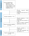
Figure 2
The process of study selection for older adults coinfected with SARS-CoV-2 and influenza.
Figure 3
Figure 3
Aging, senescent cells, and inflammaging in chronic inflammation. As age increases, the accumulation of senescent cells and the associated SASP lead to a persistent low-grade inflammatory state, known as inflammaging [56,89]. This inflammatory state tends to affect the myeloid skewing of HSCs, promoting innate immune responses while weakening adaptive immunity [56]. Innate immune cells derived from myeloid progenitor cells exhibit a decline in their migratory and antigen-presenting capabilities, indicating a weakened anti-inflammatory capacity [66]. However, their ability to secrete pro-inflammatory cytokines increases with age. On the other hand, adaptive immune cells, such as T and B cells, exhibit reduced cytotoxicity [87], leading to a weakened ability to clear senescent cells. Moreover, an increase in the subset of immune cells that secrete pro-inflammatory cytokines also contributes to elevated inflammation levels in elderly individuals [73]. The accumulation of senescent cells enhances the pro-inflammatory capacity of the immune system in elderly individuals while reducing its anti-inflammatory function, directly contributing to inflammation. Concurrently, the immune system’s diminished ability to clear senescent cells indirectly promotes inflammaging through the SASP, creating a positive feedback loop. SASP: senescence-associated secretory phenotype; HSCs: hematopoietic stem cells.
Figure 4
Figure 4
Clinical manifestations and complications of coinfection with influenza and COVID-19 in the elderly. The common symptoms associated with SARS-CoV-2 and influenza coinfection, as analyzed in a large number of cases, include fever, cough, dyspnea, nasal congestion, pharyngalgia, myalgia, fatigue, headache, and expectoration [36,40,109]. Moreover, in high-risk populations such as elderly individuals, coinfection with SARS-CoV-2 and influenza can lead to not only respiratory complications but also other severe adverse outcomes. The most common complications prior to death include ARDS, AKI, acute cardiac injury, and liver dysfunction [110].
Figure 5
Figure 5
Pathogenesis of sepsis in elderly patients with SARS-CoV-2 or influenza. The pathogenesis of sepsis primarily involves an imbalance in inflammation, immune dysfunction, mitochondrial damage, neuroendocrine–immune network dysregulation, endoplasmic reticulum stress, endothelial injury, and autophagy deficiency [138,143]. These factors interact and contribute to each other, and with increasing age, they tend to be in an imbalanced state, which is one of the important reasons why elderly individuals are more prone to developing severe and critical conditions after being infected with SARS-CoV-2 or influenza virus.
Figure 6
Figure 6
HPA axis and immune response regulation in sepsis: balancing inflammation and immunosuppression. The HPA axis modulates the immune response through the neuroendocrine pathway, releasing cortisol to maintain immune balance [154]. Concurrently, inflammatory mediators produced by immune activation can regulate the neuroendocrine system via feedback, ensuring that the immune response effectively eliminates pathogens without triggering excessive inflammation. However, in the context of sepsis, the function of the HPA axis may be compromised, along with a relative insufficiency of adrenal cortical hormones, potentially leading to systemic inflammation and immunosuppression, thereby impacting the clinical prognosis of patients. IL-1: interleukin-1; IL-6: interleukin-6; TNF-α: tumor necrosis factor-alpha.
Figure 7
Figure 7
Protein kinases PERK, ATF6, and IRE1 in the ER stress response and apoptosis. PERK is a protein kinase located on the ER membrane. When proteins are properly folded, PERK forms a stable complex with molecular chaperones such as BIP/GRP78. Misfolded proteins bind to BIP/GRP78, competing with PERK for interaction, which leads to the release and activation of PERK. Activated PERK undergoes oligomerization and autophosphorylation, which then phosphorylates eIF2α. Phosphorylated eIF2α inhibits protein translation and synthesis, reducing the load on protein folding in the ER and providing a protective effect for the cell. As the duration and intensity of the stress response increase, phosphorylated eIF2α induces the transcriptional activation of the transcription factor ATF4, which in turn promotes the expression of the apoptotic signaling molecule CHOP [148,155,156]. ATF6 is a type II transmembrane protein located on the ER membrane. During ERS, ATF6 is transported to the Golgi and is cleaved and activated by S1P and S2P. This protein subsequently migrates to the nucleus under the guidance of nuclear localization signals, where it induces the expression of CHOP and other proapoptotic signaling molecules [148,155,156]. The activation of ATF6 may also affect the function of the mitochondria by influencing the exchange of calcium ions between the ER and the mitochondria, thereby exacerbating mitochondrial damage [157]. Like PERK, IRE1 is also a protein kinase located on the ER membrane, and its activation mechanism mirrors that of PERK. Under conditions of ERS, activated IRE1 can recruit and activate JNK, which then phosphorylates and inhibits the activity of antiapoptotic proteins in the Bcl-2 family, thereby promoting apoptosis [148,155,156]. On the other hand, activated IRE1 also recruits the cytoplasmic regulatory protein TRAF-2, which in turn activates Caspase-12, initiating the caspase cascade and mediating apoptosis [158]. ER: endoplasmic reticulum; PERK: protein kinase R-like endoplasmic reticulum kinase; eIF2α: eukaryotic initiation factor 2 alpha; ATF-4: activating transcription factor 4; ATF-6: activating transcription factor 6; CHOP: C/EBP homologous protein; S1P: serine protease site-1; S2P: serine protease site-2; IRE1: inositol-requiring enzyme 1; JNK: c-Jun N-terminal kinase.
Figure 8
Figure 8
Neurological complications caused by SARS-CoV-2 and influenza virus. SARS-CoV-2 and influenza virus can cross the BBB through various means, either directly or indirectly, causing neurological damage or dysfunction, which can subsequently lead to a range of neurological complications and clinical manifestations.

Similar articles

References

    1. Hua K., Pan Y., Fang J., Wu H., Hua Y. Integrating social, climate and environmental changes to confront accelerating global aging. BMC Public Health. 2024;24:2838. doi: 10.1186/s12889-024-20346-7. - DOI - PMC - PubMed
    1. Fang E.F., Xie C., Schenkel J.A., Wu C., Long Q., Cui H., Aman Y., Frank J., Liao J., Zou H., et al. A research agenda for ageing in China in the 21st century (2nd edition): Focusing on basic and translational research, long-term care, policy and social networks. Ageing Res. Rev. 2020;64:101174. doi: 10.1016/j.arr.2020.101174. - DOI - PMC - PubMed
    1. Chen Y., Klein S.L., Garibaldi B.T., Li H., Wu C., Osevala N.M., Li T., Margolick J.B., Pawelec G., Leng S.X. Aging in COVID-19: Vulnerability, immunity and intervention. Ageing Res. Rev. 2021;65:101205. doi: 10.1016/j.arr.2020.101205. - DOI - PMC - PubMed
    1. Uyeki T.M., Hui D.S., Zambon M., Wentworth D.E., Monto A.S. Influenza. Lancet. 2022;400:693–706. doi: 10.1016/S0140-6736(22)00982-5. - DOI - PMC - PubMed
    1. Li J., Chen Y., Wang X., Yu H. Influenza-associated disease burden in mainland China: A systematic review and meta-analysis. Sci. Rep. 2021;11:2886. doi: 10.1038/s41598-021-82161-z. - DOI - PMC - PubMed

LinkOut - more resources