. 2025 Apr 16;18(8):1826.
doi: 10.3390/ma18081826.

3D Printing and Electrospinning of Drug- and Graphene-Enhanced Polycaprolactone Scaffolds for Osteochondral Nasal Repair

Affiliations

3D Printing and Electrospinning of Drug- and Graphene-Enhanced Polycaprolactone Scaffolds for Osteochondral Nasal Repair

Izabella Rajzer et al. Materials (Basel). .

Abstract

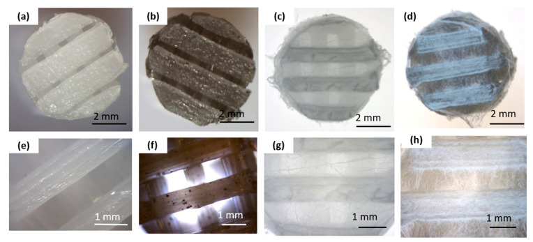
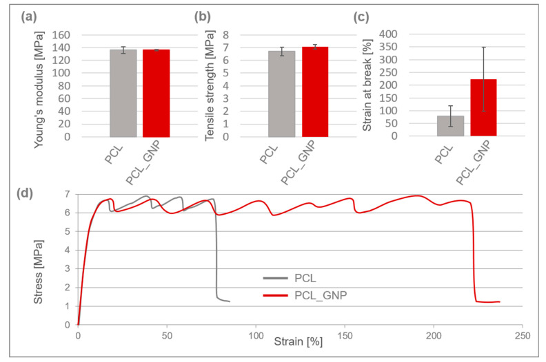
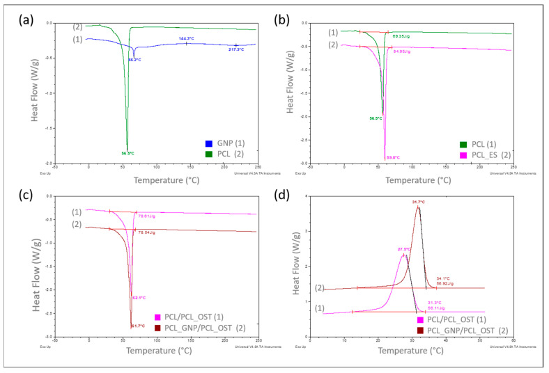
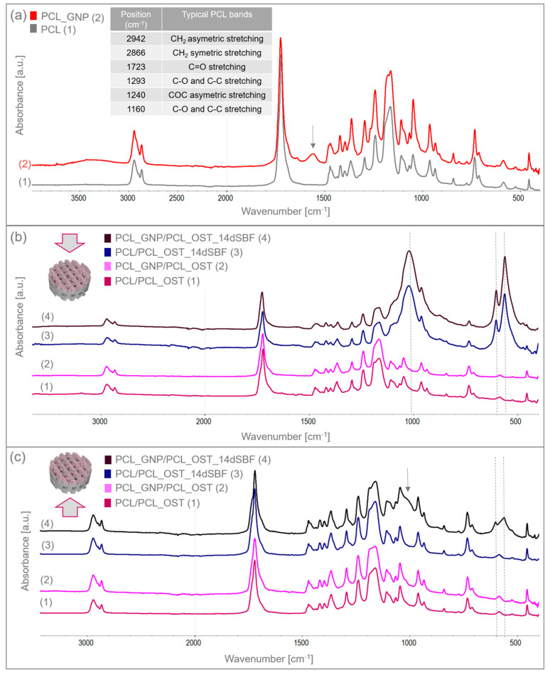
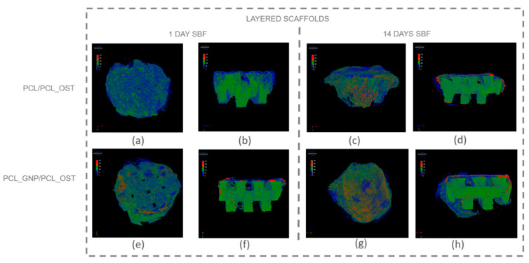
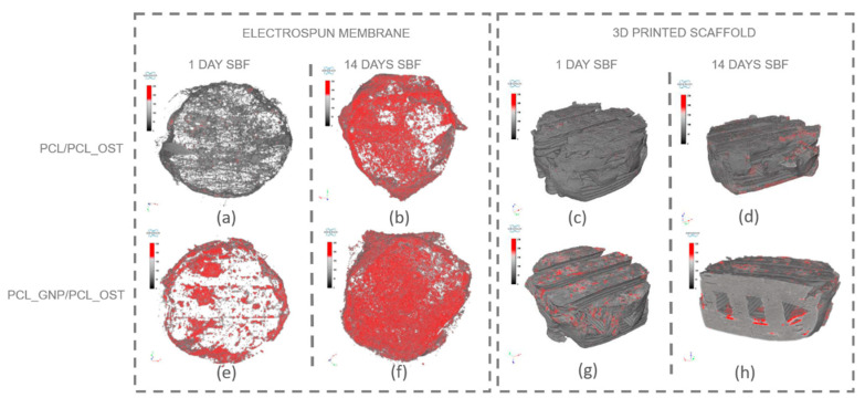
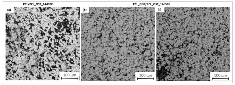
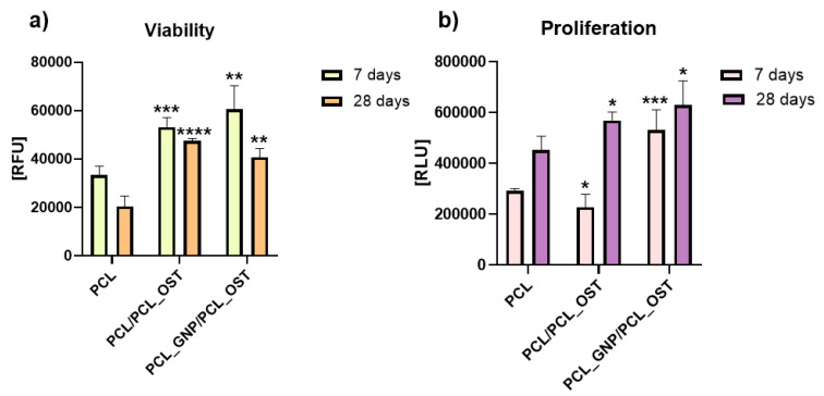
A novel bi-layered scaffold, obtained via 3D printing and electrospinning, was designed to improve osteochondral region reconstruction. The upper electrospun membrane will act as a barrier against unwanted tissue infiltration, while the lower 3D-printed layer will provide a porous structure for tissue ingrowth. Graphene was integrated into the scaffold for its antibacterial properties, and the drug Osteogenon® (OST) was added to promote bone tissue regeneration. The composite scaffolds were subjected to comprehensive physical, thermal, and mechanical evaluations. Additionally, their biological functionality was assessed by means of NHAC-kn cells. The 0.5% graphene addition to PCL significantly increased strain at break, enhancing the material ductility. GNP also acted as an effective nucleating agent, raising crystallization temperatures and supporting mineralization. The high surface area of graphene facilitated rapid apatite formation by attracting calcium and phosphate ions. This was confirmed by FTIR, µCT and SEM analyses, which highlighted the positive impact of graphene on mineral deposition. The synergistic interaction between graphene nanoplatelets and Osteogenon® created a bioactive environment that enhanced cell adhesion and proliferation, and promoted superior apatite formation. These findings highlight the scaffold's potential as a promising biomaterial for osteochondral repair and regenerative medicine.

Keywords: 3D printing; bi-layer scaffold; drug; electrospinning; graphene; polycaprolactone.


Conflict of interest statement

The authors declare no conflict of interest.

Figures

Figure 1
Figure 1
Macro- and microscopic characterization of the scaffold before and after electrospinning: (a,e) pure PCL 3D-printed scaffold; (b,f) graphene-modified 3D-printed scaffold (PCL_GNP); (c,g) 3D-printed PCL scaffold with electrospun PCL_OST membrane (PCL/PCL_OST); (d,h) 3D-printed PCL_GNP scaffold with electrospun PCL_OST membrane (PCL_GNP/PCL_OST). Stereoscopic microscope images (ad) and optical microscope images (eh).
Figure 2
Figure 2
Mechanical properties of 3D-printed PCL and PCL_GNP scaffolds: (a) Young’s modulus; (b) tensile strength; (c) strain at break; (d) typical stress–strain curves.
Figure 3
Figure 3
DSC curves recorded over a specified temperature range at a heating or cooling rate of 10°/min in a nitrogen atmosphere (flow 40 mL/min), respectively for samples: (a) PCL base polymer (2) and GNP used as a modifier (1)—heating mode; (b) PCL samples in granular (1) and ES membrane (2) form—heating mode; (c) with PCL_OST membrane applied to a scaffold printed from: PCL- (1) and GNP-modified PCL (2)—heating mode; (d) after melting 3D scaffolds printed from PCL (1) and PCL/GNP (2) with membrane PCL/OST layer applied—cooling mode.
Figure 4
Figure 4
FTIR spectra of (a) 3D-printed PCL and PCL_GNP scaffolds; (b) layered samples before and after SBF immersion, analyzed from the membrane side; (c) layered samples before and after 14-day immersion in SBF, analyzed from the scaffold side.
Figure 5
Figure 5
Micro-CT images of scaffolds after 1-day and 14-day SBF incubation: (ad) PCL/PCL_OST scaffold; (eh) PCL_GNP/PCL_OST scaffold. Images (a,c,e,g)—scaffolds top view, (b,d,f,h)—cross-sections. Bottom layer of 3D-printed scaffold marked in green, electrospun membrane marked in blue, and modifying particles, primarily distributed on the membrane, highlighted in red.
Figure 6
Figure 6
Separate micro-CT images of electrospun membranes and bottom layers of scaffolds for PCL/PCL_OST and PCL_GNP/PCL_OST samples after 1 and 14 days of incubation in SBF solution. Electrospun membranes of PCL/PCL_OST scaffold after 1 day (a) and 14 days (b); bottom layer of PCL/PCL_OST scaffold after 1 day (c) and 14 days (d). Electrospun membranes of PCL_GNP/PCL_OST scaffold after 1 day (e) and 14 days (f); bottom layer of PCL_GNP/PCL_OST scaffold after 1 day (g) and 14 days (h). Apatite formation on sample surfaces marked in red.
Figure 7
Figure 7
SEM images of samples after 14 days of immersion in SBF solution: (a) PCL/PCL_OST layered scaffold; (b,c) PCL_GNP/PCL_OST layered scaffold.
Figure 8
Figure 8
Cell viability (a) and proliferation (b) after 7 and 28 days of culture in direct contact with scaffold. Results expressed as the mean ± SD measured at least from 3 independent experiments. Significance determined with unpaired t-test. p-values of 0.05 or less considered statistically significant. * p values 0.05, ** p values 0.01, *** p values 0.001, **** p values 0.0001.

Similar articles

Cited by

References

    1. Oladele A.O., Olabanji J.K., Olekwu A.A., Awe O.O. Experience with management of nasal defects. Niger. J. Plast. Surg. 2018;14:36–44. doi: 10.4103/njps.njps_8_18. - DOI
    1. Liu Y., Zhou G., Cao Y. Recent Progress in Cartilage Tissue Engineering—Our Experience and Future Directions. Engineering. 2017;3:28–35. doi: 10.1016/J.ENG.2017.01.010. - DOI
    1. Lin G., Lawson W. Complications using grafts and implants in rhinoplasty. Oper. Tech. Otolaryngol. Head Neck Surg. 2007;18:315–323. doi: 10.1016/j.otot.2007.09.004. - DOI
    1. Rajzer I., Stręk P., Wiatr M., Skladzien J., Kurowska A., Kopeć J., Swiezy K., Wiatr A. Biomaterials in the Reconstruction of Nasal Septum Perforation. Ann. Otol. Rhinol. Laryngol. 2021;130:731–737. doi: 10.1177/0003489420970589. - DOI - PubMed
    1. Selim M., Mousa H.M., Abdel-Jaber G.T., Barhoum A., Abdal-hay A. Innovative designs of 3D scaffolds for bone tissue regeneration: Understanding principles and addressing challenges. Eur. Polym. J. 2024;215:113251. doi: 10.1016/j.eurpolymj.2024.113251. - DOI

LinkOut - more resources