. 2025 May 7;20(5):e0321145.
doi: 10.1371/journal.pone.0321145. eCollection 2025.

AAV delivery of GBA1 suppresses α-synuclein accumulation in Parkinson's disease models and restores functions in Gaucher's disease models

Affiliations

AAV delivery of GBA1 suppresses α-synuclein accumulation in Parkinson's disease models and restores functions in Gaucher's disease models

Takuro Okai et al. PLoS One. .

Abstract

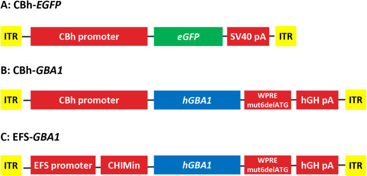
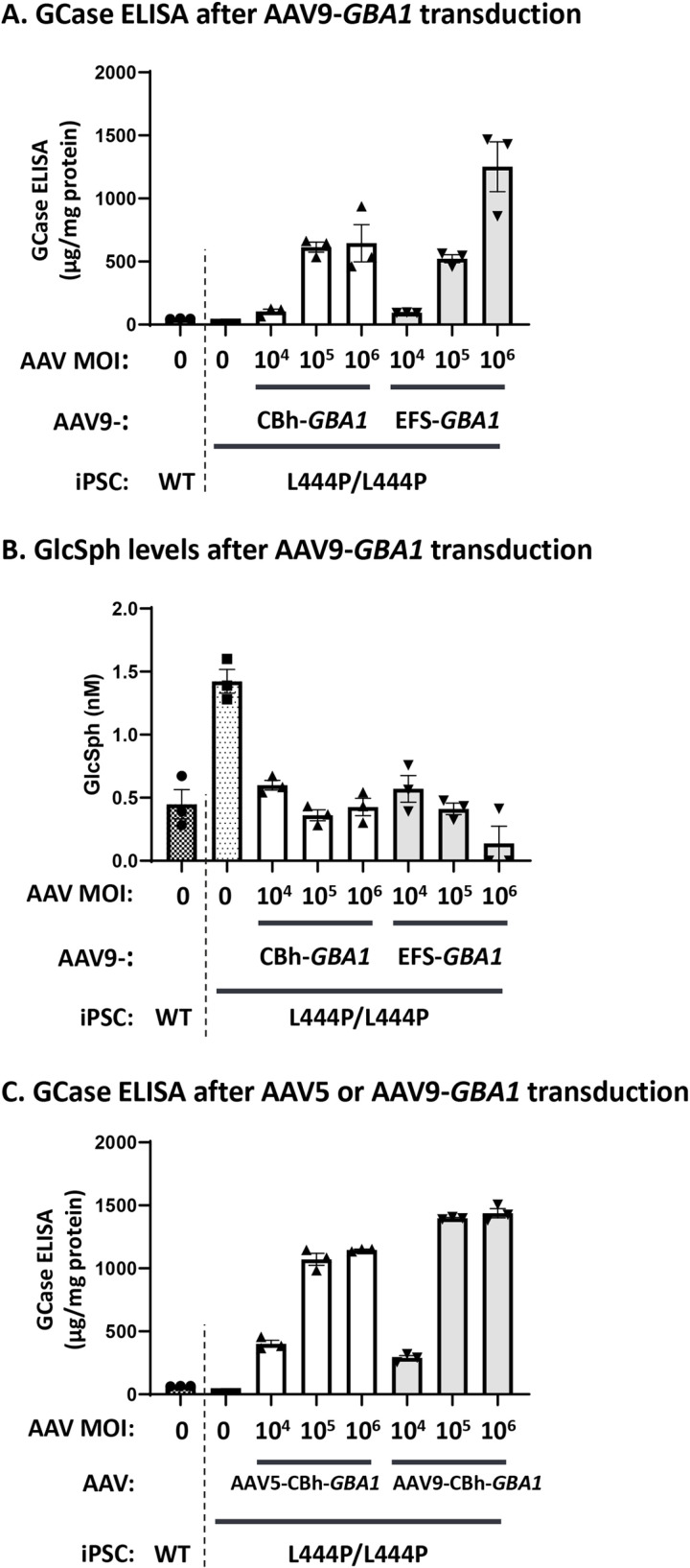
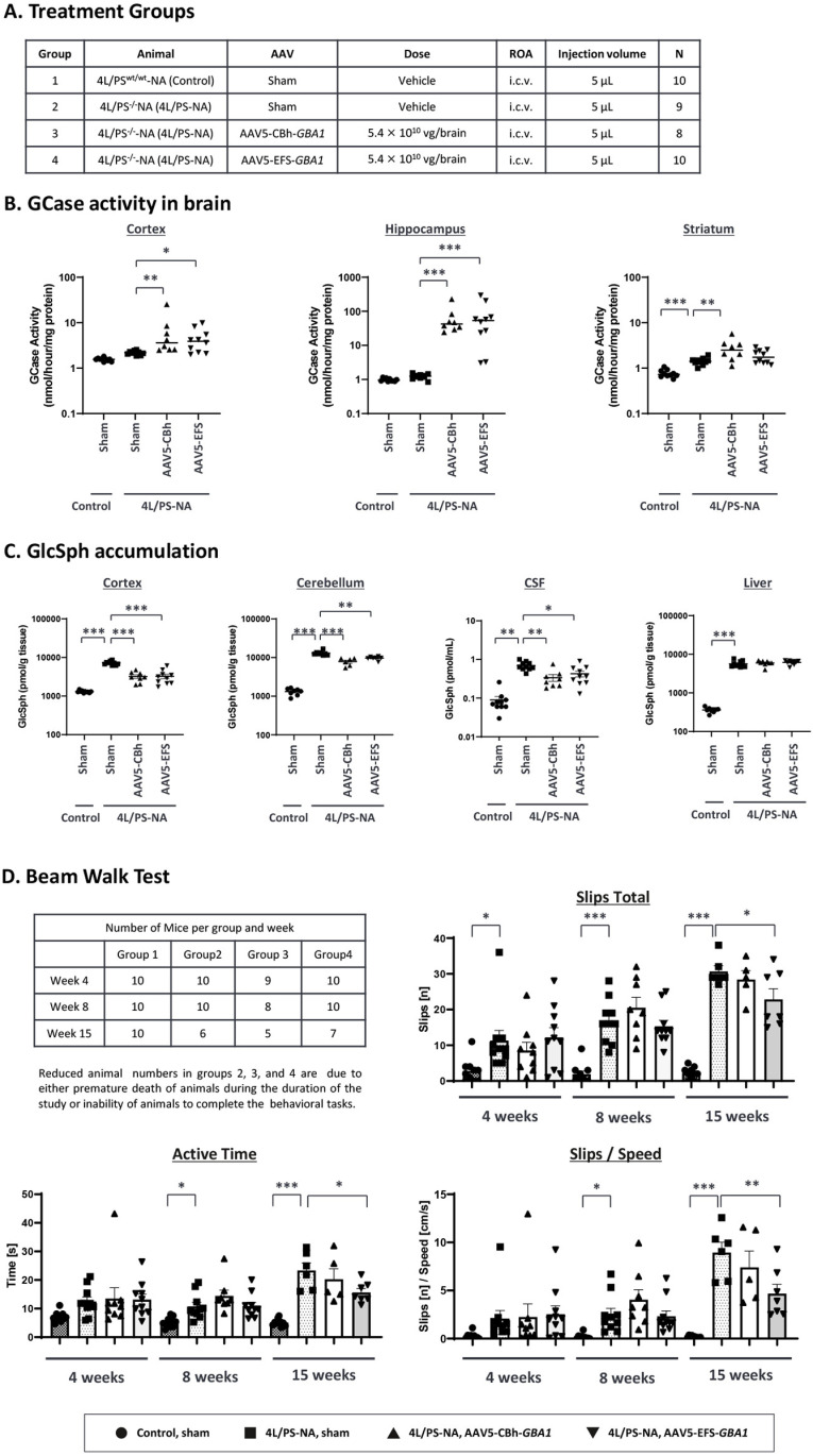
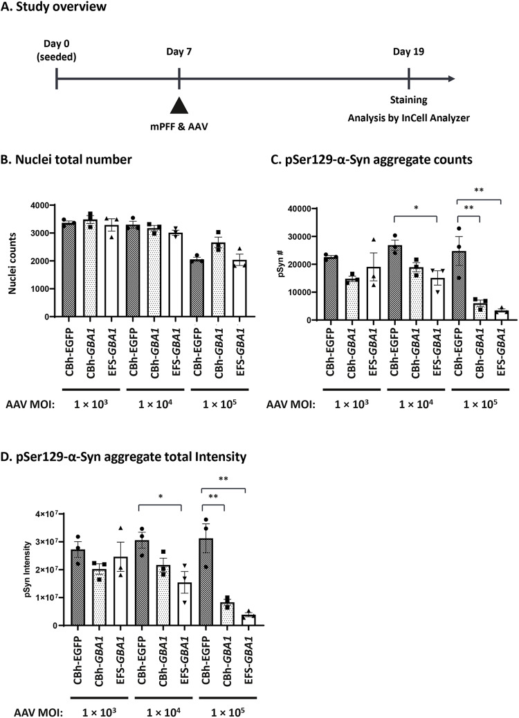
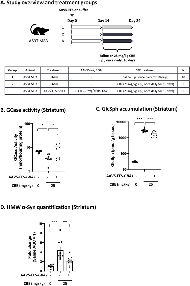
Biallelic mutations in the glucosylceramidase beta 1 (GBA1) gene are the underlying genetic cause of Gaucher's disease (GD), resulting in a deficient lysosomal hydrolase and subsequent accumulation of glycosphingolipids. More recently, GBA1 mutations have been identified as the most prevalent genetic risk factor for Parkinson's disease (PD), associated with more pronounced symptoms characterized by earlier onset and accelerated cognitive decline. In these GBA-associated PD patients the α-synuclein pathology is more prominent, and recent data suggest a link between α-synucleinopathies and GBA1 mutations. Here, we explored the effect of GBA1 gene supplementation on the GD phenotypes and α-synuclein pathology by using the adeno-associated virus (AAV) system. We have compared two AAV serotypes, AAV5 and AAV9, and two different ubiquitous promoters, and demonstrate that both promoters work efficiently albeit not the same in vitro and in vivo. GBA1 overexpression reduces the accumulation of glucosylsphingosine (GlcSph) and restores motor dysfunction in a GD mouse model. We further demonstrate that GBA1 overexpression can dissolve phospho-α-synuclein aggregation induced by the addition of α-synuclein pre-formed fibril (PFF) in a mouse primary neuron model suggesting the direct effect of β-Glucocerebrosidase (GCase) on α-synuclein accumulation. In vivo, we show that GCase inhibition can induce insoluble high-molecular-weight α-synuclein aggregation and that delivery of GBA1 achieves robust reduction of the α-synuclein aggregates in the mouse brain. In summary, GCase expression not only reduces GlcSph, but also restores GD motor dysfunction and removes α-synuclein aggregates which are the hallmark for PD and α-synucleinopathies. AAV delivery of GBA1 is a powerful approach to restore glucocerebrosidase function and to resolve misfolded α-synuclein protein, with applications for GD and PD.


Conflict of interest statement

I have read the journal's policy and the authors of this manuscript have the following competing interests: The authors declare the following financial interests/personal relationships which may be considered as potential competing interests: T. Okai, S. Sato, S. Matsumoto, M. Nakayama, S. Yamamoto, T. Hioki, and M. Tanaka report on a relationship with Takeda Pharmaceutical Company Limited that includes employment and non-financial support. M. Deshpande, B. Strack-Logue and G. Proetzel report on a relationship with Takeda Development Center Americas that includes employment and non-financial support.

Figures

Fig 1
Fig 1. Expression cassette overview.
(A) – (C) Illustration of the three expression cassettes incorporated in the AAV5 or AAV9 vectors. Abbreviations: ITR, inverted terminal repeat; WPREmut6delATG, 597 bp modified woodchuck hepatitis virus post-transcriptional regulatory element removing the ATGs; hGH poly A, human Growth hormone polyadenylation signal; CBh, chicken β-actin hybrid promoter with the CMV enhancer in reverse orientation; EFS, a truncated 255 bp short form of the human EF1α promoter; CHIMin, chimeric chicken β-actin and rabbit β-globin intron; eGFP, enhanced Green Fluorescent Protein; hGBA1, human glucosylceramidase beta 1.
Fig 2
Fig 2. GCase expression and GlcSph reduction in iPS-derived DA neurons treated with either AAV5- or AAV9-GBA1 vectors.
Human wild-type (WT) or L444P/L444P GBA1 mutant iPSC-derived DA neurons were transduced with AAV vectors at three MOIs (1 × 104, 1 × 105, and 1 × 106 VG) seven days after differentiation and analyzed two weeks after AAV transduction. In (A) and (C), GCase protein levels in the cell lysate were measured using an MSD ELISA and normalized to total protein concentration assessed by BCA protein assay (mean ± SE (n = 3)). In (B), GlcSph concentration was analyzed by LC-MS/MS (mean ± SE (n = 3)). In (A) and (B), the cells were transduced with AAV9-CBh-GBA1 or AAV9-EFS-GBA1, and in (C), cells were transduced with AAV5-CBh-GBA1 or AAV9-CBh-GBA1.
Fig 3
Fig 3. GCase activity, GlcSph, and VG analysis in Gba1 D409V KI mouse administered with either AAV5 or AAV9-GBA1.
The study outline is described in the table (A). AAV5-GBA1 or AAV9-GBA1 were delivered by bilateral i.c.v. injection of buffer (sham) or AAV (3.3 × 1010 VG/ hemisphere, total 6.6 × 1010 VG/brain) in 16-week-old male Gba1 D409V KI mice or WT littermates. Mice were sacrificed 4 weeks after injection. Brains were isolated and sliced as 2-mm-thick brain slices around the injection site, and tissues were analyzed for GCase enzymatic activity, GlcSph level, and VG. (B) GCase activity was measured in each independent study (Study 1 and Study 2). See S1 Table for mean values. (C) GlcSph concentration was analyzed by LC-MS/MS. See S2 Table for the mean values per group. (D) VG in brain and liver was measured by qPCR specific for DNA detection. VG in the liver and brain of groups 1, 2, 4, and 5 were below lower limit of quantitation (LLOQ) as expected as these were sham controls. For B, C and D, each graph represents mean ± S.E.M. (n = 4 or 8) with the Y-axis as logarithmic scale. For B and C, statistical analyses were performed by Dunnett analysis. *: < 0.05; **: < 0.01; ***: < 0.001, compared to Group 2 and Group 5 for batch 1 study and batch 2 study, respectively. For D, statistical analysis is not performed since the control value is zero.
Fig 4
Fig 4. Biochemical and behavior analysis in 4L/PS-NA mouse model treated with AAV5-GBA1.
Table (A) illustrates the study overview. 4-week-old male 4L/PSwt/wt-NA (control) and 4L/PS-/--NA (4L/PS-NA) mice were injected with buffer (sham), AAV5-CBh-GBA1 or AAV5-EFS-GBA1 (i.c.v., bilaterally, 2.7 × 1010 VG/hemisphere, total 5.4 × 1010 VG/brain). After 15 weeks, brain and liver tissues were isolated. In Group 2 one mouse died prematurely and in Group 3 two mice died prematurely, reducing the group size to 9 and 8, respectively. (B) GCase activity was measured in three different brain regions. The Y-axis is logarithmic scale. The stars indicate the statistical difference compared to the control Group 2. See S3 table for the mean values for GCase activity. (C) GlcSph concentration was measured by using MS/MS analysis. The Y-axis is logarithmic scale. The stars indicate the statistical difference compared to the control Group 2. See S4 table for the mean values for GlcSph concentration. (D) The Beam Walk Test was performed three times (at 4, 8 and 15 weeks after AAV administration). Graphs represent total slips [n], active time [s], total slips [n] per speed [cm/s] for each group on the beams tested at all time points. Data are shown as means ± SEM per group. Statistical analyses were performed by Dunnett analysis. *: < 0.05; **: < 0.01; ***: < 0.001 compared to Group 2 (4L/PS-NA, sham group) in each timepoint.
Fig 5
Fig 5. Phospho-
α-synuclein reduction in mouse primary neuron cells treated with AAV9-GBA1. (A) illustrates the study layout. Mouse primary cortical neurons were seeded on poly-D-Lysin coated 96-well plate (45,000 cells/well) on Day 0. On Day 7, mouse pre-formed fibril (mPFF; 1.1 ng/ml) and AAV9-EGFP or AAV9-GBA1 (MOI at 1 × 103, 1 × 104, or 1 × 105 VG) were added to the cells as noted on the x-axis. After 12-day culture, cells were stained with anti-phospho-α-synuclein antibody and analyzed by InCell Analyzer. Quantification of the nuclei counts is shown in (B), the pSer129 α-Syn aggregate counts in (C), and the intensity of pSer129 α-Syn aggregates in (D). Data are represented as mean ± S.E.M. (n = 3). Statistical analyses were performed by Dunnett analysis. *: < 0.05; **: < 0.01; ***: < 0.001 compared to CBh-EGFP control in each dose (MOI).
Fig 6
Fig 6. Efficacy of AAV5-GBA1 on GCase activity, GlcSph accumulation and HMW
α-synuclein in A53T M83 mouse model treated with CBE. In (A), the study outline is illustrated. 12-week-old male A53T M83 mice were administered with AAV5-EFS-GBA1 (i.c.v., bilaterally, 1.7 × 1010 VG/hemisphere, total 3.4 × 1010 VG/brain) on day 0. For Group 2 and 3 CBE treatment (25 mg/kg, i.p., once daily) started two weeks (day 14) after AAV administration and continued for 10 days (up to day 24). At the end of the study (day 24) striatum was analyzed for GCase activity (B), stars indicate statistical significance over the control Group 2. (C) shows GlcSph levels, stars indicate statistical significance over the control Group 2. In (D) the AUC was normalized with the average of Group 1 quantified by WES™. See S5 and S6 tables for mean values for GCase activity and GlcSph levels. Statistical analyses were performed by Dunnett analysis. *: < 0.05; **: < 0.01; ***: < 0.001 compared to group 2 (sham, CBE).

Similar articles

References

    1. Do J, McKinney C, Sharma P, Sidransky E. Glucocerebrosidase and its relevance to Parkinson disease. Mol Neurodegener. 2019;14(1):36. doi: 10.1186/s13024-019-0336-2 - DOI - PMC - PubMed
    1. Riboldi GM, Di Fonzo AB. GBA, gaucher disease, and Parkinson’s disease: from genetic to clinic to new therapeutic approaches. Cells. 2019;8(4):364. doi: 10.3390/cells8040364 - DOI - PMC - PubMed
    1. Velayati A, Yu WH, Sidransky E. The role of glucocerebrosidase mutations in Parkinson disease and Lewy body disorders. Curr Neurol Neurosci Rep. 2010;10(3):190–8. doi: 10.1007/s11910-010-0102-x - DOI - PMC - PubMed
    1. Ferraz MJ, Kallemeijn WW, Mirzaian M, Herrera Moro D, Marques A, Wisse P, et al.. Gaucher disease and Fabry disease: new markers and insights in pathophysiology for two distinct glycosphingolipidoses. Biochim Biophys Acta. 2014;1841(5):811–25. doi: 10.1016/j.bbalip.2013.11.004 - DOI - PubMed
    1. Charrow J, Andersson HC, Kaplan P, Kolodny EH, Mistry P, Pastores G, et al.. The Gaucher registry: demographics and disease characteristics of 1698 patients with Gaucher disease. Arch Intern Med. 2000;160(18):2835–43. doi: 10.1001/archinte.160.18.2835 - DOI - PubMed

MeSH terms