. 2025 Aug 21;76(12):3412-3426.
doi: 10.1093/jxb/eraf129.

Starting strong: development and biomechanics of the seedling-host interaction in European mistletoe (Viscum album)

Affiliations

Starting strong: development and biomechanics of the seedling-host interaction in European mistletoe (Viscum album)

Luiza Teixeira-Costa et al. J Exp Bot. .

Abstract

Attachment to a substrate is fundamental for plant growth and development. This is especially true for species that live either partially or fully off the ground, such as mistletoes, which have developed unique adaptations to anchor themselves securely to host trees from which they draw water and some nutrients. While the mechanical properties of attachment during the adult stages in many plant species have been described, the mechanical principles of the initial developmental stages are rarely investigated. Here, we focus on the parasitic European mistletoe (Viscum album L.) and its attachment to a host plant at the seedling stage. Using a combination of germination experiments, microtomography, histological analysis, and biomechanical tests, this work investigates the role of the three key attachment structures involved in this process: the seed coat, hypocotyl, and holdfast. The viscin layer, a sticky coating on the seed, provides initial adhesion before the growing hypocotyl expands towards the host surface, where it flattens and forms a holdfast that strengthens adhesion and aids tissue penetration. Tensile tests revealed that these three attachment structures withstand similar forces in the early stages, considerably higher than the weight of the seedling. Within a few months, the endophytic system interlocked with the host bark, forming a robust connection that not only transports water but also increased the mechanical strength of the structure. This work highlights the fundamental mechanisms of the initial mistletoe-host interaction, which forms the basis of their decades-long relationship.

Keywords: Haustorium; Santalaceae; microtomography; mistletoe; parasitic plant; plant biomechanics; seed attachment; seedling development; tensile test.


Conflict of interest statement

The authors have no conflict of interest to disclose.

Figures

Graphical Abstract
Graphical Abstract
Fig. 1.
Fig. 1.
Sketch of the experimental setup for the biomechanical analyses, divided according to the mistletoe organ studied. In all experiments, the host branch was gripped with the lower clamps of the testing machine. (A) For seed experiments, the mistletoe holdfast was removed (dashed line) and the seed was glued to a toothpick through which the force was applied. (B) For the hypocotyl experiments, a coated wire was threaded through the hypocotyl loop, connected to Kevlar threads and thus fixed to the upper clamps of the testing machine. (C) For the holdfast experiments, the mistletoe hypocotyl was removed (dashed line) and the holdfast was glued to a toothpick, through which the force was applied.
Fig. 2.
Fig. 2.
Seedling development. (A) Hypocotyl (arrow) protruding from the seed (s) marking successful germination. (B) Initial contact between hypocotyl (black arrows) and host branch. Notice abundant viscin (v). (C) Establishment of holdfast (arrowheads). Abundant viscin (v) still attaching the seed (s). Notice the dome-shaped holdfast (arrowheads). (D) Initial hypertrophy on host branch (Hh). Some viscin (v) is still present and the seed (s) remains attached to the host branch. (E) Seed (s) detachment from the host branch (white arrow). Notice increased host hypertrophy (Hh). (F) Cotyledon (c) release from seed coat. Notice flattening of the holdfast (arrowhead). All scale bars=1 mm.
Fig. 3.
Fig. 3.
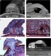
Morphology of early seedling development. (A) MicroCT scan showing a cavity (arrow) formed by the dome-shaped holdfast against the host bark (Hb). Notice high-density spots (arrowhead) visible above a high-density row of cells (stars). (B) MicroCT scan showing high-density spots (arrowheads) across the hypocotyl and holdfast visible above a high-density row of cells (stars). Penetration peg in the central area of the holdfast in contact with the host bark (Hb) forming a cavity ring (arrows). Notice viscin (v) remnants. (C) Longitudinal section through the seedling showing vessels (arrowhead) in the hypocotyl and in the upper part of the holdfast above two rows of crushed cells (stars), which flank an area of high meristematic activity (dashed circle). (D) Detail from Fig. 1C showing the holdfast with dark-stained cell wall remnants in the cavity ring (arrow) beside the area of high meristematic activity (dashed circle) and against the host bark (Hb). (E) Longitudinal section through the seedling showing initial penetration of the host bark (Hb) by the parasite endophyte (e), which has not yet reached the cambium zone (***), nor the host xylem (Hx). Notice also the elevation of the host bark (arrows) into the former cavity ring of the holdfast. (F) MicroCT scan of a Lugol-treated sample showing high density of holdfast (ho) and hypocotyl (hy) tissues indicating the presence of starch. Young endophyte (e) not yet in contact with the host cambium (***) or xylem (Hx). Low density in the region corresponding to the lines of crushed cells (black stars) indicating absence of starch. Black scale bars=1 mm; white scale bar=100 μm.
Fig. 4.
Fig. 4.
Morphology of the seedling at later developmental stages. (A) MicroCT scan showing expansion of the endophyte (e) within the host bark (Hb) prior to contact with host cambium zone (***) and xylem (Hx). Notice lines of crushed cells still visible (stars). (B) Virtual segmentation showing the three-dimensional expansion of the endophyte (e). Notice bent hypocotyl (hy) and the seed (s) still attached to host bark (Hb). (C) MicroCT showing initial contact between parasite endophyte (e) and host cambium zone (***). Lines of crushed cells (stars) become longer due to profuse endophyte proliferation. Notice host hypertrophy (Hh) in the most recent region of the host xylem (Hx). (D) Parasite sinker (Ps) embedded within the host xylem (Hx) forming vessel-to-vessel contact (dashed ellipses). (E) Virtual segmentation of a seed with two developing embryos whose endophyte (e) and sinkers (Ps) are not interconnected. (F) Virtual segmentation showing two sinkers (arrows) from the same seedling tissue within the hypertrophied (Hh) most recent annual ring on the host xylem (Hx). Black scale bar=200 μm; white scale bars=1 mm.
Fig. 5.
Fig. 5.
Maximum force data of biomechanical analysis of the three tested mistletoe organs. (A) Violin plot with individual data points of maximum forces from tensile tests on mistletoe seedling samples, divided according to the organ under load. The data points of the first batch are indicated by black symbols, and the data points of the second batch by coloured symbols. The left side of each group shows the data distribution of the first batch, with the median value marked as an asterisk. As the individual failure groups of the first batch of hypocotyl samples differ significantly from each other, they are labelled with different symbols (but were pooled for the presentation of the violin plot): white triangles indicate data points with failure at the holdfast, upright triangles indicate data points with failure at the hypocotyl, and inverted triangles indicate data points with failure at the seed. (B, C) Correlation analysis of the maximum force values of the tensile tests for the first batch of mistletoe seed samples (B) and the holdfast samples (C), with their corresponding contact area. The respective correlation coefficient of a linear regression is indicated.
Fig. 6.
Fig. 6.
Schematic representation of the forces (arrows) acting during four stages of mistletoe establishment. (A) Upon contact, the edges of the hypocotyl (hy) tip exert pressure (growth-induced forces marked by black arrows) against the host bark (Hb), generating a reaction force (white arrows) in the opposite direction along the bridge-like structure of the hypocotyl. Intermolecular adhesive forces (grey arrows) caused by viscin threads keep the seed (s) in place and in contact with the host bark. (B) At a later stage, the penetration peg grows against the host bark, causing reaction forces in the opposite direction within the holdfast (ho), forming layers of collapsed cells. (C) The endophyte (e) proliferates and causes pressure within the host bark, which becomes dislocated towards the area previously corresponding to the holdfast cavity, creating an interlocking mechanism between mistletoe and host. Note that the attachment forces between the seed and the host bark diminish as fewer viscin threads are present. (D) In the following stages, besides the formation of cortical strands (cs) and the flattening of the holdfast, additional lateral growth of the endophyte can be observed, which further strengthens the interlocking mechanism between mistletoe and host. The secondary growth of the host branch causes pressure against the main sinker, resulting in slightly irregular growth along its xylem–bark interface.

Similar articles

Cited by

References

    1. Aguilar-Venegas M, Quintana-Rodríguez E, Aguilar-Hernández V, López-García CM, Conejo-Dávila E, Brito-Argáez L, Loyola-Vargas VM, Vega-Arreguín J, Orona-Tamayo D.. 2023. Protein profiling of Psittacanthus calyculatus during mesquite infection. Plants 12, 464. - PMC - PubMed
    1. Albert M, Belastegui-Macadam X, Kaldenhoff R.. 2006. An attack of the plant parasite Cuscuta reflexa induces the expression of attAGP, an attachment protein of the host tomato. The Plant Journal 48, 548–556. - PubMed
    1. Ang SLP, Yong JWH.. 2005. A protocol for in vitro germination and sustainable growth of two tropical mistletoes. Plant Cell, Tissue and Organ Culture 80, 221–228.
    1. Aukema JE. 2003. Vectors, viscin, and Viscaceae: mistletoes as parasites, mutualists, and resources. Frontiers in Ecology and the Environment 1, 212–219.
    1. Barney CW, Hawksworth FG, Geils BW.. 1998. Hosts of Viscum album. European Journal of Forest Pathology 28, 187–208.

LinkOut - more resources