. 2025 Apr 17;30(8):1812.
doi: 10.3390/molecules30081812.

Role of Saponins from Platycodon grandiflorum in Alzheimer's Disease: DFT, Molecular Docking, and Simulation Studies in Key Enzymes

Affiliations

Role of Saponins from Platycodon grandiflorum in Alzheimer's Disease: DFT, Molecular Docking, and Simulation Studies in Key Enzymes

Ashaimaa Y Moussa et al. Molecules. .

Abstract

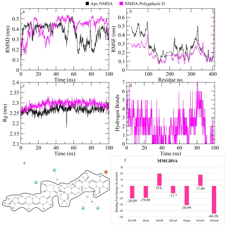
Alzheimer's disease (AD), one of the neurodegenerative disorders, afflicts negatively across the whole world. Due to its complex etiology, no available treatments are disease-altering. This study aimed to explore isolated saponins profiles from Platycodon grandiflorum in the binding pockets of six target proteins of AD using computational and quantum chemistry simulations. Initially, saponin compounds were docked to AD enzymes, such as GSK-3β and synapsin I, II, and III. The subsequent research from MD simulations of the best three docked compounds (polygalacin D2, polygalacin D, and platycodin D) suggested that their profiles match with the binding of standard active drugs like ifenprodil and donepezil to the six enzymes. Moreover, analyzing DFT quantum calculations of top-scoring compounds fully unravels their electronic and quantum properties and potential in anti-AD. The subtle differences between polygalacin D and D2, and platycodin D, were studied at the level of theory DFT/B3LYP, showing that the electron-donating effect of the hydroxy ethyl group in platycodin D rendering this compound of moderate electrophilicity and reactivity. Polygalacin D2 diglucoside substituent in position-2 contributed to its best binding and intermolecular interactions more than polygalacin D and prosapogenin D, which acted as the negative decoy drug.

Keywords: Alzheimer’s; Platycodon; density functional theory; molecular dynamics simulations; saponins.


Conflict of interest statement

The authors declare no conflicts of interest.

Figures

Figure 1
Figure 1
Chemical library of platycodin saponins subjected to computational analysis.
Figure 2
Figure 2
(A) Polygalacin D HOMO-LUMO, (B) prosapogenin D HOMO-LUMO, (C) platycodin D HOMO-LUMO, (D) polygalacin D2 HOMO-LUMO.
Figure 3
Figure 3
(a) Polygalacin D electrostatic potential, (b) prosapogenin D electrostatic potential, (c) platycodin D electrostatic potential, (d) polygalacin D2.
Figure 4
Figure 4
Polygalacin D2 in the binding site of synapsin I.
Figure 5
Figure 5
The simulation analysis. (a) The RMSD of Cα atoms of the apo protein and complex. (b) The flexibility of protein residues upon binding of the ligand. (c) Radius of gyration calculation of protein upon binding the ligand. (d) The hydrogen bonds calculation between the protein and ligand. (e) The molecular simulation result between polygalacin D2 and synapsin I. (f) The total binding free energy of energy components in the synapsin I-polygalacin D2 complex.
Figure 6
Figure 6
Docking interactions of polygalacin D2 in the binding site of synapsin II.
Figure 7
Figure 7
The simulation analysis. (a) The RMSD of Cα atoms of the apo protein and complex. (b) The flexibility of protein residues upon binding of the ligand. (c) Radius of gyration calculation of protein upon binding the ligand. (d) The hydrogen bonds calculation between the protein and ligand. (e) The molecular simulation result between polygalacin D and synapsin II. (f) The total binding free energy of energy components in the synapsin II–polygalacin D complex.
Figure 8
Figure 8
(a) The docked complex of synapsin III receptor–polygalacin D. (b) Docking interactions of polygalacin D in the binding site of synapsin III.
Figure 9
Figure 9
The simulation analysis. (a) The RMSD of Cα atoms of the apo protein and complex. (b) The flexibility of protein residues upon binding of the ligand. (c) Radius of gyration calculation of protein upon binding the ligand. (d) The hydrogen bonds calculation between the protein and ligand. (e) The molecular simulation result between polygalacin D and synapsin III. (f) The total binding free energy of energy components in the synapsin III–polygalacin D complex.
Figure 10
Figure 10
Polygalacin D2 in the binding site of the NMDA receptor.
Figure 11
Figure 11
The simulation analysis. (a) The RMSD of Cα atoms of the apo protein and complex. (b) The flexibility of protein residues upon binding of the ligand. (c) Radius of gyration calculation of protein upon binding the ligand. (d) The hydrogen bonds calculation between the protein and ligand. (e) The molecular simulation result between polygalacin D and NMDA. (f) The total binding free energy of energy components in the NMDA–polygalacin D complex.
Figure 12
Figure 12
Docking interactions of platycodin D in the binding pocket of GSK-3β.
Figure 13
Figure 13
MD simulation measurements of the GSK-3β–polygalacin D complex. (a) The RMSD of Cα atoms of the apo protein and complex. (b) The flexibility of protein residues upon binding of the ligand. (c) Radius of gyration calculation of protein upon binding the ligand. (d) The hydrogen bonds calculation between the protein and ligand. (e) The molecular simulation result between polygalacin D and GSK-3β. (f) The total binding free energy of energy components in the GSK-3β–polygalacin D complex.
Figure 14
Figure 14
Redocking of the co-crystallized native ligand in the BACE1 enzyme. (a) Superimposition of the docked and co-crystallized native ligand (b) Docking interactions of platycodin D in the binding pocket of BACE1. (c) Polygalacin D2 in the binding pocket of BACE1.
Figure 15
Figure 15
MD simulation data of the BACE1– polygalacin D complex. (a) The RMSD of Cα atoms of the apo protein and complex. (b) The flexibility of protein residues upon binding of the ligand. (c) Radius of gyration calculation of protein upon binding the ligand. (d) The hydrogen bonds calculation between the protein and ligand. (e) The molecular simulation result between polygalacin D and BACE1. (f) The total binding free energy of energy components in the BACE1–polygalacin D complex.
Figure 16
Figure 16
The molecular interactions of polygalacin D with protein targets. (a) GSK-3β, (b) NMDA receptor, (c) BACE1, (d) Synapsin I, (e) Synapsin II, (f) Synapsin III.

References

    1. Elhawary E.A., Moussa A.Y., Singab A.N.B. Genus Curcuma: Chemical and ethnopharmacological role in aging process. BMC Complement. Med. Ther. 2024;24:31. doi: 10.1186/s12906-023-04317-w. - DOI - PMC - PubMed
    1. Better M.A. 2023 Alzheimer’s disease facts and figures. Alzheimers Dement. 2023;19:1598–1695. - PubMed
    1. Limantoro J., de Liyis B.G., Sutedja J.C. Akt signaling pathway: A potential therapy for Alzheimer’s disease through glycogen synthase kinase 3 beta inhibition. Egypt. J. Neurol. Psychiatry Neurosurg. 2023;59:147. doi: 10.1186/s41983-023-00751-2. - DOI
    1. Abeysinghe A.A.D.T., Deshapriya R.D.U.S., Udawatte C. Alzheimer’s disease; a review of the pathophysiological basis and therapeutic interventions. Life Sci. 2020;256:117996. doi: 10.1016/j.lfs.2020.117996. - DOI - PubMed
    1. Hampel H., Mesulam M.M., Cuello A.C., Farlow M.R., Giacobini E., Grossberg G.T., Khachaturian A.S., Vergallo A., Cavedo E., Snyder P.J., et al. The cholinergic system in the pathophysiology and treatment of Alzheimer’s disease. Brain. 2018;141:1917–1933. doi: 10.1093/brain/awy132. - DOI - PMC - PubMed

MeSH terms

LinkOut - more resources