. 2025 May 9;11(19):eadt5388.
doi: 10.1126/sciadv.adt5388. Epub 2025 May 7.

Inhibition of placental trophoblast fusion by guanylate-binding protein 5

Affiliations

Inhibition of placental trophoblast fusion by guanylate-binding protein 5

Veronika Krchlikova et al. Sci Adv. .

Abstract

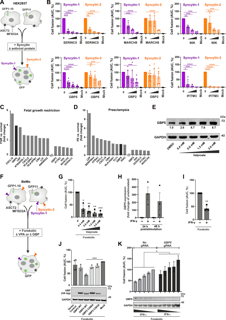
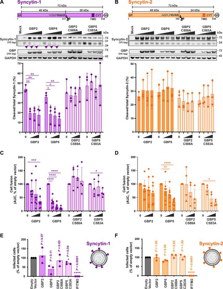
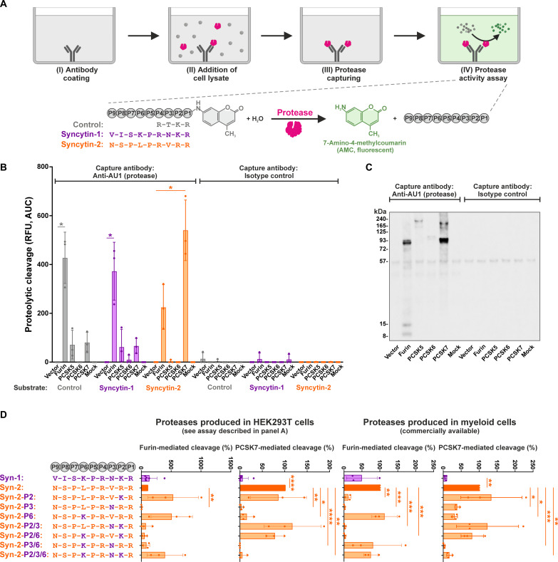
Syncytin-1 and Syncytin-2 are envelope glycoproteins encoded by human endogenous retroviruses that have been exapted for the fusion of cytotrophoblast cells into syncytiotrophoblasts during placental development. Pregnancy complications like preeclampsia are associated with altered expression of interferon-stimulated genes, including guanylate-binding protein 5 (GBP5). Here, we show that misdirected antiviral activity of GBP5 impairs processing and activation of Syncytin-1. In contrast, the proteolytic activation of Syncytin-2 is not affected by GBP5, and its fusogenic activity is only modestly reduced. Mechanistic analyses revealed that Syncytin-1 is mainly cleaved by the GBP5 target furin, whereas Syncytin-2 is also efficiently processed by the proprotein convertase subtilisin/kexin type 7 (PCSK7) and thus resistant to GBP5-mediated restriction. Mutational analyses mapped PCSK7 processing of Syncytin-2 to a leucine residue upstream of the polybasic cleavage site. In summary, we identified an innate immune mechanism that impairs the activity of a co-opted endogenous retroviral envelope protein during pregnancy and may potentially contribute to the pathogenesis of pregnancy disorders.


Figures

Fig. 1.
Fig. 1.. Antiviral proteins inhibit Syncytin-mediated cell fusion.
(A) HEK293T split-GFP assay. Two subclones of HEK293T cells express one part of GFP each. Upon Syncytin overexpression, the cells fuse and GFP is complemented. (B) HEK239T split-GFP cells were cotransfected with Syncytin and antiviral protein. An empty vector was used to adjust total DNA amounts. Fusion was monitored by quantifying GFP fluorescence for 48 hours. Means ± SD, n = 4 to 8. (C and D) Average n fold changes of the 10 most strongly up-regulated genes in placentas from patients with FGR or severe PE compared to healthy controls (n = 8 per group), and the genes analyzed in Fig. 1B were derived from (24). (E) GBP5 protein levels in BeWo cells 72 hours after valproate stimulation. (F) BeWo split-GFP reporter assay. Upon forskolin stimulation, the cells fuse and GFP is complemented. (G) BeWo cell fusion upon valproate stimulation. Means ± SD, n = 4. (H) GBP5 expression upon IFN-γ stimulation (100 ng/ml) in BeWo cells, quantified by quantitative PCR. Means ± SEM, n = 3. (I) BeWo cell fusion upon IFN-γ stimulation (100 ng/ml). Means ± SD, n = 4. (J) BeWo cell fusion after GBP2/5 overexpression. BeWo split-GFP cells were transduced with GBP2/5 and treated with forskolin, and GFP fluorescence was monitored for 72 hours. Means ± SD, n = 3 to 4. (K) Cell fusion after GBP5 depletion. BeWo split-GFP cells were transduced with CRISPR/Cas lentiviruses expressing GBP5-targeting single guide RNA (sgRNA) or a negative control. Three days later, cells were stimulated with IFN-γ, before fusion was induced by forskolin 2 days later. GFP fluorescence was monitored for 44 hours. Means ± SEM are shown on top, n = 3. One representative Western blot is shown at the bottom. A one-way analysis of variance (ANOVA) with multiple comparisons (Dunnett’s test) was performed for (G), (J), and (K). A paired t test was performed for (I) (*P < 0.05, **P < 0.01, ***P < 0.001, and ****P < 0.0001). AUC, area under the curve; DMSO, dimethyl sulfoxide; h, hours.
Fig. 2.
Fig. 2.. GBP2 and GBP5 inhibit the proteolytic activation of Syncytin-1, but not Syncytin-2.
(A and B) Syncytin cleavage in the presence of GBPs. Top illustrates the topologies of Syncytin-1 and Syncytin-2, including their polybasic cleavage sites. To monitor Syncytin cleavage, HEK293T cells were cotransfected with Syncytin expression plasmids and increasing amounts of GBP expression plasmids. Two days posttransfection, cells were harvested, and cleavage was analyzed by Western blotting. Syncytin maturation was determined by calculating the amount of cleaved to total Syncytin. Representative Western blots are shown in the middle. Purple arrowheads indicate the shift in the electrophoretic mobility of Syncytin-1. Bottom shows quantifications of three independent Western blots ± SD. SP, signal peptide; TMD, transmembrane domain; CYT, cytoplasmic tail; V5, V5 tag. (C and D) Syncytin-mediated cell fusion in the presence of GBPs. HEK239T cells stably expressing the N-terminal portion of GFP were cotransfected with expression plasmids for Syncytin-1 or Syncytin-2 and increasing amounts of plasmids expressing GBP2 (C) or GBP5 (D) and mixed with HEK293T cells expressing C-terminal part of GFP. GFP fluorescence was quantified over a period of 48 hours as a marker for syncytia formation. Mean values of four to eight independent experiments ± SD are shown. (E and F) Infectivity of Syncytin-carrying pseudovirions produced in the presence of GBPs. MFSD2A knockout HEK293T cells were cotransfected with plasmids for Syncytin-1 or Syncytin-2 together with ASLV gag-pol, mScarlet minigenome, and GBP or IFITM3. Two days later, supernatants were collected and used for the infection of HEK293T cells overexpressing MFSD2A. Three days postinfection, the percentage of infected cells was analyzed by flow cytometry. IFITM3 served as a positive control. Mean values of three independent experiments ± SD are shown. A one-way ANOVA with multiple comparisons (Dunnett’s test) was performed (*P < 0.05, **P < 0.01, ***P < 0.001, and ****P < 0.0001).
Fig. 3.
Fig. 3.. Syncytin-2 is cleaved by PCSK7.
(A) Principle of the PCSK capture assay. (I) A 96-well plate is coated with antibodies against the AU1 tag or the respective isotype control. (II) Lysates of HEK293T cells overexpressing individual, AU1-tagged PCSKs are added, and (III) proteases are captured and washed. (IV) Captured proteases are incubated with AMC reporter substrates harboring the cleavage sites of Syncytin-1, Syncytin-2, or a furin consensus cleavage site, and fluorescence signal is integrated with a plate reader. (B) PCSK-mediated cleavage of Syncytins. The ability of furin, PCSK5, PCSK6, and PCSK7 to cleave Syncytin-1 or Syncytin 2 was determined as described in (A). Mean values of three independent experiments ± SD are shown. A paired t test was performed (*P < 0.05). RFU, relative fluorescence units. (C) Western blot analysis of captured proteases. Capture efficiency of PCSKs was monitored by Western blotting. One exemplary Western blot of the experiments shown in (B) is shown. (D) Furin- and PCSK7-mediated cleavage of mutated Syncytin substrates. AMC reporter substrates harboring the cleavage sites (P1 to P9) of Syncytin-1 or Syncytin-2 are shown on the top left. The chimeras that were generated to map determinants of PCSK7 cleavage are shown below. Amino acids found in Syncytin-1 and Syncytin-2 are shown in purple and orange, respectively. The AMC reporter substrates shown on the left were either incubated with proteases captured from transfected HEK293T cells as described in Fig. 3A (middle) or commercially available proteases produced in myeloid cells (right). Substrate cleavage was monitored over time, and the area under the curve was calculated. Mean values of three to six independent experiments ± SD are shown. A one-way ANOVA with multiple comparisons (Dunnett’s test) was performed (*P < 0.05, **P < 0.01, ***P < 0.001, and ****P < 0.0001).
Fig. 4.
Fig. 4.. PCSK7 is resistant to GBP-mediated restriction.
(A) Interaction of furin or PCSK7 with GBPs. HEK293T cells were cotransfected with expression plasmids for the indicated PCSKs and GBPs or the respective vector controls. One day posttransfection, cells were harvested, and furin or PCSK7 were pulled down via their C-terminal AU1 tag. Western blotting was performed to detect PCSKs and GBPs in whole cell lysates (left) and the pull down (right). The results of one of three independent experiments are shown. IP, immunoprecipitation; #, protein size marker. (B and C) Furin- and PCSK7-mediated Syncytin cleavage in the presence of GBPs. Furin and PCSK7 were captured from cell lysates of transfected HEK293T cells essentially as described in Fig. 3A. However, cells were additionally cotransfected with expression plasmids for GBP2 or GBP5 (WT or isoprenlyation mutant) or the respective vector control. The ability of captured furin/PCSK7 to process a consensus furin cleavage site (B) or the Syncytin-1 and Syncytin-2 cleavage sites (C) were analyzed using the AMC reporter assay described in Fig. 3A. Mean values of three independent experiments ± SD are shown on top. A one-way ANOVA with multiple comparisons (Dunnett’s test) was performed (*P < 0.05, **P < 0.01, and ***P < 0.001). Capture efficiency of PCSKs was monitored by Western blotting and is shown at the bottom.

References

    1. Johnson W. E., Origins and evolutionary consequences of ancient endogenous retroviruses. Nat. Rev. Microbiol. 17, 355–370 (2019). - PubMed
    1. Imakawa K., Nakagawa S., Miyazawa T., Baton pass hypothesis: Successive incorporation of unconserved endogenous retroviral genes for placentation during mammalian evolution. Genes Cells 20, 771–788 (2015). - PubMed
    1. Blaise S., de Parseval N., Bénit L., Heidmann T., Genomewide screening for fusogenic human endogenous retrovirus envelopes identifies syncytin 2, a gene conserved on primate evolution. Proc. Natl. Acad. Sci. U.S.A. 100, 13013–13018 (2003). - PMC - PubMed
    1. Blond J. L., Lavillette D., Cheynet V., Bouton O., Oriol G., Chapel-Fernandes S., Mandrand B., Mallet F., Cosset F. L., An envelope glycoprotein of the human endogenous retrovirus HERV-W is expressed in the human placenta and fuses cells expressing the type D mammalian retrovirus receptor. J. Virol. 74, 3321–3329 (2000). - PMC - PubMed
    1. Chen C.-P., Chen L.-F., Yang S.-R., Chen C.-Y., Ko C.-C., Chang G.-D., Chen H., Functional characterization of the human placental fusogenic membrane protein syncytin 2. Biol. Reprod. 79, 815–823 (2008). - PubMed

LinkOut - more resources