

ACL rupture from medial meniscus tear and high tibial slope: Case report
- PMID: 40334444
- PMCID: PMC12138557
- DOI: 10.1016/j.ijscr.2025.111352
ACL rupture from medial meniscus tear and high tibial slope: Case report
Abstract
Introduction and importance: Tears in the posterior medial meniscus leading to anterior cruciate ligament (ACL) rupture are rare. While the ACL is the primary structure responsible for anterior knee stability, its function can also be influenced by the medial meniscus and the posterior tibial slope.
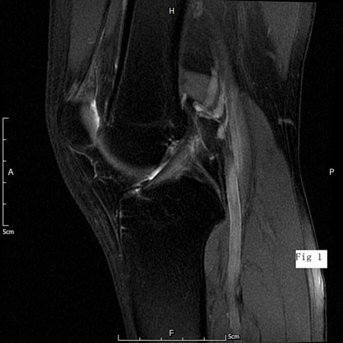
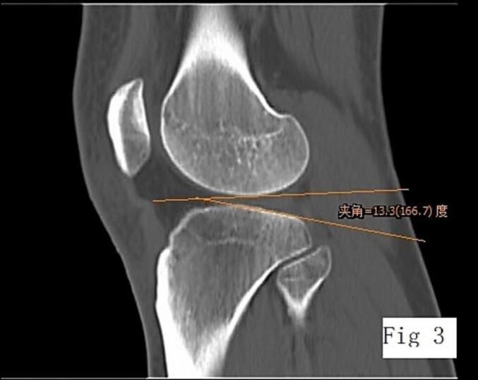
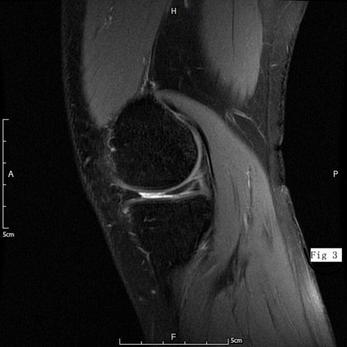
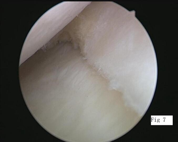
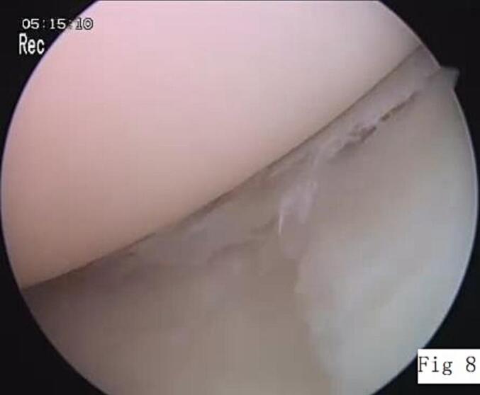
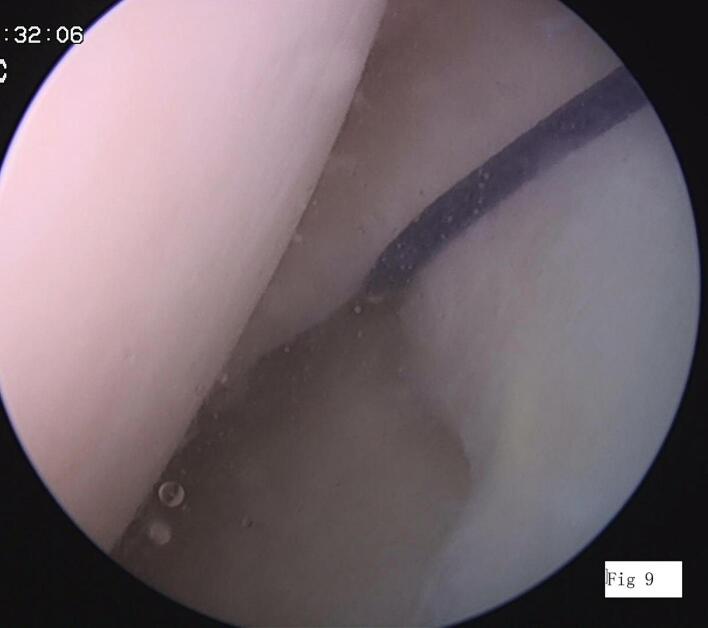
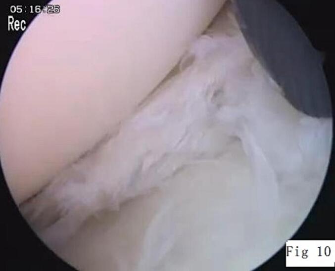
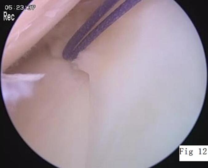
Case presentation: We report a case of a patient with a rupture of the posterior medial meniscus accompanied by a significantly elevated posterior tibial slope. Despite undergoing two arthroscopic meniscectomies, the patient failed to recover. Four years postoperatively, the anterior cruciate ligament degenerated and ruptured.
Clinical discussion: We hypothesize that damage to the posterior medial meniscus may increase stress on the anterior cruciate ligament, particularly in the presence of a high posterior tibial slope.
Conclusion: Early surgical repair improved outcomes in medial meniscal posterior horn tears. Tibial slope correction reduced revision rates in patients with >10°tibial slope undergoing ACL reconstruction.
Keywords: Anterior cruciate ligament; Arthroscopy; Case report; Medial meniscus; Posterior tibial slope.
Copyright © 2025 The Authors. Published by Elsevier Ltd.. All rights reserved.
Conflict of interest statement
Conflict of interest statement All authors had no financial and personal relationships with other people or organisations that could inappropriately influence (bias) their work. (Include employment, consultancies, stock ownership, honoraria, paid expert testimony, patent applications/registrations, and grants or other funding.)
Figures


References
-
- Van de Velde S.K., Telfer S., van Arkel E.R.A., Schmale G.A. A lateral extra-articular tenodesis without additional hardware: surgical technique and biomechanical comparison with an anatomic anterolateral ligament reconstruction in the augmentation of anterior cruciate ligament reconstruction. Knee. 2024;47:112–120. - PubMed
-
- Ekinci M., Demir T.B., Sahinkaya T., Yakal S., Polat G., Bayraktar B. The effect of gracilis tendon preservation on postoperative knee joint stability and muscle strength in arthroscopic anterior cruciate ligament reconstruction surgery. J. Knee Surg. 2024;37(12):843–850. - PubMed
-
- Kawashima I., Tsukahara T., Sakai T., et al. Delayed anterior cruciate ligament reconstruction increases the incidence of medial meniscal bucket handle tears and medial compartment chondral injuries in patients aged 40 years and older. Arch. Orthop. Trauma Surg. 2021;141(6):971–975. - PubMed
-
- Okazaki Y., Furumatsu T., Kodama Y., et al. Steep posterior slope and shallow concave shape of the medial tibial plateau are risk factors for medial meniscus posterior root tears. Knee Surg. Sports Traumatol. Arthrosc. 2021;29(1):44–50. - PubMed
Publication types
LinkOut - more resources
Full Text Sources
