Review
. 2025 Sep 5;72(9):967-978.
doi: 10.1507/endocrj.EJ25-0051. Epub 2025 May 3.

Biased antibodies and beyond: a new era in the diagnosis of PTH-dependent hypercalcemia

Affiliations
Review

Biased antibodies and beyond: a new era in the diagnosis of PTH-dependent hypercalcemia

Noriko Makita et al. Endocr J. .

Abstract

Hypercalcemia, a common electrolyte imbalance, requires accurate differential diagnosis to guide appropriate management. PTH-dependent hypercalcemia, predominantly caused by primary hyperparathyroidism (PHPT) and rarely by familial hypocalciuric hypercalcemia (FHH)-mainly due to heterozygous loss-of-function mutations in the CASR gene encoding the calcium-sensing receptor (CaSR)-now includes acquired hypocalciuric hypercalcemia (AHH) as an emerging disease entity. Initially identified as analogous to FHH, AHH was characterized by blocking antibodies targeting the CaSR. However, our research has identified unique autoantibodies, termed biased antibodies, that paradoxically regulate signaling by enhancing Gq activity while suppressing Gi activity. Investigating their mechanisms has not only provided insights into specific treatments for AHH but also suggested novel activation mechanisms and binding sites of the CaSR, offering a fresh perspective on the regulation of PTH secretion. In clinical practice, recognizing AHH is crucial. A key diagnostic feature is fluctuating serum calcium levels, making a wait-and-see approach viable for mild hypercalcemia. Conversely, hypercalcemic crises necessitate immediate diagnostic and therapeutic interventions. The most important diagnostic clue to differentiate AHH from PHPT is hypermagnesemia. Additionally, AHH is less likely to involve AVP resistance (i.e., nephrogenic diabetes insipidus) and acute kidney injury (AKI), owing to preserved medullary hyperosmolarity and minimal interference with AVP signaling. Finally, a relatively low PTH level serves as another distinguishing feature. Based on these observations, we propose a novel diagnostic guide for PTH-dependent hypercalcemia. We anticipate that this guide will help identify previously undiagnosed AHH cases in routine practice, enabling timely and effective management of this rare condition.

Keywords: Acquired hypocalciuric hypercalcemia (AHH); Biased antibodies; Calcimimetics; Calcium-sensing receptor (CaSR); PTH-dependent hypercalcemia.


Conflict of interest statement

This study did not receive any specific grant from funding agencies in the public, commercial, or not-for-profit sectors.

Noriko Makita is a member of Endocrine Journal’s Editorial Board.

Figures

Fig. 1
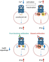
Fig. 1. AHH caused by pure blocking antibodies or biased antibodies
(Top) Inhibition of CaSR signaling enhances PTH secretion, while activation of CaSR signaling suppresses PTH secretion. (Bottom) Pure blocking antibodies inhibit both CaSR signals, namely Gq- and Gi-dependent signaling, whereas biased antibodies detected in AHH patients regulate these two signals in opposite directions.
Fig. 2
Fig. 2. Characteristics of hypercalcemia in AHH, compared to FHH and neonatal severe hyperparathyroidism (NSHPT)
The clinical presentation of AHH forms a continuum, ranging from mild hypercalcemia resembling FHH to severe hypercalcemia resembling NSHPT, depending on the antibody titer. FHH: familial hypocalciuric hypercalcemia NSHPT: neonatal severe hyperparathyroidism
Fig. 3
Fig. 3. Electrolyte movement in the thick ascending limb of Henle’s loop in hypercalcemia, FHH, and AHH
a) When serum calcium levels are elevated, the CaSR expressed on the basolateral side of the thick ascending limb is activated. This activation inhibits ROMK activity, reducing the positive charge in the tubular lumen, diminishing the reabsorption of Ca2+ and Mg2+, and increasing urinary calcium excretion. b) In FHH or AHH, CaSR signaling is suppressed or altered, relieving the inhibition of ROMK. This leads to increased potassium recycling, enhancing paracellular reabsorption of Ca2+ and Mg2+ in the thick ascending limb of Henle’s loop. Consequently, urinary calcium excretion is reduced, resulting in hypocalciuria and hypermagnesemia. ROMK: renal outer medullary potassium channel
Fig. 4
Fig. 4. Mechanism of AVP resistance (nephrogenic diabetes insipidus) in hypercalcemia
There are two mechanisms: a) Impairment of medullary hyperosmolarity formation via CaSR activation on the basolateral side of the thick ascending limb of Henle’s loop: In hypercalcemia, CaSR activation on the basolateral side of the thick ascending limb of Henle’s loop inhibits the activity of ROMK. This inhibition reduces the upstream reabsorption of Na+, K+, and Cl via the NKCC2. As a result, sodium and chloride reabsorption into the vasculature through the Na+-K+-ATPase and Cl channel is also diminished, impairing the formation of medullary hyperosmolarity, which is essential for AVP action. This disruption ultimately leads to a reduced urine concentration capacity. b) Suppression of AVP-V2 receptor (V2R) signaling via CaSR activation on the apical side of the collecting duct: Hypercalcemia typically induces hypercalciuria, which activates the CaSR expressed on the apical side of the collecting duct. This activation, likely mediated through Gi/o signaling, disrupts the AVP-V2R-Gs-cAMP pathway, reducing AVP action and further impairing the reabsorption of free water. ROMK: renal outer medullary potassium channel NKCC2: Na+/K+/2Cl cotransporter type 2
Fig. 5
Fig. 5. A vicious cycle of hypercalcemia and acute kidney injury (AKI)
Hypercalcemia leads to anorexia and AVP resistance, resulting in dehydration. Dehydration causes a pre-renal decline in GFR, which is further exacerbated by hypercalcemia-induced vasoconstriction of the afferent arterioles, leading to pre-renal AKI. The resulting GFR reduction decreases calcium filtration by the kidneys, reducing renal calcium excretion and worsening hypercalcemia. Additionally, dehydration enhances sodium reabsorption in the proximal tubules, which in turn increases passive calcium reabsorption, further exacerbating hypercalcemia.
Fig. 6
Fig. 6. A Proposed new diagnostic guide for PTH-dependent hypercalcemia with examples of long-term calcium trends in untreated PHPT and AHH cases
a) When encountering PTH-dependent hypercalcemia, evaluate the CCCR using a 24-hour urine collection. In FHH/AHH, CCCR is typically <0.01, but values between 0.01 and 0.02 cannot rule out these conditions. On the contrary, in mild PHPT, CCCR is sometimes <0.01. Assess family history, past calcium levels, and imaging findings for further clarification. In cases of mild hypercalcemia, observe for a while: if calcium levels remain stable, suspect FHH or PHPT; if they fluctuate, suspect AHH. In cases of moderate to severe hypercalcemia, it is recommended to pay attention to magnesium and creatinine levels. If there is hypomagnesemia and worsening kidney function with elevated PTH levels, PHPT should be suspected. Conversely, if there is hypermagnesemia and preserved kidney function with relatively low PTH levels, AHH should be suspected. CCCR: calcium-to-creatinine clearance ratio CCCR is calculated as (urinary calcium (mg/dL) × serum creatinine (mg/dL))/(urinary creatinine (mg/dL) × serum calcium (mg/dL)). b) Calcium-trends are shown for more than 10 years in two untreated cases of PHPT and one untreated case of AHH.
Graphical Abstract
Graphical Abstract

References

    1. Renaghan AD, Rosner MH (2018) Hypercalcemia: etiology and management. Nephrol Dial Transplant 33: 549–551.
    1. Walker MD, Shane E (2022) Hypercalcemia: A Review. JAMA 328: 1624–1636. - PubMed
    1. Tonon CR, Silva T, Pereira FWL, Queiroz DAR, Junior ELF, et al. (2022) A review of current clinical concepts in the pathophysiology, etiology, diagnosis, and management of hypercalcemia. Med Sci Monit 28: e935821. - PMC - PubMed
    1. Potts JT, Jüppner H (2022) Disorders of the parathyroid gland and calcium homeostasis. In: Loscalzo J, Fauci A, Kasper D, Hauser S, Longo D, et al. (eds) Harrison’s principles of internal medicine (21st) McGraw-Hill Education, New York, USA: 3169–3190.
    1. Motlaghzadeh Y, Bilezikian JP, Sellmeyer DE (2021) Rare causes of hypercalcemia: 2021 update. J Clin Endocrinol Metab 106: 3113–3128. - PubMed

MeSH terms