. 2025 May 8;13(1):50.
doi: 10.1038/s41413-025-00422-3.

Sirt6 deficiency promotes senescence and age-associated intervertebral disc degeneration in mice

Affiliations

Sirt6 deficiency promotes senescence and age-associated intervertebral disc degeneration in mice

Pranay Ramteke et al. Bone Res. .

Abstract

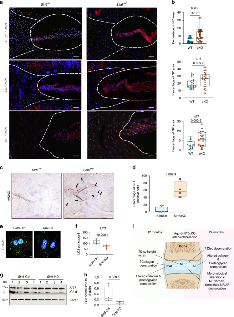
Intervertebral disc degeneration is a major risk factor contributing to chronic low back and neck pain. While the etiological factors for disc degeneration vary, age is still one of the most important risk factors. Recent studies have shown the promising role of SIRT6 in mammalian aging and skeletal tissue health, however its role in the intervertebral disc health remains unexplored. We investigated the contribution of SIRT6 to disc health by studying the age-dependent spinal phenotype of mice with conditional deletion of Sirt6 in the disc (AcanCreERT2; Sirt6fl/fl). Histological studies showed a degenerative phenotype in knockout mice compared to Sirt6fl/fl control mice at 12 months, which became pronounced at 24 months. RNA-Seq analysis of NP and AF tissues, in vitro quantitative histone analysis, and RNA-seq with ATAC-seq multiomic studies revealed that SIRT6-loss resulted in changes in acetylation and methylation status of specific Histone 3 lysine residues and affected DNA accessibility and transcriptomic landscape. A decrease in autophagy and an increase in DNA damage were also noted in Sirt6-deficient cells. Further mechanistic insights revealed that loss of SIRT6 increased senescence and SASP burden in the disc characterized by increased p21, p19, γH2AX, IL-6, IL-1β, and TGF-β abundance. Taken together, our study highlights the contribution of SIRT6 in modulating DNA damage, autophagy, and cell senescence and its importance in maintaining disc health during aging, thereby underscoring it as a potential therapeutic target to treat intervertebral disc degeneration.


Conflict of interest statement

Competing interests: The authors declare no competing interests. Ethics: All animal experiments were performed under IACUC protocols approved by the University of North Carolina at Chapel Hill and Thomas Jefferson University.

Figures

Fig. 1
Fig. 1
Conditional deletion of SIRT6 in intervertebral disc accelerates age-associated degeneration. a Schematic showing Sirt6 floxed allele which following Cre-mediate recombination generates a functionally null mutant allele. b Experimental design showing the timeline of tamoxifen injection and analysis of control (Sirt6fl/fl) and sirt6 loss (Sirt6AcanCreERT2/Sirt6cKO) mouse cohorts. c Immunofluorescence staining for H3K9ac shows a robust increase in NP, AF and EP compartment of the lumbar disc confirming the deletion of SIRT6. d Safranin-O/Fast Green staining of Sirt6fl/fl and Sirt6cKO lumbar discs at 12- and 24 months. Scale bar 1A: row 1 = 200 μm; row 2 = 50 μm. e, f Distribution of and (e’, f’) average modified thompson’s grades of lumbar discs of Sirt6fl/fl and Sirt6cKO mice analyzed at 12 and 24 months. 12 months: n = 4–6 mice/group, 3–4 discs/animal. 24 months: n = 3–6 animals/mice, 3–4 discs/animal. g-g”’ Level by level average modified thompson grading scores for NP and AF compartments of lumbar discs analyzed from 12 and 24 months Sirt6fl/fl and Sirt6cKO mice. h μCT analysis showing (i-i”) disc height (DH), vertebral height (VB) and disc height index (DHI) measured at 12 months and 24 months. j TUNEL staining images and (k-k”) TUNEL quantitation. Statistical difference between grade distributions (e, f) was tested using chi-square test, all other quantitative data was compared using unpaired t-test, *P < 0.05
Fig. 2
Fig. 2
SIRT6 deletion dysregulates disc matrix homeostasis. a Representative immunofluorescence images of lumbar disc sections and (b) respective quantitation for FCHP, COL1, COLX, aggrecan (ACAN), chondroitin sulfate (CS) in 12 months and 24 months old Sirt6fl/fl and Sirt6cKO mice, scale bar = 50 μmol/L. n = 4–6 mice/genotype and 3-4 discs/animal. White dotted lines demarcate disc compartments. Significance was determined using unpaired t-test
Fig. 3
Fig. 3
SIRT6 loss causes changes in transcriptomic landscape of NP tissues. Microarray analysis of NP tissue transcripts from Sirt6fl/fl and Sirt6cKO represented as (a) Three-dimensional Principal component analysis (PCA) showing discrete clustering based on genotype (n = 4 mice/genotype, 5-6 pooled discs/animal). b Heat map and hierarchical clustering of Z-score of differentially expressed genes (DEGs) between Sirt6fl/fl and Sirt6cKO (P ≤ 0.05, FC ≥ 1.75). c Volcano plot of DEGs in the NP showing P-value versus magnitude of change (fold change). d CompBIO analysis of Upregulated DEGs in NP tissue of 24 months Sirt6cKO represented in a ball and stick model. The enrichment of themes is shown by the size of the ball and connectedness is shown based on thickness of the lines between them. Themes of interest are colored, and superclusters comprised of related themes are highlighted. e Top thematic DEGs plotted based on CompBio entity enrichment score
Fig. 4
Fig. 4
Loss of SIRT6 causes changes in histone modifications and alters chromatin accessibility. a Schematic of experimental design. b Immunoblotting analysis of SIRT6 and TXNIP and (c) densitometric quantitation normalized to β-actin of Sirt6-Ctrl and Sirt6-KD NP cells. (n = 4 independent cell isolations). d Quantitative ELISA for H3 lysine modifications from Sirt6-Ctrl and Sirt6-KD NP cells (n = 3 independent cell isolations). Statistical significance was tested by unpaired t-test
Fig. 5
Fig. 5
Sirt6 knockdown in NP cells causes transcriptomic changes in senescence and ECM related pathways. RNA-Sequencing of Sirt6-Ctrl and Sirt6-KD represented as (a) three-dimensional PCA showing discrete clustering of samples based on the genotypes (n = 4 independent samples/ group). b Volcano plot of DEGs from Sirt6-KD vs Sirt6-Ctrl. c CompBio analysis for upregulated DEGs (FDR < 0.05, FC > 1.5) represented as a ball and stick model. Themes of interest are colored, and superclusters comprised of related themes are highlighted. d Top thematic DEGs plotted based on CompBio entity enrichment score
Fig. 6
Fig. 6
Loss of SIRT6 results in changes in chromatin accessibility. a Heatmap and (b) Quadrant map of commonly upregulated and downregulated DEGs (FDR < 0.05, FC > 1.5) between ATAC-seq and RNA-seq mapped to one of the thematic superclusters in RNA-Seq data. c Gene tracks showing the enriched peaks for a select group of genes from ATAC-seq experiment. (n = 4 independent samples/group)
Fig. 7
Fig. 7
SIRT6 deletion increases DNA damage, senescence and SASP burden in Sirt6cKO discs and modulates NP cell autophagy. a Representative immunofluorescence images and (b) quantitative analysis of SASP markers IL-6, TGF-β, p21. n = 6 animals/genotype, 3 discs/mouse. Scale bar = 50 μmol/L. c Representative immunostaining images and (d) quantitation of yH2AX in Sirt6cKO and Sirt6fl/fl intervertebral discs, n = 3 animals/genotype, 1-2 discs/mouse. e Immunofluorescence analysis and (f) quantitation of LC3 puncta in Sirt6-KD and Sirt6-Ctrl cells cultured in hypoxia. Significance was tested with unpaired t-test (g) immunoblot of LC3 and (h) densitometric quantification showing LC3II/actin from Sirt6-KD and Sirt6-Ctrl cells under hypoxia. i Schematic showing age dependent consequences of SIRT6 loss in the spine

Update of

References

    1. Ferreira, M. L. et al. Global, regional, and national burden of low back pain, 1990–2020, its attributable risk factors, and projections to 2050: a systematic analysis of the Global Burden of Disease Study 2021. Lancet Rheumatol.5, e316–e329 (2023). - PMC - PubMed
    1. Kalichman, L. & Hunter, D. J. Diagnosis and conservative management of degenerative lumbar spondylolisthesis. Eur. Spine J.17, 327–335 (2008). - PMC - PubMed
    1. Farsetti, A., Illi, B. & Gaetano, C. How epigenetics impacts on human diseases. Eur. J. Intern. Med.114, 15–22 (2023). - PubMed
    1. Levine, D. C. et al. NADH inhibition of SIRT1 links energy state to transcription during time-restricted feeding. Nat. Metab.3, 1621–1632 (2021). - PMC - PubMed
    1. Kane, A. E. & Sinclair, D. A. Sirtuins and NAD+ in the development and treatment of metabolic and cardiovascular diseases. Circ. Res.123, 868–885 (2018). - PMC - PubMed